Tag Archives: Institute for Basic Science (IBS)

Striking similarity between memory processing of artificial intelligence (AI) models and hippocampus of the human brain

A December 18, 2023 news item on ScienceDaily shifted my focus from hardware to software when considering memory in brainlike (neuromorphic) computing,

An interdisciplinary team consisting of researchers from the Center for Cognition and Sociality and the Data Science Group within the Institute for Basic Science (IBS) [Korea] revealed a striking similarity between the memory processing of artificial intelligence (AI) models and the hippocampus of the human brain. This new finding provides a novel perspective on memory consolidation, which is a process that transforms short-term memories into long-term ones, in AI systems.

A November 28 (?), 2023 IBS press release (also on EurekAlert but published December 18, 2023, which originated the news item, describes how the team went about its research,

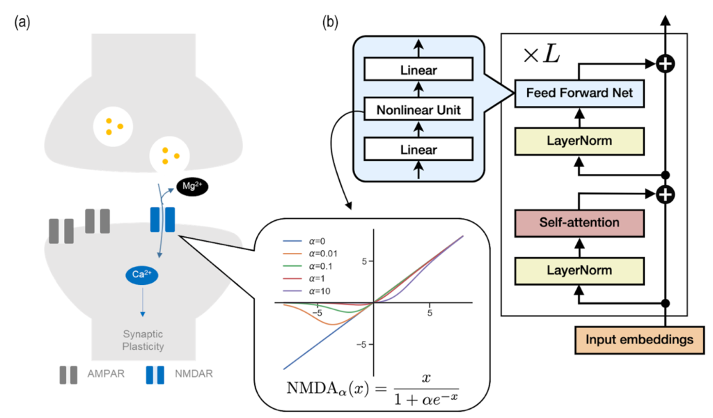
In the race towards developing Artificial General Intelligence (AGI), with influential entities like OpenAI and Google DeepMind leading the way, understanding and replicating human-like intelligence has become an important research interest. Central to these technological advancements is the Transformer model [Figure 1], whose fundamental principles are now being explored in new depth.

The key to powerful AI systems is grasping how they learn and remember information. The team applied principles of human brain learning, specifically concentrating on memory consolidation through the NMDA receptor in the hippocampus, to AI models.

The NMDA receptor is like a smart door in your brain that facilitates learning and memory formation. When a brain chemical called glutamate is present, the nerve cell undergoes excitation. On the other hand, a magnesium ion acts as a small gatekeeper blocking the door. Only when this ionic gatekeeper steps aside, substances are allowed to flow into the cell. This is the process that allows the brain to create and keep memories, and the gatekeeper’s (the magnesium ion) role in the whole process is quite specific.

The team made a fascinating discovery: the Transformer model seems to use a gatekeeping process similar to the brain’s NMDA receptor [see Figure 1]. This revelation led the researchers to investigate if the Transformer’s memory consolidation can be controlled by a mechanism similar to the NMDA receptor’s gating process.

In the animal brain, a low magnesium level is known to weaken memory function. The researchers found that long-term memory in Transformer can be improved by mimicking the NMDA receptor. Just like in the brain, where changing magnesium levels affect memory strength, tweaking the Transformer’s parameters to reflect the gating action of the NMDA receptor led to enhanced memory in the AI model. This breakthrough finding suggests that how AI models learn can be explained with established knowledge in neuroscience.

C. Justin LEE, who is a neuroscientist director at the institute, said, “This research makes a crucial step in advancing AI and neuroscience. It allows us to delve deeper into the brain’s operating principles and develop more advanced AI systems based on these insights.”

CHA Meeyoung, who is a data scientist in the team and at KAIST [Korea Advanced Institute of Science and Technology], notes, “The human brain is remarkable in how it operates with minimal energy, unlike the large AI models that need immense resources. Our work opens up new possibilities for low-cost, high-performance AI systems that learn and remember information like humans.”

What sets this study apart is its initiative to incorporate brain-inspired nonlinearity into an AI construct, signifying a significant advancement in simulating human-like memory consolidation. The convergence of human cognitive mechanisms and AI design not only holds promise for creating low-cost, high-performance AI systems but also provides valuable insights into the workings of the brain through AI models.

Fig. 1: (a) Diagram illustrating the ion channel activity in post-synaptic neurons. AMPA receptors are involved in the activation of post-synaptic neurons, while NMDA receptors are blocked by magnesium ions (Mg²⁺) but induce synaptic plasticity through the influx of calcium ions (Ca²⁺) when the post-synaptic neuron is sufficiently activated. (b) Flow diagram representing the computational process within the Transformer AI model. Information is processed sequentially through stages such as feed-forward layers, layer normalization, and self-attention layers. The graph depicting the current-voltage relationship of the NMDA receptors is very similar to the nonlinearity of the feed-forward layer. The input-output graph, based on the concentration of magnesium (α), shows the changes in the nonlinearity of the NMDA receptors. Courtesy: IBS

This research was presented at the 37th Conference on Neural Information Processing Systems (NeurIPS 2023) before being published in the proceedings, I found a PDF of the presentation and an early online copy of the paper before locating the paper in the published proceedings.

PDF of presentation: Transformer as a hippocampal memory consolidation model based on NMDAR-inspired nonlinearity

PDF copy of paper:

Transformer as a hippocampal memory consolidation model based on NMDAR-inspired nonlinearity by Dong-Kyum Kim, Jea Kwon, Meeyoung Cha, C. Justin Lee.

This paper was made available on OpenReview.net:

OpenReview is a platform for open peer review, open publishing, open access, open discussion, open recommendations, open directory, open API and open source.

It’s not clear to me if this paper is finalized or not and I don’t know if its presence on OpenReview constitutes publication.

Finally, the paper published in the proceedings,

Transformer as a hippocampal memory consolidation model based on NMDAR-inspired nonlinearity by Dong Kyum Kim, Jea Kwon, Meeyoung Cha, C. Justin Lee. Part of Advances in Neural Information Processing Systems 36 (NeurIPS 2023) Main Conference Track

This link will take you to the abstract, access the paper by clicking on the Paper tab.

Nanoparticle treatment for rheumatoid arthritis

An October 26, 2023 news item on phys.org announced a possible new treatment for rheumatoid arthritis (RA), Note: Links have been removed,

A team of scientists [Korea] has developed a new solution for the treatment of rheumatoid arthritis (RA). The work has been published in Nature Nanotechnology.

RA is a chronic disease that, unfortunately, has no cure. The disease triggers a mix of troublesome symptoms like inflamed joints, harmful cytokines, and immune system imbalances, which work together to create a relentless cycle of worsening symptoms. While targeting some of these factors can provide short-term relief, others remain unresolved, leading to a frustrating cycle of remission and flare-ups.

One of the major hurdles in RA treatment is the inability to restore the immune system to its healthy state. This leaves the body unable to control the continuous production of harmful substances like reactive oxygen species (ROS) and inflammatory cytokines, leading to persistent inflammation and discomfort.

In essence, the ideal treatment for RA should not only provide immediate relief from inflammation and symptoms but also address the root cause by restoring the immune system to its normal, balanced state.

New nanoparticle-based system as a solution

The new platform involves immobilizing ceria nanoparticles (Ce NPs) onto mesenchymal stem cell-derived nanovesicles (MSCNVs). Both of these components can hinder different pathogenic factors, allowing them to work both individually and cooperatively to achieve a comprehensive treatment.

Caption: Schematic illustration of comprehensive and combination RA therapy by Ce-MSCNV nanoparticles. Ce-MSCNVs scavenge the over-produced ROS in an RA knee joint, induce M1 to M2 macrophage polarization for immediate relief of inflammation and symptoms, modulate DCs into tDCs, and finally induce Tregs. Credit: Institute for Basic Science

An October 27, 2023 Institute for Basic Science (IBS) press release (also on EurekAlert but published October 26, 2023), which originated the news item, provides more details about the proposed treatment,

Ce NPs – can scavenge the overproduced ROS in RA-inflicted knee joints. They also induce polarization of M1 macrophages into M2, achieving immediate relief of inflammation and symptoms.

MSCNVs – deliver immunomodulatory cytokines, which turn dendritic cells (DC) into tolerogenic dendritic cells (tDCs). This consequently generates regulatory T cells for long-term immune tolerance.

In short, this approach aims to bridge both innate and adaptive immunity to achieve both short-term pain relief, as well as convert the tissue environment into an immune-tolerant state to prevent the recurrence of symptoms.

Researchers confirmed the efficacy of this approach using a collagen-induced arthritis mouse model. The Ce-MSCNV system was able to comprehensively treat and prevent RA by simultaneously relieving the immediate and restoring T cell immunity. Supporting data suggest that improvement in conditions can be achieved after only a single-dose treatment.

The mice treated with the Ce-MSCNV combination fared far better compared to the ones only treated using the Ce NP or MSCNV group. This clearly demonstrates the synergy between anti-inflammation and immunomodulation and underlines the importance of the combined therapy for effective RA treatment. In addition, Ce-MSCNV administration prior to booster injection markedly reduced the incidence and severity of symptoms, supporting the prophylactic potential of these nanoparticles.

First author KOO Sagang stated, “One of the hardest decisions in intractable disease therapy is determining how long the treatment should be carried on. For RA, it would not be appropriate to stop treatment just because the target marker is stabilized. A safer indicator should be that the innate and adaptive components of the collapsed immune system are normalized to protect the body.”

Koo believes that the strategy adopted by Ce-MSCNVs, where different treatment mechanisms work together, provides a unique advantage in this regard. Furthermore, she predicts that a similar approach would also be applicable to other intractable, inflammatory, and autoimmune diseases for this purpose. The components within the system may also be modified. For example, other catalysts for generating ROS or other cell-derived nanovesicles could be utilized depending on the types of diseases. Overall, this study proves the potential of a hybrid nanoparticle system for the comprehensive treatment of autoimmune disease and modulation of the immune system.

Here’s a link to and a citation for the paper,

Ceria-vesicle nanohybrid therapeutic for modulation of innate and adaptive immunity in a collagen-induced arthritis model by Sagang Koo, Hee Su Sohn, Tae Hee Kim, Siyeon Yang, Se Youn Jang, Seongryeol Ye, Boomin Choi, Soo Hyeon Kim, Kyoung Sun Park, Hyun Mu Shin, Ok Kyu Park, Cheesue Kim, Mikyung Kang, Min Soh, Jin Yoo, Dokyoon Kim, Nohyun Lee, Byung-Soo Kim, Youngmee Jung & Taeghwan Hyeon. Nature Nanotechnology (2023) DOI: https://doi.org/10.1038/s41565-023-01523-y Published: 26 October 2023

This paper is behind a paywall.

World’s smallest magnetic resonance imaging (MRI) of a single atom

While not science’s sleekest machine, this microscope was able to capture M.R.I. scans of single atoms. Credit: IBM Research

Such a messy looking thing—it makes me feel better about my housekeeping. In any event, it’s fascinating to think this scanning tunneling microscope as seen in the above can actually act as an MRI device and create an image of a single atom.

There’s a wonderful article in the New York Times about the work but I’m starting first with a July 1, 2019 news item on Nanowerk,

Researchers at the Center for Quantum Nanoscience (QNS) within the Institute for Basic Science (IBS) at Ewha Womans University [Seoul, South Korea) have made a major scientific breakthrough by performing the world’s smallest magnetic resonance imaging (MRI). In an international collaboration with colleagues from the US, QNS scientists used their new technique to visualize the magnetic field of single atoms.

A July 2, 2019 IBS news release (also on EurekAlert but published July 1, 2019), which originated the news item, provides some insight into the research,

An MRI is routinely done in hospitals nowadays as a part of imaging for diagnostics. MRI’s detect the density of spins – the fundamental magnets in electrons and protons – in the human body. Traditionally, billions and billions of spins are required for an MRI scan. The new findings, published today [July 1, 2019] in the journal Nature Physics, show that this process is now also possible for an individual atom on a surface. To do this, the team used a Scanning Tunneling Microscope, which consists of an atomically sharp metal tip that allows researchers to image and probe single atoms by scanning the tip across the surface.

The two elements that were investigated in this work, iron and titanium, are both magnetic. Through precise preparation of the sample, the atoms were readily visible in the microscope. The researchers then used the microscope’s tip like an MRI machine to map the three-dimensional magnetic field created by the atoms with unprecedented resolution. In order to do so, they attached another spin cluster to the sharp metal tip of their microscope. Similar to everyday magnets, the two spins would attract or repel each other depending on their relative position. By sweeping the tip spin cluster over the atom on the surface, the researchers were able to map out the magnetic interaction. Lead author, Dr. Philip Willke of QNS says: “It turns out that the magnetic interaction we measured depends on the properties of both spins, the one on the tip and the one on the sample. For example, the signal that we see for iron atoms is vastly different from that for titanium atoms. This allows us to distinguish different kinds of atoms by their magnetic field signature and makes our technique very powerful.”

The researchers plan to use their single-atom MRI to map the spin distribution in more complex structures such as molecules and magnetic materials. “Many magnetic phenomena take place on the nanoscale, including the recent generation of magnetic storage devices.” says Dr. Yujeong Bae also of QNS, a co-author in this study. “We now plan to study a variety of systems using our microscopic MRI.” The ability to analyze the magnetic structure on the nanoscale can help to develop new materials and drugs. Moreover, the research team wants to use this kind of MRI to characterize and control quantum systems. These are of great interest for future computation schemes, also known as quantum computing

“I am very excited about these results. It is certainly a milestone in our field and has very promising implications for future research.” says Prof. Andreas Heinrich, Director of QNS. “The ability to map spins and their magnetic field with previously unimaginable precision, allows us to gain deeper knowledge about the structure of matter and opens new fields of basic research.”

The Center for Quantum Nanoscience, on the campus of Ewha Womans University in Seoul, South Korea, is a world-leading research center merging quantum and nanoscience to engineer the quantum future through basic research. Backed by Korea’s Institute for Basic Science, which was founded in 2011, the Center for Quantum Nanoscience draws on decades of QNS Director Andreas J. Heinrich’s (A Boy and His Atom, IBM, 2013) scientific leadership to lay the foundation for future technology by exploring the use of quantum behavior atom-by-atom on surfaces with highest precision.

You may have noticed that other than a brief mention in the first paragraph (in the Nanowerk news item excerpt), there’s no mention of the US researchers and their contribution to the work.

Interestingly, the July 1, 2019 New York Time article by Knvul Sheikh returns the favour by focusing almost entirely on US researchers while giving the Korean researchers a passing mention (Note: Links have been removed),

Different microscopy techniques allow scientists to see the nucleotide-by-nucleotide genetic sequences in cells down to the resolution of a couple atoms as seen in an atomic force microscopy image. But scientists at the IBM Almaden Research Center in San Jose, Calif., and the Institute for Basic Sciences in Seoul, have taken imaging a step further, developing a new magnetic resonance imaging technique that provides unprecedented detail, right down to the individual atoms of a sample.

When doctors want to detect tumors, measure brain function or visualize the structure of joints, they employ huge M.R.I. machines, which apply a magnetic field across the human body. This temporarily disrupts the protons spinning in the nucleus of every atom in every cell. A subsequent, brief pulse of radio-frequency energy causes the protons to spin perpendicular to the pulse. Afterward, the protons return to their normal state, releasing energy that can be measured by sensors and made into an image.

But to gather enough diagnostic data, traditional hospital M.R.I.s must scan billions and billions of protons in a person’s body, said Christopher Lutz, a physicist at IBM. So he and his colleagues decided to pack the power of an M.R.I. machine into the tip of another specialized instrument known as a scanning tunneling microscope to see if they could image individual atoms.

The tip of a scanning tunneling microscope is just a few atoms wide. And it moves along the surface of a sample, it picks up details about the size and conformation of molecules.

The researchers attached magnetized iron atoms to the tip, effectively combining scanning-tunneling microscope and M.R.I. technologies.

When the magnetized tip swept over a metal wafer of iron and titanium, it applied a magnetic field to the sample, disrupting the electrons (rather than the protons, as a typical M.R.I. would) within each atom. Then the researchers quickly turned a radio-frequency pulse on and off, so that the electrons would emit energy that could be visualized. …

Here’s a link to and a citation for the paper,

Magnetic resonance imaging of single atoms on a surface by Philip Willke, Kai Yang, Yujeong Bae, Andreas J. Heinrich & Christopher P. Lutz. Nature Physics (2019) DOI: https://doi.org/10.1038/s41567-019-0573-x Published 01 July 2019

This paper is behind a paywall.

A pumpkin-shaped molecule for the first real-time methamphetamine and amphetamine sensor

A Sept. 28,2017 news item on Nanowerk announces a portable, inexpensive sensor for drugs (Note: A link has been renewed),

Speed, uppers, chalk, glass, crystal, or whatever you prefer to call them, can be instantly detected from biological fluids with a new portable kit that costs as little as $50. Scientists at the Center for Self-Assembly and Complexity, within the Institute for Basic Science (IBS, South Korea), in collaboration with Pohang University of Science and Technology (POSTECH), have devised the first methamphetamine and amphetamine sensor that can detect minute concentrations of these drugs from a single drop of urine in real-time.

Published in the journal Chem (“Point-of-Use Detection of Amphetamine-Type Stimulants with Host-Molecule-Functionalized Organic Transistors”), this simple and flexible sensor, which can be attached to a wristband and connected to an Android app via Bluetooth, could move drug screening from the labs to the streets.

A Sept. 28 (?), 2017 IBS press release by Letizia Diamante (also on EurekAlert), which originated the news item, expands on the theme,

Easy to synthesize and cheaper than heroin or cocaine, amphetamine-based drugs are the most abused drugs in the world, after cannabis. Conventional drug detection methods require a long time, as the sample must be taken into a lab for the analysis. It also needs experts to run the expensive equipment. The technology reported in this study is instead small, portable, cheap, fast and easy to use.

The idea for this technology came from the IBS chemist HWANG Ilha: “I was watching a TV news report on the usage of illegal drugs, and I thought to check what the chemical structure of methamphetamine looks like.” Soon after, the scientist anticipated that the drug would form a tight complex with a family of hollow pumpkin-shaped molecules, called cucurbituril (CB) members. The team then discovered that cucurbit[7]uril (CB[7])’s empty cavity binds well with amphetamine-based drugs and can be used as the drug recognition unit of a sensor. Cucurbiturils’ hollow chamber has already been studied for various technological uses, but this is the first device application in amphetamine-based drug detection.


▲ Figure 1: Wireless sensor for amphetamine-based drug detection.The kit is made of an organic field-effect transistor (OFET) device, an electric circuit board with a rechargeable battery and an antenna. The OFET device surface is coated with CB[7], whose function is to bind amphetamine and methamphetamine drugs in solution. The binding event is instantly converted to current, whose magnitude is proportional to the concentration of the drug. The app on the smartphone shows a peak as soon as a drop of urine with the drug is applied to the device. Moreover the entire kit can fit in a handy wristband.


▲ Video 1: The detector in action.
[Click text not image]
As soon as a drop of water with 0.0001 ng/mL (1 pM) of amphetamine is applied to the kit, the app shows a peak in current proportional to the concentration of drug. When the liquid is removed, the current level goes back to baseline, and the sensor can be reused. (Modified from Jang et al, Chem 2017)

Combining a transistor coated with CB[7], flexible materials, rechargeable batteries and a Bluetooth antenna, the research team developed a detector wristband connected to an app. In the presence of the drug, the molecular recognition between CB[7] and the drug molecule triggers an electrical signal which appears as a peak on the smartphone screen.

Current drug detection based on immunoassay or liquid chromatography/mass spectrometry techniques has a detection limit of about 10 ng/mL. On the contrary, the sensitivity of this new sensor is about 0.0001 ng/mL in water and 0.1 ng/mL in urine. Therefore, it is expected that this method will allow the detection of drug molecules in biological fluids, like urine and sweat, for a longer time after drug consumption.


▲ Figure 2: Graphic representation of the drug detection platform.Binding of drug molecules to the hollow cucurbit[7]uril (CB[7])’s cavity changes the current signal flowing in the transistor and therefore can be used as a detection system. The molecular structure of amphetamine and methamphetamine bound to cucurbit[7]uril (CB[7]) was confirmed with X-ray crystallography. Each color indicates a different atom (blue: nitrogen, red: oxygen, gray: carbon, and white: hydrogen). CB[7]’s hydrogen atoms have been omitted for clarity.


▲ Figure 3: Humorous view of the pumpkin-shaped molecule, cucurbit[7]uril (CB[7]), able to bind and detect amphetamine and methamphetamine molecules.(Credits: Modified from Titusurya – Freepik.com)

“Real time detection of amphetamine drugs on location would bring a big change to society,” explains another corresponding author KIM Kimoon. “In the same way as police can use a breathalyzer to detect alcohol on the spot, we aim to achieve the same with this device.”

False positives cannot be excluded yet, as urine contains a rich mixture of proteins and other metabolites that could affect the reading. Therefore, before commercializing it, clinical trials with drug users’ biological fluids are necessary. The researchers have patented the technology and they will continue to do further research in the near future.s

“Combining basic science with the latest technology, we can expect that this research will also lead to other new sensors, useful for our daily life,” concludes the third corresponding author OH Joon Hak. Indeed, the team is also keen on developing sensors for other kinds of drugs, as well as kits for the detection of dangerous substances, environmental monitoring, healthcare and safety.

Here’s a link to and a citation for the paper,

Point-of-Use Detection of Amphetamine-Type Stimulants with Host-Molecule-Functionalized Organic Transistors by Yoonjung Jang, Moonjeong Jang, Hyoeun Kim, Sang Jin Lee, Eunyeong Jin, Jin Young Koo, In-Chul Hwang, Yonghwi Kim, Young Ho Ko, Ilha Hwang., Joon Hak Oh, Kimoon Kim. Chem (2017). DOI: 10.1016/j.chempr.2017.08.015 Publication stage: In Press Corrected Proof

This paper appears to be behind a paywall.

Wearable device to monitor and control diabetes is based on graphene

The research comes from Korea’s Institute of Basic Science and was announced in a March 22, 2016 news article by Lee Chi-dong for Yonhap News Agency,

A team of South Korean scientists announced Tuesday [March 22, 2016] that they have developed a wearable device, based on nanotechnology, for more convenient diabetes monitoring and therapy.

The graphene-using “smart patch” has improved the accuracy of blood sugar level measurements as it checks not only glucose in sweat but also temperature and acidity, according to the Institute for Basic Science (IBS) located in Daejeon, some 160 kilometers south of Seoul.

Existing smart patches gauge blood sugar merely in sweat.

Google is working on “smart contact lens” with an ultra-tiny super sensitive glucose sensor for tear fluid. Its accuracy remains a question amid concerns about adverse effects on eye health.

A March 21, 2016 IBS press release on EurekAlert provides more details about the work,

A scientific team from the Center for Nanoparticle Research at IBS has created a wearable GP [graphene]-based patch that allows accurate diabetes monitoring and feedback therapy by using human sweat. The researchers improved the device’s detecting capabilities by integrating electrochemically active and soft functional materials on the hybrid of gold-doped graphene and a serpentine-shape gold mesh. The device’s pH and temperature monitoring functions enable systematic corrections of sweat glucose measurements as the enzyme-based glucose sensor is affected by pH (blood acidity levels) and temperature.

Diabetes and regulating glucose levels

Insulin is produced in the pancreas and regulates the use of glucose, maintaining a balance in blood sugar levels. Diabetes causes an imbalance: insufficient amounts of insulin results in high blood glucose levels, known as hyperglycemia. Type 2 diabetes is the most common form of diabetes with no known cure. It affects some 3 million Koreans with the figure increasing due to dietary patterns and an aging society. The current treatments available to diabetics are painful, inconvenient and costly; regular visits to a doctor and home testing kits are needed to record glucose levels. Patients also have to inject uncomfortable insulin shots to regulate glucose levels. There is a significant need for non-invasive, painless, and stress-free monitoring of important markers of diabetes using multifunctional wearable devices. The IBS device facilitates this and thereby reduces the lengthy and expensive cycles of visiting doctors and pharmacies.

Components of the graphene-based wearable device

KIM Dae-Hyeong, a scientist from the Center for Nanoparticle Research, describes the vast array of components: “Our wearable GP-based device is capable of not only sweat-based glucose and pH monitoring but also controlled transcutaneous drug delivery through temperature-responsive microneedles. Precise measurement of sweat glucose concentrations are used to estimate the levels of glucose in the blood of a patient. The device retains its original sensitivity after multiple uses, thereby allowing for multiple treatments. The connection of the device to a portable/ wireless power supply and data transmission unit enables the point-of-care treatment of diabetes.” The professor went on to describe how the device works, “The patch is applied to the skin where sweat-based glucose monitoring begins on sweat generation. The humidity sensor monitors the increase in relative humidity (RH). It takes an average of 15 minutes for the sweat-uptake layer of the patch to collect sweat and reach a RH over 80% at which time glucose and pH measurements are initiated.”

Merits of the device and drug administration

The device shows dramatic advances over current treatment methods by allowing non-invasive treatments. During the team’s research, two healthy males participated in tests to demonstrate the sweat-based glucose sensing of the device. Glucose and pH levels of both subjects were recorded; a statistical analysis confirmed the reliable correlation between sweat glucose data from the diabetes patch and those from commercial glucose tests. If abnormally high levels of glucose are detected, a drug is released into a patient’s bloodstream via drug loaded microneedles. The malleable, semi-transparent skin-like appearance of the GP device provides easy and comfortable contact with human skin, allowing the sensors to remain unaffected by any skin deformations. This enables stable sensing and efficient drug delivery.

The scientific team also demonstrated the therapeutic effects by experimenting on diabetic (db/db) mice. Treatment began by applying the device near the abdomen of the db mouse. Microneedles pierced the skin of the mouse and released Metformin, an insulin regulating drug, into the bloodstream. The group treated with microneedles showed a significant suppression of blood glucose concentrations with respect to control groups. “One can easily replace the used microneedles with new ones. Treatment with Metformin through the skin is more efficient than that through the digestive system because the drug is directly introduced into metabolic circulation through the skin,” commented KIM Dae-Hyeong. He went on: “These advances using nanomaterials and devices provide new opportunities for the treatment of chronic diseases like diabetes.”

The researchers have made an image illustrating their work available,

Caption: Optical image of the GP-hybrid electrochemical device array on the human skin Credit: IBS

Caption: Optical image of the GP-hybrid electrochemical device array on the human skin Credit: IBS

Here’s a link to and a citation for the paper,

A graphene-based electrochemical device with thermoresponsive microneedles for diabetes monitoring and therapy by Hyunjae Lee, Tae Kyu Choi, Young Bum Lee, Hye Rim Cho, Roozbeh Ghaffari, Liu Wang, Hyung Jin Choi, Taek Dong Chung, Nanshu Lu, Taeghwan Hyeon, Seung Hong Choi, & Dae-Hyeong Kim. Nature Nanotechnology (2016) doi:10.1038/nnano.2016.38 Published online 21 March 2016

This paper is behind a paywall.

The birth of a molecule

This research comes from Korea’s Institute of Basic Science in a Feb. 27, 2015 news item on Azonano,

The research team of the Center for Nanomaterials and Chemical Reactions at the Institute for Basic Science (IBS) has successfully visualized the entire process of bond formation in solution by using femtosecond time-resolved X-ray liquidography (femtosecond TRXL) for the first time in the world.

A Feb. 18, 2015 IBS press release, which originated the news item, provides more details,

Every researcher’s longstanding dream to observe real-time bond formation in chemical reactions has come true. Since this formation takes less than one picosecond, researchers have not been able to visualize the birth of molecules.

The research team has used femtosecond TRXL in order to visualize the formation of a gold trimer complex in real time without being limited by slow diffusion.

They have focused on the process of photoinduced bond formation between gold (Au) atoms dissolved in water. In the ground (S0) state, Au atoms are weakly bound to each other in a bent geometry by van der Waals interactions. On photoexcitation, the S0 state rapidly converts into an excited (S1) state, leading to the formation of covalent Au-Au bonds and bent-to-linear transition. Then, the S1 state changes to a triplet (T1) state with a time constant of 1.6 picosecond, accompanying further bond contraction by 0.1 Å. Later, the T1 state of the trimer transforms to a tetramer on nanosecond time scale, and Au atoms return to their original bent structure.

“By using femtosecond TRXL, we will be able to observe molecular vibration and rotation in the solution phase in real time,” says Hyotcherl Ihee, the group leader of the Center for Nanomaterials at IBS, as well as the professor of the Department of Chemistry at Korea Advanced Institute of Science and Technology.

Here’s a link to and a citation for the paper,

Direct observation of bond formation in solution with femtosecond X-ray scattering by Kyung Hwan Kim, Jong Goo Kim, Shunsuke Nozawa, Tokushi Sato, Key Young Oang, Tae Wu Kim, Hosung Ki, Junbeom Jo, Sungjun Park, Changyong Song, Takahiro Sato, Kanade Ogawa, Tadashi Togashi, Kensuke Tono, Makina Yabashi, Tetsuya Ishikawa, Joonghan Kim, Ryong Ryoo, Jeongho Kim, Hyotcherl Ihee & Shin-ichi Adachi. Nature 518, 385–389 (19 February 2015) doi:10.1038/nature14163 Published online 18 February 2015

This paper is behind a paywall although there is a free preview via ReadCube access.