Tag Archives: iron

How do nanoscale crystals make volcanoes explode?

This research may have the answer as to why a supposedly peaceful volcano will suddenly explode violently. From a September 24, 2020 University of Bayreuth press release (also on EurekAlert),

Tiny crystals, ten thousand times thinner than a human hair, can cause explosive volcanic eruptions. This surprising connection has recently been discovered by a German-British research team led by Dr. Danilo Di Genova from the Bavarian Research Institute of Experimental Geochemistry & Geophysics (BGI) at the University of Bayreuth. The crystals increase the viscosity of the underground magma. As a result, a build-up of rising gases occurs. The continuously rising pressure finally discharges in massive eruptions. The scientists present the results of their nanogeoscientific research in the journal “Science Advances“.

“Exactly what causes the sudden and violent eruption of apparently peaceful volcanoes has always been a mystery in geology research. Nanogeoscience research has now allowed us to find an explanation. Tiny crystal grains containing mostly iron, silicon, and aluminium are the first link in a chain of cause and effect that can end in catastrophe for people living in the vicinity of a volcano. The most powerful volcanic eruption in human history was Mount Tambora in Indonesia in 1815”, says Dr. Danilo Di Genova. For the recently published study, he worked closely with scientists from the University of Bristol, the Clausthal University of Technology, and two European synchrotron radiation facilities.

Because of their diameter of a few nanometres, the crystals are also known as nanolites. Using spectroscopic and electron microscopy methods, the researchers have detected traces of these particles, invisible to the eye, in the ashes of active volcanoes. In the BGI’s laboratory, they were then able to describe these crystals and finally to demonstrate how they influence the properties of volcanic magma. The investigations focused on magma of low silicon oxide content cooling to form basalt on the earth’s surface after a volcanic eruption. Low silica magma is known for its low viscosity: It forms a thin lava that flows quickly and easily. The situation is different, however, if it contains a large number of nanolites. This makes the magma viscous – and far less permeable to gases rising from the earth’s interior. Instead of continuously escaping from the volcanic cone, the gases in the depths of the volcano become trapped in the hot magma. As a result, the magma is subjected to increasing pressure until it is finally ejected explosively from the volcano.

“Constant light plumes of smoke above a volcanic cone need not necessarily be interpreted as a sign of an imminent dangerous eruption. Conversely, however, the inactivity of apparently peaceful volcanoes can be deceptive. Rock analyses, written and archaeological sources suggest, for example, that people in the vicinity of Vesuvius were surprised by an extremely violent eruption of the volcano in 79 AD. Numerous fatalities and severe damage to buildings were the result”, says Di Genova. In his further research, the Bayreuth scientist hopes to use high-pressure facilites and computer simulation to model the geochemical processes that lead to such unexpected violent eruptions. The aim is to better understand these processes and thus also to reduce the risks for the population in the vicinity of volcanoes.

The researchers have included a nanocrystal image to illustrate their work,

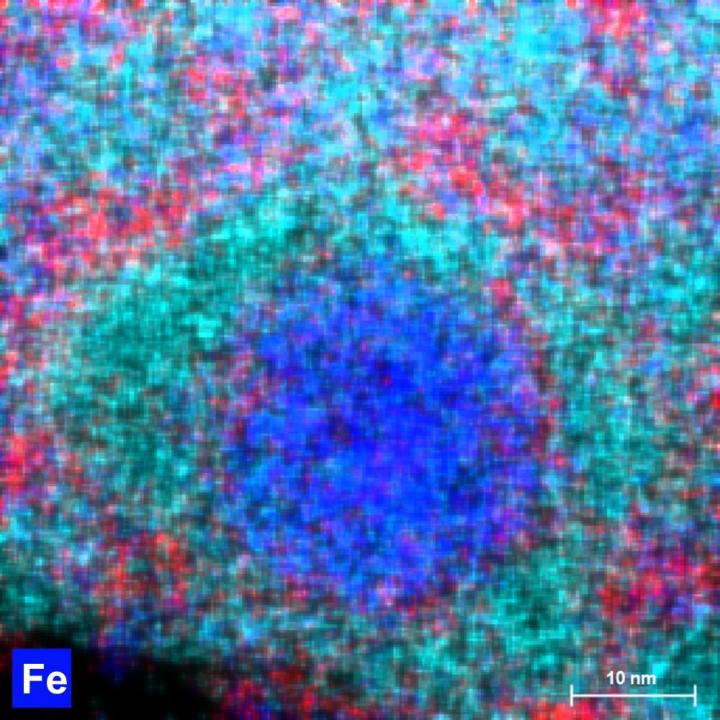
Caption: A transmission electron microscopy image of a nano crystal (ca 25 nm in diameter) in a basaltic magma from Mt. Etna (Italy). The nano crystal is enriched in iron (Fe) and it was produced in a laboratory during at BGI. Credit Image: Nobuyoshi Miyajima.

Here’s a link to and a citation for the paper,

In situ observation of nanolite growth in volcanic melt: A driving force for explosive eruptions by Danilo Di Genova, Richard A. Brooker, Heidy M. Mader, James W. E. Drewitt, Alessandro Longo, Joachim Deubener, Daniel R. Neuville, Sara Fanara, Olga Shebanova, Simone Anzellini, Fabio Arzilli, Emily C. Bamber, Louis Hennet, Giuseppe La Spina and Nobuyoshi Miyajima. Science Advances DOI: 10.1126/sciadv.abb0413 Vol. 6, no. 39, eabb0413 Published: 23 Sep 2020

This paper appears to be open access.

Make electricity by flowing water over nanolayers of metal

Scientists at Northwestern University (Chicago, Illinois) and the California Institute of Technology (CalTech) have developed what could be a more sustainable way to produce electricity. From a July 31, 2019 news item on Nanowerk,

Scientists from Northwestern University and Caltech have produced electricity by simply flowing water over extremely thin layers of inexpensive metals, including iron, that have oxidized. These films represent an entirely new way of generating electricity and could be used to develop new forms of sustainable power production.

A July 31, 2019 Northwester University news release (also on EurekAlert) by Megan Fellman, which originated the news item, provides details that suggest this discovery could prove beneficial in medical implants, as well as, in solar cells,

The films have a conducting metal nanolayer (10 to 20 nanometers thick) that is insulated with an oxide layer (2 nanometers thick). Current is generated when pulses of rainwater and ocean water alternate and move across the nanolayers. The difference in salinity drags the electrons along in the metal below.

“It’s the oxide layer over the nanometal that really makes this device go,” said Franz M. Geiger, the Dow Professor of Chemistry in Northwestern’s Weinberg College of Arts and Sciences. “Instead of corrosion, the presence of the oxides on the right metals leads to a mechanism that shuttles electrons.”

The films are transparent, a feature that could be taken advantage of in solar cells. The researchers intend to study the method using other ionic liquids, including blood. Developments in this area could lead to use in stents and other implantable devices.

“The ease of scaling up the metal nanolayer to large areas and the ease with which plastics can be coated gets us to three-dimensional structures where large volumes of liquids can be used,” Geiger said. “Foldable designs that fit, for instance, into a backpack are a possibility as well. Given how transparent the films are, it’s exciting to think about coupling the metal nanolayers to a solar cell or coating the outside of building windows with metal nanolayers to obtain energy when it rains.”

The study, titled “Energy Conversion via Metal Nanolayers,” was published this week [on July 29, 2019] in the journal Proceedings of the National Academy of Sciences (PNAS).

Geiger is the study’s corresponding author; his Northwestern team conducted the experiments. Co-author Thomas Miller, professor of chemistry at Caltech, led a team that conducted atomistic simulations to study the device’s behavior at the atomic level.

The new method produces voltages and currents comparable to graphene-based devices reported to have efficiencies of around 30% — similar to other approaches under investigation (carbon nanotubes and graphene) but with a single-step fabrication from earth-abundant elements instead of multistep fabrication. This simplicity allows for scalability, rapid implementation and low cost. Northwestern has filed for a provisional patent.

Of the metals studied, the researchers found that iron, nickel and vanadium worked best. They tested a pure rust sample as a control experiment; it did not produce a current.

The mechanism behind the electricity generation is complex, involving ion adsorption and desorption, but it essentially works like this: The ions present in the rainwater/saltwater attract electrons in the metal beneath the oxide layer; as the water flows, so do those ions, and through that attractive force, they drag the electrons in the metal along with them, generating an electrical current.

“There are interesting prospects for a variety of energy and sustainability applications, but the real value is the new mechanism of oxide-metal electron transfer,” Geiger said. “The underlying mechanism appears to involve various oxidation states.”

The team used a process called physical vapor deposition (PVD), which turns normally solid materials into a vapor that condenses on a desired surface. PVD allowed them to deposit onto glass metal layers only 10 to 20 nanometers thick. An oxide layer then forms spontaneously in air. It grows to a thickness of 2 nanometers and then stops growing.

“Thicker films of metal don’t succeed — it’s a nano-confinement effect,” Geiger said. “We have discovered the sweet spot.”

When tested, the devices generated several tens of millivolts and several microamps per centimeter squared.

“For perspective, plates having an area of 10 square meters each would generate a few kilowatts per hour — enough for a standard U.S. home,” Miller said. “Of course, less demanding applications, including low-power devices in remote locations, are more promising in the near term.”

Here’s a link to and a citation for the paper,

Energy conversion via metal nanolayers by Mavis D. Boamah, Emilie H. Lozier, Jeongmin Kim, Paul E. Ohno, Catherine E. Walker, Thomas F. Miller III, and Franz M. Geiger. PNAS DOI: https://doi.org/10.1073/pnas.1906601116 First published July 29, 2019

This paper is behind a paywall.

Rijksmuseum’s ‘live’ restoration of Rembrandt’s masterpiece: The Nightwatch: is it or isn’t it like watching paint dry?

Somewhere in my travels, I saw ‘like watching paint dry’ as a description for the experience of watching researchers examining Rembrandt’s Night Watch. Granted it’s probably not that exciting but there has to be something to be said for being present while experts undertake an extraordinary art restoration effort. The Night Watch is not only a masterpiece—it’s huge.

This posting was written closer to the time the ‘live’ restoration first began. I have an update at the end of this posting.

A July 8, 2019 news item on the British Broadcasting Corporation’s (BBC) news online sketches in some details,

The masterpiece, created in 1642, has been placed inside a specially designed glass chamber so that it can still be viewed while being restored.

Enthusiasts can follow the latest on the restoration work online.

The celebrated painting was last restored more than 40 years ago after it was slashed with a knife.

The Night Watch is considered Rembrandt’s most ambitious work. It was commissioned by the mayor and leader of the civic guard of Amsterdam, Frans Banninck Cocq, who wanted a group portrait of his militia company.

The painting is nearly 4m tall and 4.5m wide (12.5 x 15 ft) and weighs 337kg (743lb) [emphasis mine]. As well as being famous for its size, the painting is acclaimed for its use of dramatic lighting and movement.

But experts at Amsterdam’s Rijksmuseum are concerned that aspects of the masterpiece are changing, pointing as an example to the blanching of the figure of a small dog. The museum said the multi-million euro research and restoration project under way would help staff gain a better understanding of the painting’s condition.

An October 16, 2018 Rijksmuseum press release announced the restoration work months prior to the start (Note: Some of the information is repetitive;),

Before the restoration begins, The Night Watch will be the centrepiece of the Rijksmuseum’s display of their entire collection of more than 400 works by Rembrandt in an exhibition to mark the 350th anniversary of the artist’s death opening on 15 February 2019.

Commissioned in 1642 by the mayor and leader of the civic guard of Amsterdam, Frans Banninck Cocq, to create a group portrait of his shooting company, The Night Watch is recognised as one of the most important works of art in the world today and hangs in the specially designed “Gallery of Honour” at the Rijksmuseum. It is more than 40 years since The Night Watch underwent its last major restoration, following an attack on the painting in 1975.

The Night Watch will be encased in a state-of-the-art clear glass chamber designed by the French architect Jean Michel Wilmotte. This will ensure that the painting can remain on display for museum visitors. A digital platform will allow viewers from all over the world to follow the entire process online [emphasis mine] continuing the Rijksmuseum innovation in the digital field.

Taco Dibbits, General Director Rijksmuseum: The Night Watch is one of the most famous paintings in the world. It belongs to us all, and that is why we have decided to conduct the restoration within the museum itself – and everyone, wherever they are, will be able to follow the process online.

The Rijksmuseum continually monitors the condition of The Night Watch, and it has been discovered that changes are occurring, such as the blanching [emphasis mine] on the dog figure at the lower right of the painting. To gain a better understanding of its condition as a whole, the decision has been taken to conduct a thorough examination. This detailed study is necessary to determine the best treatment plan, and will involve imaging techniques, high-resolution photography and highly advanced computer analysis. Using these and other methods, we will be able to form a very detailed picture of the painting – not only of the painted surface, but of each and every layer, from varnish to canvas.

A great deal of experience has been gained in the Rijksmuseum relating to the restoration of Rembrandt’s paintings. Last year saw the completion of the restoration of Rembrandt’s spectacular portraits of Marten Soolmans and Oopjen Coppit. The research team working on The Night Watch is made up of researchers, conservators and restorers from the Rijksmuseum, which will conduct this research in close collaboration with museums and universities in the Netherlands and abroad.

The Night Watch

The group portrait of the officers and other members of the militia company of District II, under the command of Captain Frans Banninck Cocq and Lieutenant Willem van Ruytenburch, now known as The Night Watch, is Rembrandt’s most ambitious painting. This 1642 commission by members of Amsterdam’s civic guard is Rembrandt’s first and only painting of a militia group. It is celebrated particularly for its bold and energetic composition, with the musketeers being depicted ‘in motion’, rather than in static portrait poses. The Night Watch belongs to the city of Amsterdam, and it been the highlight of the Rijksmuseum collection since 1808. The architect of the Rijksmuseum building Pierre Cuypers (1827-1921) even created a dedicated gallery of honour for The Night Watch, and it is now admired there by more than 2.2 million people annually.

2019, The Year of Rembrandt

The Year of Rembrandt, 2019, marks the 350th anniversary of the artist’s death with two major exhibitions honouring the great master painter. All the Rembrandts of the Rijksmuseum (15 February to 10 June 2019) will bring together the Rijksmuseum’s entire collection of Rembrandt’s paintings, drawings and prints, for the first time in history. The second exhibition, Rembrandt-Velázquez (11 October 2019 to 19 January 2020), will put the master in international context by placing 17th-century Spanish and Dutch masterpieces in dialogue with each another.

First, the restoration work is not being livestreamed; the digital platform Operation Night Watch is a collection of resources, which are being updated constantly, For example, the first scan was placed online in Operation Night Watch on July 16, 2019.

Second, ‘blanching’ reminded me of a June 22, 2017 posting where I featured research into why masterpieces were turning into soap, (Note: The second paragraph should be indented to indicated that it’s an excerpt fro the news release. Unfortunately, the folks at WordPress appear to have removed the tools that would allow me to do that and more),

This piece of research has made a winding trek through the online science world. First it was featured in an April 20, 2017 American Chemical Society news release on EurekAlert

A good art dealer can really clean up in today’s market, but not when some weird chemistry wreaks havoc on masterpieces. Art conservators started to notice microscopic pockmarks forming on the surfaces of treasured oil paintings that cause the images to look hazy. It turns out the marks are eruptions of paint caused, weirdly, by soap that forms via chemical reactions. Since you have no time to watch paint dry, we explain how paintings from Rembrandts to O’Keefes are threatened by their own compositions — and we don’t mean the imagery.

….

Getting back to the Night Watch, there’s a July 8, 2019 Rijksmuseum press release which provides some technical details,

On 8 July 2019 the Rijksmuseum starts Operation Night Watch. It will be the biggest and most wide-ranging research and conservation project in the history of Rembrandt’s masterpiece. The goal of Operation Night Watch is the long-term preservation of the painting. The entire operation will take place in a specially designed glass chamber so the visiting public can watch.

Never before has such a wide-ranging and thorough investigation been made of the condition of The Night Watch. The latest and most advanced research techniques will be used, ranging from digital imaging and scientific and technical research, to computer science and artificial intelligence. The research will lead to a better understanding of the painting’s original appearance and current state, and provide insight into the many changes that The Night Watch has undergone over the course of the last four centuries. The outcome of the research will be a treatment plan that will form the basis for the restoration of the painting.

Operation Night Watch can also be followed online from 8 July 2019 at rijksmuseum.nl/nightwatch

From art historical research to artificial intelligence

Operation Night Watch will look at questions regarding the original commission, Rembrandt’s materials and painting technique, the impact of previous treatments and later interventions, as well as the ageing, degradation and future of the painting. This will involve the newest and most advanced research methods and technologies, including art historical and archival research, scientific and technical research, computer science and artificial intelligence.

During the research phase The Night Watch will be unframed and placed on a specially designed easel. Two platform lifts will make it possible to study the entire canvas, which measures 379.5 cm in height and 454.5 cm in width.

Advanced imaging techniques

Researchers will make use of high resolution photography, as well as a variety of advanced imaging techniques, such as macro X-ray fluorescence scanning (macro-XRF) and hyperspectral imaging, also called infrared reflectance imaging spectroscopy (RIS), to accurately determine the condition of the painting.

56 macro-XRF scans

The Night Watch will be scanned millimetre by millimetre using a macro X-ray fluorescence scanner (macro-XRF scanner). This instrument uses X-rays to analyse the different chemical elements in the paint, such as calcium, iron, potassium and cobalt. From the resulting distribution maps of the various chemical elements in the paint it is possible to determine which pigments were used. The macro-XRF scans can also reveal underlying changes in the composition, offering insights into Rembrandt’s painting process. To scan the entire surface of the The Night Watch it will be necesary to make 56 scans, each one of which will take 24 hours.

12,500 high-resolution photographs

A total of some 12,500 photographs will be taken at extremely high resolution, from 180 to 5 micrometres, or a thousandth of a millimetre. Never before has such a large painting been photographed at such high resolution. In this way it will be possible to see details such as pigment particles that normally would be invisible to the naked eye. The cameras and lamps will be attached to a dynamic imaging frame designed specifically for this purpose.

Glass chamber

Operation Night Watch is for everyone to follow and will take place in full view of the visiting public in an ultra-transparent glass chamber designed by the French architect Jean Michel Wilmotte.

Research team

The Rijksmuseum has extensive experience and expertise in the investigation and treatment of paintings by Rembrandt. The conservation treatment of Rembrandt’s portraits of Marten Soolmans and Oopjen Coppit was completed in 2018. The research team working on The Night Watch is made up of more than 20 Rijksmuseum scientists, conservators, curators and photographers. For this research, the Rijksmuseum is also collaborating with museums and universities in the Netherlands and abroad, including the Dutch Cultural Heritage Agency (RCE), Delft University of Technology (TU Delft), the University of Amsterdam (UvA), Amsterdam University Medical Centre (AUMC), University of Antwerp (UA) and National Gallery of Art, Washington DC.

The Night Watch

Rembrandt’s Night Watch is one of the world’s most famous works of art. The painting is the property of the City of Amsterdam, and it is the heart of Amsterdam’s Rijksmuseum, where it is admired by more than two million visitors each year. The Night Watch is the Netherland’s foremost national artistic showpiece, and a must-see for tourists.

Rembrandt’s group portrait of officers and other civic guardsmen of District 2 in Amsterdam under the command of Captain Frans Banninck Cocq and Lieutenant Willem van Ruytenburch has been known since the 18th century as simply The Night Watch. It is the artist’s most ambitious painting. One of Amsterdam’s 20 civic guard companies commissioned the painting for its headquarters, the Kloveniersdoelen, and Rembrandt completed it in 1642. It is Rembrandt’s only civic guard piece, and it is famed for the lively and daring composition that portrays the troop in active poses rather than the traditional static ones.

Donors and partners

AkzoNobel is main partner of Operation Night Watch.

Operation Night Watch is made possible by The Bennink Foundation, PACCAR Foundation, Piet van der Slikke & Sandra Swelheim, American Express Foundation, Familie De Rooij, Het AutoBinck Fonds, Segula Technologies, Dina & Kjell Johnsen, Familie D. Ermia, Familie M. van Poecke, Henry M. Holterman Fonds, Irma Theodora Fonds, Luca Fonds, Piek-den Hartog Fonds, Stichting Zabawas, Cevat Fonds, Johanna Kast-Michel Fonds, Marjorie & Jeffrey A. Rosen, Stichting Thurkowfonds and the Night Watch Fund.

With the support of the Ministry of Education, Culture and Science, the City of Amsterdam, Founder Philips and main sponsors ING, BankGiro Loterij and KPN every year more than 2 million people visit the Rijksmuseum and The Night Watch.

Details:
Rembrandt van Rijn (1606-1669)
The Night Watch, 1642
oil on canvas
Rijksmuseum, on loan from the Municipality of Amsterdam

Update as of November 22, 2019

I just clicked on the Operation Night Watch link and found a collection of resources including videos of live updates from October 2019. As noted earlier, they’re not livestreaming the restoration. The October 29, 2019 ‘live update’ features a host speaking in Dutch (with English subtitles in the version I was viewing) and interviews with the scientists conducting the research necessary before they start actually restoring the painting.
World’s smallest magnetic resonance imaging (MRI) of a single atom

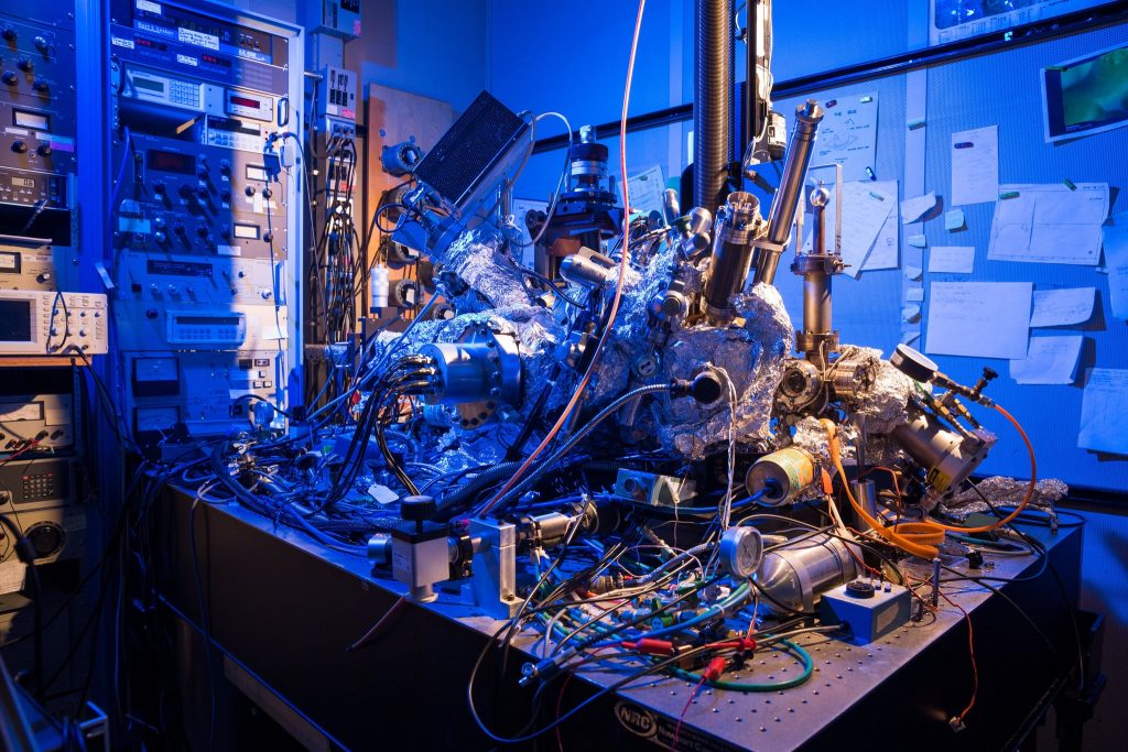
While not science’s sleekest machine, this microscope was able to capture M.R.I. scans of single atoms. Credit: IBM Research

Such a messy looking thing—it makes me feel better about my housekeeping. In any event, it’s fascinating to think this scanning tunneling microscope as seen in the above can actually act as an MRI device and create an image of a single atom.

There’s a wonderful article in the New York Times about the work but I’m starting first with a July 1, 2019 news item on Nanowerk,

Researchers at the Center for Quantum Nanoscience (QNS) within the Institute for Basic Science (IBS) at Ewha Womans University [Seoul, South Korea) have made a major scientific breakthrough by performing the world’s smallest magnetic resonance imaging (MRI). In an international collaboration with colleagues from the US, QNS scientists used their new technique to visualize the magnetic field of single atoms.

A July 2, 2019 IBS news release (also on EurekAlert but published July 1, 2019), which originated the news item, provides some insight into the research,

An MRI is routinely done in hospitals nowadays as a part of imaging for diagnostics. MRI’s detect the density of spins – the fundamental magnets in electrons and protons – in the human body. Traditionally, billions and billions of spins are required for an MRI scan. The new findings, published today [July 1, 2019] in the journal Nature Physics, show that this process is now also possible for an individual atom on a surface. To do this, the team used a Scanning Tunneling Microscope, which consists of an atomically sharp metal tip that allows researchers to image and probe single atoms by scanning the tip across the surface.

The two elements that were investigated in this work, iron and titanium, are both magnetic. Through precise preparation of the sample, the atoms were readily visible in the microscope. The researchers then used the microscope’s tip like an MRI machine to map the three-dimensional magnetic field created by the atoms with unprecedented resolution. In order to do so, they attached another spin cluster to the sharp metal tip of their microscope. Similar to everyday magnets, the two spins would attract or repel each other depending on their relative position. By sweeping the tip spin cluster over the atom on the surface, the researchers were able to map out the magnetic interaction. Lead author, Dr. Philip Willke of QNS says: “It turns out that the magnetic interaction we measured depends on the properties of both spins, the one on the tip and the one on the sample. For example, the signal that we see for iron atoms is vastly different from that for titanium atoms. This allows us to distinguish different kinds of atoms by their magnetic field signature and makes our technique very powerful.”

The researchers plan to use their single-atom MRI to map the spin distribution in more complex structures such as molecules and magnetic materials. “Many magnetic phenomena take place on the nanoscale, including the recent generation of magnetic storage devices.” says Dr. Yujeong Bae also of QNS, a co-author in this study. “We now plan to study a variety of systems using our microscopic MRI.” The ability to analyze the magnetic structure on the nanoscale can help to develop new materials and drugs. Moreover, the research team wants to use this kind of MRI to characterize and control quantum systems. These are of great interest for future computation schemes, also known as quantum computing

“I am very excited about these results. It is certainly a milestone in our field and has very promising implications for future research.” says Prof. Andreas Heinrich, Director of QNS. “The ability to map spins and their magnetic field with previously unimaginable precision, allows us to gain deeper knowledge about the structure of matter and opens new fields of basic research.”

The Center for Quantum Nanoscience, on the campus of Ewha Womans University in Seoul, South Korea, is a world-leading research center merging quantum and nanoscience to engineer the quantum future through basic research. Backed by Korea’s Institute for Basic Science, which was founded in 2011, the Center for Quantum Nanoscience draws on decades of QNS Director Andreas J. Heinrich’s (A Boy and His Atom, IBM, 2013) scientific leadership to lay the foundation for future technology by exploring the use of quantum behavior atom-by-atom on surfaces with highest precision.

You may have noticed that other than a brief mention in the first paragraph (in the Nanowerk news item excerpt), there’s no mention of the US researchers and their contribution to the work.

Interestingly, the July 1, 2019 New York Time article by Knvul Sheikh returns the favour by focusing almost entirely on US researchers while giving the Korean researchers a passing mention (Note: Links have been removed),

Different microscopy techniques allow scientists to see the nucleotide-by-nucleotide genetic sequences in cells down to the resolution of a couple atoms as seen in an atomic force microscopy image. But scientists at the IBM Almaden Research Center in San Jose, Calif., and the Institute for Basic Sciences in Seoul, have taken imaging a step further, developing a new magnetic resonance imaging technique that provides unprecedented detail, right down to the individual atoms of a sample.

When doctors want to detect tumors, measure brain function or visualize the structure of joints, they employ huge M.R.I. machines, which apply a magnetic field across the human body. This temporarily disrupts the protons spinning in the nucleus of every atom in every cell. A subsequent, brief pulse of radio-frequency energy causes the protons to spin perpendicular to the pulse. Afterward, the protons return to their normal state, releasing energy that can be measured by sensors and made into an image.

But to gather enough diagnostic data, traditional hospital M.R.I.s must scan billions and billions of protons in a person’s body, said Christopher Lutz, a physicist at IBM. So he and his colleagues decided to pack the power of an M.R.I. machine into the tip of another specialized instrument known as a scanning tunneling microscope to see if they could image individual atoms.

The tip of a scanning tunneling microscope is just a few atoms wide. And it moves along the surface of a sample, it picks up details about the size and conformation of molecules.

The researchers attached magnetized iron atoms to the tip, effectively combining scanning-tunneling microscope and M.R.I. technologies.

When the magnetized tip swept over a metal wafer of iron and titanium, it applied a magnetic field to the sample, disrupting the electrons (rather than the protons, as a typical M.R.I. would) within each atom. Then the researchers quickly turned a radio-frequency pulse on and off, so that the electrons would emit energy that could be visualized. …

Here’s a link to and a citation for the paper,

Magnetic resonance imaging of single atoms on a surface by Philip Willke, Kai Yang, Yujeong Bae, Andreas J. Heinrich & Christopher P. Lutz. Nature Physics (2019) DOI: https://doi.org/10.1038/s41567-019-0573-x Published 01 July 2019

This paper is behind a paywall.

Mixing the unmixable for all new nanoparticles

This news comes out of the University of Maryland and the discovery could led to nanoparticles that have never before been imagined. From a March 29, 2018 news item on ScienceDaily,

Making a giant leap in the ‘tiny’ field of nanoscience, a multi-institutional team of researchers is the first to create nanoscale particles composed of up to eight distinct elements generally known to be immiscible, or incapable of being mixed or blended together. The blending of multiple, unmixable elements into a unified, homogenous nanostructure, called a high entropy alloy nanoparticle, greatly expands the landscape of nanomaterials — and what we can do with them.

This research makes a significant advance on previous efforts that have typically produced nanoparticles limited to only three different elements and to structures that do not mix evenly. Essentially, it is extremely difficult to squeeze and blend different elements into individual particles at the nanoscale. The team, which includes lead researchers at University of Maryland, College Park (UMD)’s A. James Clark School of Engineering, published a peer-reviewed paper based on the research featured on the March 30 [2018] cover of Science.

A March 29, 2018 University of Maryland press release (also on EurekAlert), which originated the news item, delves further (Note: Links have been removed),

“Imagine the elements that combine to make nanoparticles as Lego building blocks. If you have only one to three colors and sizes, then you are limited by what combinations you can use and what structures you can assemble,” explains Liangbing Hu, associate professor of materials science and engineering at UMD and one of the corresponding authors of the paper. “What our team has done is essentially enlarged the toy chest in nanoparticle synthesis; now, we are able to build nanomaterials with nearly all metallic and semiconductor elements.”

The researchers say this advance in nanoscience opens vast opportunities for a wide range of applications that includes catalysis (the acceleration of a chemical reaction by a catalyst), energy storage (batteries or supercapacitors), and bio/plasmonic imaging, among others.

To create the high entropy alloy nanoparticles, the researchers employed a two-step method of flash heating followed by flash cooling. Metallic elements such as platinum, nickel, iron, cobalt, gold, copper, and others were exposed to a rapid thermal shock of approximately 3,000 degrees Fahrenheit, or about half the temperature of the sun, for 0.055 seconds. The extremely high temperature resulted in uniform mixtures of the multiple elements. The subsequent rapid cooling (more than 100,000 degrees Fahrenheit per second) stabilized the newly mixed elements into the uniform nanomaterial.

“Our method is simple, but one that nobody else has applied to the creation of nanoparticles. By using a physical science approach, rather than a traditional chemistry approach, we have achieved something unprecedented,” says Yonggang Yao, a Ph.D. student at UMD and one of the lead authors of the paper.

To demonstrate one potential use of the nanoparticles, the research team used them as advanced catalysts for ammonia oxidation, which is a key step in the production of nitric acid (a liquid acid that is used in the production of ammonium nitrate for fertilizers, making plastics, and in the manufacturing of dyes). They were able to achieve 100 percent oxidation of ammonia and 99 percent selectivity toward desired products with the high entropy alloy nanoparticles, proving their ability as highly efficient catalysts.

Yao says another potential use of the nanoparticles as catalysts could be the generation of chemicals or fuels from carbon dioxide.

“The potential applications for high entropy alloy nanoparticles are not limited to the field of catalysis. With cross-discipline curiosity, the demonstrated applications of these particles will become even more widespread,” says Steven D. Lacey, a Ph.D. student at UMD and also one of the lead authors of the paper.

This research was performed through a multi-institutional collaboration of Prof. Liangbing Hu’s group at the University of Maryland, College Park; Prof. Reza Shahbazian-Yassar’s group at University of Illinois at Chicago; Prof. Ju Li’s group at the Massachusetts Institute of Technology; Prof. Chao Wang’s group at Johns Hopkins University; and Prof. Michael Zachariah’s group at the University of Maryland, College Park.

What outside experts are saying about this research:

“This is quite amazing; Dr. Hu creatively came up with this powerful technique, carbo-thermal shock synthesis, to produce high entropy alloys of up to eight different elements in a single nanoparticle. This is indeed unthinkable for bulk materials synthesis. This is yet another beautiful example of nanoscience!,” says Peidong Yang, the S.K. and Angela Chan Distinguished Professor of Energy and professor of chemistry at the University of California, Berkeley and member of the American Academy of Arts and Sciences.

“This discovery opens many new directions. There are simulation opportunities to understand the electronic structure of the various compositions and phases that are important for the next generation of catalyst design. Also, finding correlations among synthesis routes, composition, and phase structure and performance enables a paradigm shift toward guided synthesis,” says George Crabtree, Argonne Distinguished Fellow and director of the Joint Center for Energy Storage Research at Argonne National Laboratory.

More from the research coauthors:

“Understanding the atomic order and crystalline structure in these multi-element nanoparticles reveals how the synthesis can be tuned to optimize their performance. It would be quite interesting to further explore the underlying atomistic mechanisms of the nucleation and growth of high entropy alloy nanoparticle,” says Reza Shahbazian-Yassar, associate professor at the University of Illinois at Chicago and a corresponding author of the paper.

“Carbon metabolism drives ‘living’ metal catalysts that frequently move around, split, or merge, resulting in a nanoparticle size distribution that’s far from the ordinary, and highly tunable,” says Ju Li, professor at the Massachusetts Institute of Technology and a corresponding author of the paper.

“This method enables new combinations of metals that do not exist in nature and do not otherwise go together. It enables robust tuning of the composition of catalytic materials to optimize the activity, selectivity, and stability, and the application will be very broad in energy conversions and chemical transformations,” says Chao Wang, assistant professor of chemical and biomolecular engineering at Johns Hopkins University and one of the study’s authors.

Here’s a link to and a citation for the paper,

Carbothermal shock synthesis of high-entropy-alloy nanoparticles by Yonggang Yao, Zhennan Huang, Pengfei Xie, Steven D. Lacey, Rohit Jiji Jacob, Hua Xie, Fengjuan Chen, Anmin Nie, Tiancheng Pu, Miles Rehwoldt, Daiwei Yu, Michael R. Zachariah, Chao Wang, Reza Shahbazian-Yassar, Ju Li, Liangbing Hu. Science 30 Mar 2018: Vol. 359, Issue 6383, pp. 1489-1494 DOI: 10.1126/science.aan5412

This paper is behind a paywall.

Seaweed supercapacitors

I like munching on seaweed from time to time but it seems that seaweed may be more than just a foodstuff according to an April 5, 2017 news item on Nanowerk,

Seaweed, the edible algae with a long history in some Asian cuisines, and which has also become part of the Western foodie culture, could turn out to be an essential ingredient in another trend: the development of more sustainable ways to power our devices. Researchers have made a seaweed-derived material to help boost the performance of superconductors, lithium-ion batteries and fuel cells.

The team will present the work today [April 5, 2017] at the 253rd National Meeting & Exposition of the American Chemical Society (ACS). ACS, the world’s largest scientific society, is holding the meeting here through Thursday. It features more than 14,000 presentations on a wide range of science topics.

An April 5, 2017 American Chemical Society news release on EurekAlert), which originated the news item, gives more details about the presentation,

“Carbon-based materials are the most versatile materials used in the field of energy storage and conversion,” Dongjiang Yang, Ph.D., says. “We wanted to produce carbon-based materials via a really ‘green’ pathway. Given the renewability of seaweed, we chose seaweed extract as a precursor and template to synthesize hierarchical porous carbon materials.” He explains that the project opens a new way to use earth-abundant materials to develop future high-performance, multifunctional carbon nanomaterials for energy storage and catalysis on a large scale.

Traditional carbon materials, such as graphite, have been essential to creating the current energy landscape. But to make the leap to the next generation of lithium-ion batteries and other storage devices, an even better material is needed, preferably one that can be sustainably sourced, Yang says.

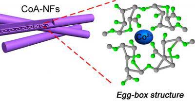
With these factors in mind, Yang, who is currently at Qingdao University (China), turned to the ocean. Seaweed is an abundant algae that grows easily in salt water. While Yang was at Griffith University in Australia, he worked with colleagues at Qingdao University and at Los Alamos National Laboratory in the U.S. to make porous carbon nanofibers from seaweed extract. Chelating, or binding, metal ions such as cobalt to the alginate molecules resulted in nanofibers with an “egg-box” structure, with alginate units enveloping the metal ions. This architecture is key to the material’s stability and controllable synthesis, Yang says.

Testing showed that the seaweed-derived material had a large reversible capacity of 625 milliampere hours per gram (mAhg-1), which is considerably more than the 372 mAhg-1 capacity of traditional graphite anodes for lithium-ion batteries. This could help double the range of electric cars if the cathode material is of equal quality. The egg-box fibers also performed as well as commercial platinum-based catalysts used in fuel-cell technologies and with much better long-term stability. They also showed high capacitance as a superconductor material at 197 Farads per gram, which could be applied in zinc-air batteries and supercapacitors. The researchers published their initial results in ACS Central Science in 2015 and have since developed the materials further.

For example, building on the same egg-box structure, the researchers say they have suppressed defects in seaweed-based, lithium-ion battery cathodes that can block the movement of lithium ions and hinder battery performance. And recently, they have developed an approach using red algae-derived carrageenan and iron to make a porous sulfur-doped carbon aerogel with an ultra-high surface area. The structure could be a good candidate to use in lithium-sulfur batteries and supercapacitors.

More work is needed to commercialize the seaweed-based materials, however. Yang says currently more than 20,000 tons of alginate precursor can be extracted from seaweed per year for industrial use. But much more will be required to scale up production.

Here’s an image representing the research,

Scientists have created porous ‘egg-box’ structured nanofibers using seaweed extract. Credit: American Chemical Society

I’m not sure that looks like an egg-box but I’ll take their word for it.

Watching rust turn into iron

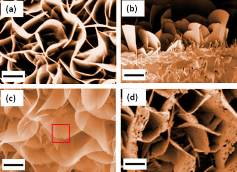
a) Colorized SEM images of iron oxide nanoblades used in the experiment. b) Colorized cross-section of SEM image of the nanoblades. c) Colorized SEM image of nanoblades after 1 hour of reduction reaction at 500 °C in molecular hydrogen, showing the sawtooth shape along the edges (square). d) Colorized SEM image showing the formation of holes after 2 hours of reduction. The scale bar is 1 micrometer. Credit: W. Zhu et al./ACS Nano and K. Irvine/NIST

Here’s more about being able to watch iron transition from one state to the next according to an April 5, 2017 news item on phys.org

Using a state-of-the-art microscopy technique, experimenters at the National Institute of Standards and Technology (NIST) and their colleagues have witnessed a slow-motion, atomic-scale transformation of rust—iron oxide—back to pure iron metal, in all of its chemical steps.

An April 4, 2017 NIST news release describes the role iron plays in modern lifestyles and the purpose of this research,

Among the most abundant minerals on Earth, iron oxides play a leading role in magnetic data storage, cosmetics, the pigmentation of paints and drug delivery. These materials also serve as catalysts for several types of chemical reactions, including the production of ammonia for fertilizer.

To fine-tune the properties of these minerals for each application, scientists work with nanometer-scale particles of the oxides. But to do so, researchers need a detailed, atomic-level understanding of reduction, a key chemical reaction that iron oxides undergo. That knowledge, however, is often lacking because reduction—a process that is effectively the opposite of rusting—proceeds too rapidly for many types of probes to explore at such a fine level.

In a new effort to study the microscopic details of metal oxide reduction, researchers used a specially adapted transmission electron microscope (TEM) at NIST’s NanoLab facility to document the step-by-step transformation of nanocrystals of the iron oxide hematite (Fe2O3) to the iron oxide magnetite (Fe3O4), and finally to iron metal.

“Even though people have studied iron oxide for many years, there have been no dynamic studies at the atomic scale,” said Wenhui Zhu of the State University of New York at Binghamton, who worked on her doctorate in the NanoLab in 2015 and 2016. “We are seeing what’s actually happening during the entire reduction process instead of studying just the initial steps.”

That’s critical, added NIST’s Renu Sharma, “if you want to control the composition or properties of iron oxides and understand the relationships between them.”

By lowering the temperature of the reaction and decreasing the pressure of the hydrogen gas that acted as the reducing agent, the scientists slowed down the reduction process so that it could be captured with an environmental TEM—a specially configured TEM that can study both solids and gas. The instrument enables researchers to perform atomic-resolution imaging of a sample under real-life conditions—in this case the gaseous environment necessary for iron oxides to undergo reduction–rather than under the vacuum needed in ordinary TEMs.

“This is the most powerful tool I’ve used in my research and one of the very few in the United States,” said Zhu. She, Sharma and their colleagues describe their findings in a recent issue of ACS Nano.

The team examined the reduction process in a bicrystal of iron oxide, consisting of two identical iron oxide crystals rotated at 21.8 degrees with respect to each other. The bicrystal structure also served to slow down the reduction process, making it easier to follow with the environmental TEM.

In studying the reduction reaction, the researchers identified a previously unknown intermediate state in the transformation from magnetite to hematite. In the middle stage, the iron oxide retained its original chemical structure, Fe2O3, but changed the crystallographic arrangement of its atoms from rhombohedral (a diagonally stretched cube) to cubic.

This intermediate state featured a defect in which oxygen atoms fail to populate some of the sites in the crystal that they normally would. This so-called oxygen vacancy defect is not uncommon and is known to strongly influence the electrical and catalytic properties of oxides. But the researchers were surprised to find that the defects occurred in an ordered pattern, which had never been found before in the reduction of Fe2O3 to Fe3O4, Sharma said.

The significance of the intermediate state remains under study, but it may be important for controlling the reduction rate and other properties of the reduction process, she adds. “The more we understand, the better we can manipulate the microstructure of these oxides,” said Zhu. By manipulating the microstructure, researchers may be able to enhance the catalytic activity of iron oxides.

Even though a link has already been provided for the paper, I will give it again along with a citation,

In Situ Atomic-Scale Probing of the Reduction Dynamics of Two-Dimensional Fe2O3 Nanostructures by Wenhui Zhu, Jonathan P. Winterstein, Wei-Chang David Yang, Lu Yuan, Renu Sharma, and Guangwen Zhou. ACS Nano, 2017, 11 (1), pp 656–664 DOI: 10.1021/acsnano.6b06950 Publication Date (Web): December 13, 2016

Copyright © 2016 American Chemical Society

This paper is behind a paywall.

Split some water molecules and save solar and wind (energy) for a future day

Professor Ted Sargent’s research team at the University of Toronto has a developed a new technique for saving the energy harvested by sun and wind farms according to a March 28, 2016 news item on Nanotechnology Now,

We can’t control when the wind blows and when the sun shines, so finding efficient ways to store energy from alternative sources remains an urgent research problem. Now, a group of researchers led by Professor Ted Sargent at the University of Toronto’s Faculty of Applied Science & Engineering may have a solution inspired by nature.

The team has designed the most efficient catalyst for storing energy in chemical form, by splitting water into hydrogen and oxygen, just like plants do during photosynthesis. Oxygen is released harmlessly into the atmosphere, and hydrogen, as H2, can be converted back into energy using hydrogen fuel cells.

Discovering a better way of storing energy from solar and wind farms is “one of the grand challenges in this field,” Ted Sargent says (photo above by Megan Rosenbloom via flickr) Courtesy: University of Toronto

Discovering a better way of storing energy from solar and wind farms is “one of the grand challenges in this field,” Ted Sargent says (photo above by Megan Rosenbloom via flickr) Courtesy: University of Toronto

A March 24, 2016 University of Toronto news release by Marit Mitchell, which originated the news item, expands on the theme,

“Today on a solar farm or a wind farm, storage is typically provided with batteries. But batteries are expensive, and can typically only store a fixed amount of energy,” says Sargent. “That’s why discovering a more efficient and highly scalable means of storing energy generated by renewables is one of the grand challenges in this field.”

You may have seen the popular high-school science demonstration where the teacher splits water into its component elements, hydrogen and oxygen, by running electricity through it. Today this requires so much electrical input that it’s impractical to store energy this way — too great proportion of the energy generated is lost in the process of storing it.

This new catalyst facilitates the oxygen-evolution portion of the chemical reaction, making the conversion from H2O into O2 and H2 more energy-efficient than ever before. The intrinsic efficiency of the new catalyst material is over three times more efficient than the best state-of-the-art catalyst.

Details are offered in the news release,

The new catalyst is made of abundant and low-cost metals tungsten, iron and cobalt, which are much less expensive than state-of-the-art catalysts based on precious metals. It showed no signs of degradation over more than 500 hours of continuous activity, unlike other efficient but short-lived catalysts. …

“With the aid of theoretical predictions, we became convinced that including tungsten could lead to a better oxygen-evolving catalyst. Unfortunately, prior work did not show how to mix tungsten homogeneously with the active metals such as iron and cobalt,” says one of the study’s lead authors, Dr. Bo Zhang … .

“We invented a new way to distribute the catalyst homogenously in a gel, and as a result built a device that works incredibly efficiently and robustly.”

This research united engineers, chemists, materials scientists, mathematicians, physicists, and computer scientists across three countries. A chief partner in this joint theoretical-experimental studies was a leading team of theorists at Stanford University and SLAC National Accelerator Laboratory under the leadership of Dr. Aleksandra Vojvodic. The international collaboration included researchers at East China University of Science & Technology, Tianjin University, Brookhaven National Laboratory, Canadian Light Source and the Beijing Synchrotron Radiation Facility.

“The team developed a new materials synthesis strategy to mix multiple metals homogeneously — thereby overcoming the propensity of multi-metal mixtures to separate into distinct phases,” said Jeffrey C. Grossman, the Morton and Claire Goulder and Family Professor in Environmental Systems at Massachusetts Institute of Technology. “This work impressively highlights the power of tightly coupled computational materials science with advanced experimental techniques, and sets a high bar for such a combined approach. It opens new avenues to speed progress in efficient materials for energy conversion and storage.”

“This work demonstrates the utility of using theory to guide the development of improved water-oxidation catalysts for further advances in the field of solar fuels,” said Gary Brudvig, a professor in the Department of Chemistry at Yale University and director of the Yale Energy Sciences Institute.

“The intensive research by the Sargent group in the University of Toronto led to the discovery of oxy-hydroxide materials that exhibit electrochemically induced oxygen evolution at the lowest overpotential and show no degradation,” said University Professor Gabor A. Somorjai of the University of California, Berkeley, a leader in this field. “The authors should be complimented on the combined experimental and theoretical studies that led to this very important finding.”

Here’s a link to and a citation for the paper,

Homogeneously dispersed, multimetal oxygen-evolving catalysts by Bo Zhang, Xueli Zheng, Oleksandr Voznyy, Riccardo Comin, Michal Bajdich, Max García-Melchor, Lili Han, Jixian Xu, Min Liu, Lirong Zheng, F. Pelayo García de Arquer, Cao Thang Dinh, Fengjia Fan, Mingjian Yuan, Emre Yassitepe, Ning Chen, Tom Regier, Pengfei Liu, Yuhang Li, Phil De Luna, Alyf Janmohamed, Huolin L. Xin, Huagui Yang, Aleksandra Vojvodic, Edward H. Sargent. Science  24 Mar 2016: DOI: 10.1126/science.aaf1525

This paper is behind a paywall.

Nanodiamonds detect the iron in your blood

Too little iron in the blood can lead to anemia and too much can signal problems with the immune system; German researchers have devised a promising new technique for detecting the amount of iron in the blood according to an Oct. 2, 2013 news item on ScienceDaily,

Lack of iron — caused by malnutrition — can lead to anemia while an increased level of iron may signal the presence of an acute inflammatory response. Therefore, the blood iron level is an important medical diagnostic agent. Researchers at Ulm University [Germany], led by experimental physicist Fedor Jelezko, theoretical physicist Martin Plenio and chemist Tanja Weil, have developed a novel biosensor for determination of iron content that is based on nanodiamonds.

Here’s an image of microscopic diamonds before they’ve been ground down to the nanoscale,

(Photo: Fedor Jelezko): Microscope picture of small diamonds, 100 microns in diameter. Specific lattice defects do not only impart colour on the diamonds but also provide the basis for the magnetic field sensor. In their experiments the team at Ulm ground down these diamonds to a size of 20 nanometers (as a comparison, a human hair has a diameter of 70 microns and is therefore 3000 times thicker than the nanodiamonds).

(Photo: Fedor Jelezko): Microscope picture of small diamonds, 100 microns in diameter. Specific lattice defects do not only impart colour on the diamonds but also provide the basis for the magnetic field sensor. In their experiments the team at Ulm ground down these diamonds to a size of 20 nanometers (as a comparison, a human hair has a diameter of 70 microns and is therefore 3000 times thicker than the nanodiamonds).

The Oct. 2, 2013 University of Ulm news release (on the Alpha Galileo Foundation website,) which originated the news item, describes the problem the scientists were addressing and their solution,

“Standard blood tests do not capture — as one might expect — free iron ions in the blood, because free iron is toxic and is therefore hardly detectable in blood,” explains Professor Tanja Weil, director of the Institute for Organic Chemistry III, University of Ulm. These methods are based on certain proteins instead that are responsible for the storage and transport of iron. One of these proteins is Ferritin that can contain up to 4,500 magnetic iron ions. Most standard tests are based on immunological techniques and estimate the iron concentration indirectly based on different markers. Results from different tests may however lead to inconsistent results in some clinical situations.

The Ulm scientists have developed a completely new approach to detect Ferritin. This required a combination of several new ideas. First, each ferritin-bound iron atom generates a magnetic field but as there are only 4,500 of them, the total magnetic field they generate is very small indeed and therefore hard to measure. This indeed, posed the second challenge for the team: to develop a method that is sufficiently sensitive to detect such weak magnetic fields. This they achieved by making use of a completely new, innovative technology based on tiny artificial diamonds of nanometer size. Crucially these diamonds are not perfect —colorless and transparent — but contain lattice defects which are optically active and thus provide the color of diamonds.

“These color centers allow us to measure the orientation of electron spins in external fields and thus measure their strength” explains Professor Fedor Jelezko, director of the Ulm Institute of Quantum Optics. Thirdly, the team had to find a way to adsorb ferritin on the surface of the diamond. “This we achieved with the help of electrostatic interactions between the tiny diamond particles and ferritin proteins,” adds Weil. Finally, “Theoretical modeling was essential to ensure that the signal measured is in fact consistent with the presence of ferritin and thus to validate the method,” states Martin Plenio, director of the Institute for Theoretical Physics. Future plans of the Ulm team include the precise determination of the number of ferritin proteins and the average iron load of individual proteins.

As the news release notes, this research is part of a larger project,

The demonstration of this innovative method, reported in Nano Letters [journal], represents a first step towards the goals of their recently awarded BioQ Synergy Grant. [10.3 million Euro which the scientists were awarded last December 2012 by the European Research Council] The focus of this project is the exploration of quantum properties in biology and the creation of self-organized diamond structures.

“Diamond sensors can thus be applied in biology and medicine,” say the Ulm scientists. But their new invention has its limits “. Whether the children have actually eaten their spinach cannot be detected with the diamond sensor, that’s still the prerogative of parents “, confesses quantum physicist Plenio

Here’s a link to and a citation for the paper,

Detection of a Few Metallo-Protein Molecules Using Color Centers in Nanodiamonds by A. Ermakova, G. Pramanik, J.-M. Cai, G. Algara-Siller,  U. Kaiser, T. Weil, Y.-K. Tzeng, H. C. Chang, L. P. McGuinness, M. B. Plenio, B. Naydenov, and F. Jelezko. Nano Lett., 2013, 13 (7), pp 3305–3309 DOI: 10.1021/nl4015233 Publication Date (Web): June 5, 2013
Copyright © 2013 American Chemical Society

This paper is behind a paywall.

Enriching food with nanoparticles?

There’s a team of Swiss researchers addressing the problem of anemia (iron deficiency) and zinc deficiency by adding iron and/or zinc nanoparticles to food. According to the article by Eric Bland on the Discovery News website,

“Iron and zinc deficiencies are common around the world,” said Michael Zimmermann, a scientist at ETH Zurich and a co-author of a recent Nature Nanotechnology article. “Yet many compounds used in food fortification are either absorbed poorly or, when they have high absorption, change the color, taste and smell of food.”

Anemia, or a lack of iron, affects more than 2 billion people worldwide and is arguably the most widespread micronutrient deficiency. Without enough iron the the body can become lethargic and cognitively impaired. For some pregnant women, the lack of iron can kill them during childbirth. Some economists have even speculated that a nation’s gross domestic product is depressed because of anemic and lethargic workers, said Zimmermann.

Lack of zinc impairs a person’s normal growth and can lead to diarrhea, pneumonia, anorexia and other conditions.

Standard ways of fortifying food with zinc and/or iron present various challenges including this one as noted by Zimmermann only a limited amount of iron can be added as it affects the food’s taste, smell, and/or appearance (this and other challenges are detailed in Bland’s article). So scientists continue to work on better ways to fortify food so that more people on the planet can benefit. The Swiss team’s approach,

The new research solves this conundrum. To create the nanoparticles the Swiss scientists dissolved iron in water, then sprayed the solution over very hot fire. The intense heat quickly evaporates the water, leaving tiny iron or zinc crystals, each one about 10 nanometers across. Those nanocrystals then clump together.

The large clumps do not change the taste, color or smell of food. When the clumps drop into the stomach acid, however, they break apart into tiny particles, which are easily absorbed by the body.

These zinc and/or iron nanoparticles, which do not affect the food’s taste, smell, or appearance, have been tested on rats. (I wonder how they figured out that taste isn’t affected since there haven’t been any human clinical trials.) More research needs to be done before humans get a chance to try these nanotechnology-enabled foods but this does seem promising.

By the way, the rats were fed chocolate milk and banana smoothies.