Tag Archives: Jiping Yue

You mean Fitbit makes mistakes? More accuracy with ‘drawn-on-skin’ electronics

A July 30, 2020 news item on ScienceDaily announces news about more accurate health monitoring with electronics applied directly to your skin,

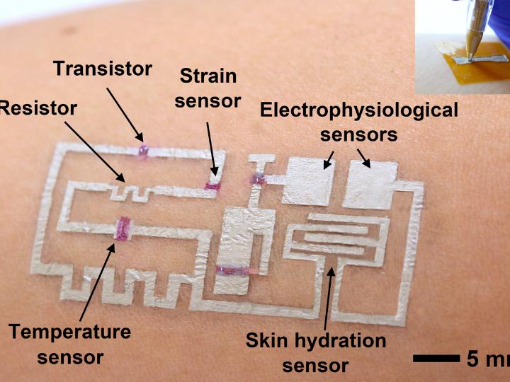
A team of researchers led by Cunjiang Yu, Bill D. Cook Associate Professor of Mechanical Engineering at the University of Houston, has developed a new form of electronics known as “drawn-on-skin electronics,” allowing multifunctional sensors and circuits to be drawn on the skin with an ink pen.

The advance, the researchers report in Nature Communications, allows for the collection of more precise, motion artifact-free health data, solving the long-standing problem of collecting precise biological data through a wearable device when the subject is in motion.

The imprecision may not be important when your FitBit registers 4,000 steps instead of 4,200, but sensors designed to check heart function, temperature and other physical signals must be accurate if they are to be used for diagnostics and treatment.

A July 30, 2020 University of Houston news release (also on EurekAlert) by Jeannie Kever, which originated the news item, goes on to explain why you might want to have electronics ‘drawn on your skin’,

The drawn-on-skin electronics are able to seamlessly collect data, regardless of the wearer’s movements.  

They also offer other advantages, including simple fabrication techniques that don’t require dedicated equipment.

“It is applied like you would use a pen to write on a piece of paper,” said Yu. “We prepare several electronic materials and then use pens to dispense them. Coming out, it is liquid. But like ink on paper, it dries very quickly.”

Wearable bioelectronics – in the form of soft, flexible patches attached to the skin – have become an important way to monitor, prevent and treat illness and injury by tracking physiological information from the wearer. But even the most flexible wearables are limited by motion artifacts, or the difficulty that arises in collecting data when the sensor doesn’t move precisely with the skin.

The drawn-on-skin electronics can be customized to collect different types of information, and Yu said it is expected to be especially useful in situations where it’s not possible to access sophisticated equipment, including on a battleground.

The electronics are able to track muscle signals, heart rate, temperature and skin hydration, among other physical data, he said. The researchers also reported that the drawn-on-skin electronics have demonstrated the ability to accelerate healing of wounds.

In addition to Yu, researchers involved in the project include Faheem Ershad, Anish Thukral, Phillip Comeaux, Yuntao Lu, Hyunseok Shim, Kyoseung Sim, Nam-In Kim, Zhoulyu Rao, Ross Guevara, Luis Contreras, Fengjiao Pan, Yongcao Zhang, Ying-Shi Guan, Pinyi Yang, Xu Wang and Peng Wang, all from the University of Houston, and Jiping Yue and Xiaoyang Wu from the University of Chicago.

The drawn-on-skin electronics are actually comprised of three inks, serving as a conductor, semiconductor and dielectric.

“Electronic inks, including conductors, semiconductors, and dielectrics, are drawn on-demand in a freeform manner to develop devices, such as transistors, strain sensors, temperature sensors, heaters, skin hydration sensors, and electrophysiological sensors,” the researchers wrote.

This research is supported by the Office of Naval Research and National Institutes of Health.

Caption: A new form of electronics known as “drawn-on-skin electronics” allows multifunctional sensors and circuits to be drawn on the skin with an ink pen. Credit: University of Houston

Here’s a link to and a citation for the paper,

Ultra-conformal drawn-on-skin electronics for multifunctional motion artifact-free sensing and point-of-care treatment by Faheem Ershad, Anish Thukral, Jiping Yue, Phillip Comeaux, Yuntao Lu, Hyunseok Shim, Kyoseung Sim, Nam-In Kim, Zhoulyu Rao, Ross Guevara, Luis Contreras, Fengjiao Pan, Yongcao Zhang, Ying-Shi Guan, Pinyi Yang, Xu Wang, Peng Wang, Xiaoyang Wu & Cunjiang Yu. Nature Communications volume 11, Article number: 3823 (2020) DOI: https://doi.org/10.1038/s41467-020-17619-1

This paper is open access.

The roles mathematics and light play in cellular communication

These are two entirely different types of research but taken together they help build a picture about how the cells in our bodies function.

Cells and light

An April 30, 2018 news item on phys.org describes work on controlling biology with light,

Over the past five years, University of Chicago chemist Bozhi Tian has been figuring out how to control biology with light.

A longterm science goal is devices to serve as the interface between researcher and body—both as a way to understand how cells talk among each other and within themselves, and eventually, as a treatment for brain or nervous system disorders [emphasis mine] by stimulating nerves to fire or limbs to move. Silicon—a versatile, biocompatible material used in both solar panels and surgical implants—is a natural choice.

In a paper published April 30 in Nature Biomedical Engineering, Tian’s team laid out a system of design principles for working with silicon to control biology at three levels—from individual organelles inside cells to tissues to entire limbs. The group has demonstrated each in cells or mice models, including the first time anyone has used light to control behavior without genetic modification.

“We want this to serve as a map, where you can decide which problem you would like to study and immediately find the right material and method to address it,” said Tian, an assistant professor in the Department of Chemistry.
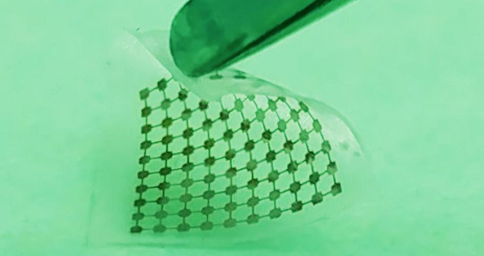
Researchers built this thin layer of silicon lace to modulate neural signals when activated by light. Courtesy of Yuanwen Jiang and Bozhi Tian

An April 30, 2018 University of Chicago news release by Louise Lerner, which originated the news item, describes the work in greater detail,

The scientists’ map lays out best methods to craft silicon devices depending on both the intended task and the scale—ranging from inside a cell to a whole animal.

For example, to affect individual brain cells, silicon can be crafted to respond to light by emitting a tiny ionic current, which encourages neurons to fire. But in order to stimulate limbs, scientists need a system whose signals can travel farther and are stronger—such as a gold-coated silicon material in which light triggers a chemical reaction.

The mechanical properties of the implant are important, too. Say researchers would like to work with a larger piece of the brain, like the cortex, to control motor movement. The brain is a soft, squishy substance, so they’ll need a material that’s similarly soft and flexible, but can bind tightly against the surface. They’d want thin and lacy silicon, say the design principles.

The team favors this method because it doesn’t require genetic modification or a power supply wired in, since the silicon can be fashioned into what are essentially tiny solar panels. (Many other forms of monitoring or interacting with the brain need to have a power supply, and keeping a wire running into a patient is an infection risk.)

They tested the concept in mice and found they could stimulate limb movements by shining light on brain implants. Previous research tested the concept in neurons.

“We don’t have answers to a number of intrinsic questions about biology, such as whether individual mitochondria communicate remotely through bioelectric signals,” said Yuanwen Jiang, the first author on the paper, then a graduate student at UChicago and now a postdoctoral researcher at Stanford. “This set of tools could address such questions as well as pointing the way to potential solutions for nervous system disorders.”

Other UChicago authors were Assoc. Profs. Chin-Tu Chen and Chien-Min Kao, Asst. Prof Xiaoyang, postdoctoral researchers Jaeseok Yi, Yin Fang, Xiang Gao, Jiping Yue, Hsiu-Ming Tsai, Bing Liu and Yin Fang, graduate students Kelliann Koehler, Vishnu Nair, and Edward Sudzilovsky, and undergraduate student George Freyermuth.

Other researchers on the paper hailed from Northwestern University, the University of Illinois at Chicago and Hong Kong Polytechnic University.

The researchers have also made this video illustrating their work,

via Gfycat Tiny silicon nanowires (in blue), activated by light, trigger activity in neurons. (Courtesy Yuanwen Jiang and Bozhi Tian)

Here’s a link to and a citation for the paper,

Rational design of silicon structures for optically controlled multiscale biointerfaces by Yuanwen Jiang, Xiaojian Li, Bing Liu, Jaeseok Yi, Yin Fang, Fengyuan Shi, Xiang Gao, Edward Sudzilovsky, Ramya Parameswaran, Kelliann Koehler, Vishnu Nair, Jiping Yue, KuangHua Guo, Yin Fang, Hsiu-Ming Tsai, George Freyermuth, Raymond C. S. Wong, Chien-Min Kao, Chin-Tu Chen, Alan W. Nicholls, Xiaoyang Wu, Gordon M. G. Shepherd, & Bozhi Tian. Nature Biomedical Engineering (2018) doi:10.1038/s41551-018-0230-1 Published: 30 April 2018

This paper is behind a paywall.

Mathematics and how living cells ‘think’

This May 2, 2018 Queensland University of Technology (QUT; Australia) press release is also on EurekAlert,

How does the ‘brain’ of a living cell work, allowing an organism to function and thrive in changing and unfavourable environments?

Queensland University of Technology (QUT) researcher Dr Robyn Araujo has developed new mathematics to solve a longstanding mystery of how the incredibly complex biological networks within cells can adapt and reset themselves after exposure to a new stimulus.

Her findings, published in Nature Communications, provide a new level of understanding of cellular communication and cellular ‘cognition’, and have potential application in a variety of areas, including new targeted cancer therapies and drug resistance.

Dr Araujo, a lecturer in applied and computational mathematics in QUT’s Science and Engineering Faculty, said that while we know a great deal about gene sequences, we have had extremely limited insight into how the proteins encoded by these genes work together as an integrated network – until now.

“Proteins form unfathomably complex networks of chemical reactions that allow cells to communicate and to ‘think’ – essentially giving the cell a ‘cognitive’ ability, or a ‘brain’,” she said. “It has been a longstanding mystery in science how this cellular ‘brain’ works.

“We could never hope to measure the full complexity of cellular networks – the networks are simply too large and interconnected and their component proteins are too variable.

“But mathematics provides a tool that allows us to explore how these networks might be constructed in order to perform as they do.

“My research is giving us a new way to look at unravelling network complexity in nature.”

Dr Araujo’s work has focused on the widely observed function called perfect adaptation – the ability of a network to reset itself after it has been exposed to a new stimulus.

“An example of perfect adaptation is our sense of smell,” she said. “When exposed to an odour we will smell it initially but after a while it seems to us that the odour has disappeared, even though the chemical, the stimulus, is still present.

“Our sense of smell has exhibited perfect adaptation. This process allows it to remain sensitive to further changes in our environment so that we can detect both very feint and very strong odours.

“This kind of adaptation is essentially what takes place inside living cells all the time. Cells are exposed to signals – hormones, growth factors, and other chemicals – and their proteins will tend to react and respond initially, but then settle down to pre-stimulus levels of activity even though the stimulus is still there.

“I studied all the possible ways a network can be constructed and found that to be capable of this perfect adaptation in a robust way, a network has to satisfy an extremely rigid set of mathematical principles. There are a surprisingly limited number of ways a network could be constructed to perform perfect adaptation.

“Essentially we are now discovering the needles in the haystack in terms of the network constructions that can actually exist in nature.

“It is early days, but this opens the door to being able to modify cell networks with drugs and do it in a more robust and rigorous way. Cancer therapy is a potential area of application, and insights into how proteins work at a cellular level is key.”

Dr Araujo said the published study was the result of more than “five years of relentless effort to solve this incredibly deep mathematical problem”. She began research in this field while at George Mason University in Virginia in the US.

Her mentor at the university’s College of Science and co-author of the Nature Communications paper, Professor Lance Liotta, said the “amazing and surprising” outcome of Dr Araujo’s study is applicable to any living organism or biochemical network of any size.

“The study is a wonderful example of how mathematics can have a profound impact on society and Dr Araujo’s results will provide a set of completely fresh approaches for scientists in a variety of fields,” he said.

“For example, in strategies to overcome cancer drug resistance – why do tumours frequently adapt and grow back after treatment?

“It could also help understanding of how our hormone system, our immune defences, perfectly adapt to frequent challenges and keep us well, and it has future implications for creating new hypotheses about drug addiction and brain neuron signalling adaptation.”

Hre’s a link to and a citation for the paper,

The topological requirements for robust perfect adaptation in networks of any size by Robyn P. Araujo & Lance A. Liotta. Nature Communicationsvolume 9, Article number: 1757 (2018) doi:10.1038/s41467-018-04151-6 Published: 01 May 2018

This paper is open access.
Small, soft, and electrically functional: an injectable biomaterial

This development could be looked at as a form of synthetic biology without the genetic engineering. From a July 1, 2016 news item on ScienceDaily,

Ideally, injectable or implantable medical devices should not only be small and electrically functional, they should be soft, like the body tissues with which they interact. Scientists from two UChicago labs set out to see if they could design a material with all three of those properties.

The material they came up with, published online June 27, 2016, in Nature Materials, forms the basis of an ingenious light-activated injectable device that could eventually be used to stimulate nerve cells and manipulate the behavior of muscles and organs.

“Most traditional materials for implants are very rigid and bulky, especially if you want to do electrical stimulation,” said Bozhi Tian, an assistant professor in chemistry whose lab collaborated with that of neuroscientist Francisco Bezanilla on the research.

The new material, in contrast, is soft and tiny — particles just a few micrometers in diameter (far less than the width of a human hair) that disperse easily in a saline solution so they can be injected. The particles also degrade naturally inside the body after a few months, so no surgery would be needed to remove them.

A July 1, 2016 University of Chicago news release (also on EurekAlert) by , which originated the news item, provides more detail,

Each particle is built of two types of silicon that together form a structure full of nano-scale pores, like a tiny sponge. And like a sponge, it is squishy — a hundred to a thousand times less rigid than the familiar crystalline silicon used in transistors and solar cells. “It is comparable to the rigidity of the collagen fibers in our bodies,” said Yuanwen Jiang, Tian’s graduate student. “So we’re creating a material that matches the rigidity of real tissue.”

The material constitutes half of an electrical device that creates itself spontaneously when one of the silicon particles is injected into a cell culture, or, eventually, a human body. The particle attaches to a cell, making an interface with the cell’s plasma membrane. Those two elements together — cell membrane plus particle — form a unit that generates current when light is shined on the silicon particle.

“You don’t need to inject the entire device; you just need to inject one component,” João L. Carvalho-de-Souza , Bezanilla’s postdoc said. “This single particle connection with the cell membrane allows sufficient generation of current that could be used to stimulate the cell and change its activity. After you achieve your therapeutic goal, the material degrades naturally. And if you want to do therapy again, you do another injection.”

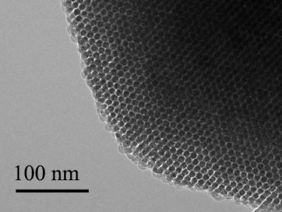
The scientists built the particles using a process they call nano-casting. They fabricate a silicon dioxide mold composed of tiny channels — “nano-wires” — about seven nanometers in diameter (less than 10,000 times smaller than the width of a human hair) connected by much smaller “micro-bridges.” Into the mold they inject silane gas, which fills the pores and channels and decomposes into silicon.

And this is where things get particularly cunning. The scientists exploit the fact the smaller an object is, the more the atoms on its surface dominate its reactions to what is around it. The micro-bridges are minute, so most of their atoms are on the surface. These interact with oxygen that is present in the silicon dioxide mold, creating micro-bridges made of oxidized silicon gleaned from materials at hand. The much larger nano-wires have proportionately fewer surface atoms, are much less interactive, and remain mostly pure silicon. [I have a note regarding ‘micro’ and ‘nano’ later in this posting.]

“This is the beauty of nanoscience,” Jiang said. “It allows you to engineer chemical compositions just by manipulating the size of things.”

Web-like nanostructure

Finally, the mold is dissolved. What remains is a web-like structure of silicon nano-wires connected by micro-bridges of oxidized silicon that can absorb water and help increase the structure’s softness. The pure silicon retains its ability to absorb light.

Transmission electron microscopy image shows an ordered nanowire array. The 100-nanometer scale bar is 1,000 times narrower than a hair. Courtesy of Tian Lab

Transmission electron microscopy image shows an ordered nanowire array. The 100-nanometer scale bar is 1,000 times narrower than a hair. Courtesy of
Tian Lab

The scientists have added the particles onto neurons in culture in the lab, shone light on the particles, and seen current flow into the neurons which activates the cells. The next step is to see what happens in living animals. They are particularly interested in stimulating nerves in the peripheral nervous system that connect to organs. These nerves are relatively close to the surface of the body, so near-infra-red wavelength light can reach them through the skin.

Tian imagines using the light-activated devices to engineer human tissue and create artificial organs to replace damaged ones. Currently, scientists can make engineered organs with the correct form but not the ideal function.

To get a lab-built organ to function properly, they will need to be able to manipulate individual cells in the engineered tissue. The injectable device would allow a scientist to do that, tweaking an individual cell using a tightly focused beam of light like a mechanic reaching into an engine and turning a single bolt. The possibility of doing this kind of synthetic biology without genetic engineering [emphasis mine] is enticing.

“No one wants their genetics to be altered,” Tian said. “It can be risky. There’s a need for a non-genetic system that can still manipulate cell behavior. This could be that kind of system.”

Tian’s graduate student Yuanwen Jiang did the material development and characterization on the project. The biological part of the collaboration was done in the lab of Francisco Bezanilla, the Lillian Eichelberger Cannon Professor of Biochemistry and Molecular Biology, by postdoc João L. Carvalho-de-Souza. They were, said Tian, the “heroes” of the work.

I was a little puzzled about the use of the word ‘micro’ in a context suggesting it was smaller than something measured at the nanoscale. Dr. Tian very kindly cleared up my confusion with this response in a July 4, 2016 email,

In fact, the definition of ‘micro’ and ’nano’ have been quite ambiguous in literature. For example, microporous materials (e.g., zeolite) usually refer to materials with pore sizes of less than 2 nm — this is defined based on IUPAC [International Union of Pure and Applied Chemistry] definition (http://goldbook.iupac.org/M03853.html). We used ‘micro-bridges’ because they come from the ‘micropores’ in the original template.

Thank you Dr. Tian for that very clear reply and Steve Koppes for forwarding my request to Dr. Tian!

Here’s a link to and a citation for the paper,

Heterogeneous silicon mesostructures for lipid-supported bioelectric interfaces by Yuanwen Jiang, João L. Carvalho-de-Souza, Raymond C. S. Wong, Zhiqiang Luo, Dieter Isheim, Xiaobing Zuo, Alan W. Nicholls, Il Woong Jung, Jiping Yue, Di-Jia Liu, Yucai Wang, Vincent De Andrade, Xianghui Xiao, Luizetta Navrazhnykh, Dara E. Weiss, Xiaoyang Wu, David N. Seidman, Francisco Bezanilla, & Bozhi Tian. Nature Materials (2016)  doi:10.1038/nmat4673 Published online 27 June 2016

This paper is behind a paywall.

I gather animal testing will be the next step as they continue to develop this exciting technology. Good luck!