Tag Archives: Johannes Gutenberg University Mainz (JGU)

Optimizing mRNA nanoparticles

The process of continuously working on scientific improvements is not always appreciated by outsiders such as myself. A December 28, 2021 news item on Nanowerk highlights research published (from 2019 and 2020) on improving delivery of mRNA used in vaccines (Note: A link has been removed),

The research neutron source Hein Maier-Leibnitz (FRM II) at the Technical University of Munich (TUM) is playing an important role in the investigation of mRNA nanoparticles similar to the ones used in the Covid-19 vaccines from vendors BioNTech and Moderna. Researchers at the Heinz Maier-Leibnitz Zentrum (MLZ) used the high neutron flux available in Garching to characterize various formulations for the mRNA vaccine and thus to lay the groundwork for improving the vaccine’s efficacy.

A December 27, 2021 TUM press release, which originated the news item, delves further into the science of improving something that already works well,

The idea of using messenger RNA (mRNA) as an active ingredient is a brilliant one: The molecule contains the specific blueprint for proteins which are then synthesize by the cell. This makes it generally possible to provide a very wide spectrum of different therapeutically effective proteins.

In the case of the Covid-19 vaccine, these are the proteins of the characteristic spikes on the surface of the Corona virus which are used for vaccination. The proteins are presented on the surface of immune cells; then the human immune system triggers defenses against these foreign proteins and thus against the Corona virus. The mRNA itself is completely broken down after only a few hours, a fact which is advantageous to the safety of these vaccines.

The road to the best packaging

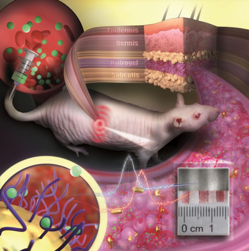
The mRNA has to be packaged appropriately in order to keep it from being broken down on the way to the cell by the ubiquitous enzymes of the human body. This is done using nanoparticles which can consist of a mixture of lipids or polymers.

The lipids are fat molecules similar to the molecules of the cell membrane and help deposit the mRNA in the interior of the cell. Lipids and biopolymers are then broken down or excreted by the body.

To this ends, the BioNTech formulation team led by Dr. Heinrich Haas worked together with the group led by Prof. Peter Langguth of the Pharmaceutical Technology department at the Johannes Gutenberg University Mainz’s Institute of Pharmaceutical and Biomedical Sciences. They developed a series of formulations in which the nanoparticles consisted of various mixtures of lipids and biopolymers already proved in pharmaceuticals.

In the light of neutrons

In order to compare the properties of variously composed nanoparticles with one another, the researchers subjected the nanoparticles to a wide range of investigations. In addition to x-ray and microscopic analyses, these investigations included radiation with neutrons using the instrument KWS-2, operated by the Forschungszentrum Jülich at the FRM II of the Technical University of Munich in Garching.

The neutrons are scattered in the interior of the nanoparticles, inter alia, on the hydrogen nuclei and are deflected from their paths in a characteristic way. This is the basis for conclusions about their distribution. If the hydrogen atoms of certain components – for example of the lipids only – are exchanged with heavy hydrogen, the chemical properties and the pharmaceutical efficacy do not change, but the scattering pattern of the neutrons does.

“This method makes it possible to selectively highlight parts of a complex multi-component morphology without changing the physical chemistry of the sample,” says Dr. Aurel Radulescu of the Jülich Centre for Neutron Science (JCNS), who is responsible for the instrument KWS-2 and who led the evaluation of the measurement results. “This makes it possible to depict structural properties which other methods can only barely render visible, if at all.”

The right degree of order is the key

In these analyses the research teams were interested in how efficiently the various formulations were able to transmit the mRNA into the cell, referred to as transfection. The researchers thus found out that the highest transfection rates were achieved with nanoparticles that are characterized by a certain type of internal arrangement.

“High levels of biological activity were registered whenever ordered and less ordered areas alternated in the interior of the nanoparticles in a characteristic manner. This could be a generally valid concept of structure-activity relationship which can be applied independently of the systems investigated here,” Dr. Heinrich Haas of BioNTech points out. A similarly low degree of order had also been found previously by the research teams using x-ray radiation in other lipid nanoparticles.

An improved procedure

In order to receive the desired structural properties lipids and biopolymers had to be combined with the mRNA using exactly defined procedures. Here the research team was able to show that the nanoparticles for packaging the mRNA could be produced in a single step, which means a significant simplification compared to the two-step procedure which was originally also investigated.

Thus a simplified method for the creation of mRNA nanoparticles with improved activity was ultimately found. “Such questions of practical producibility represent an important prerequisite for the possibility of developing pharmaceutical products,” says Prof. Langguth. In the future such concepts could be taken into account in the development of new mRNA-based therapeutic agents.

Here are links to and citations for the papers (Note: This is not my usual way of setting the links),

Hybrid Biopolymer and Lipid Nanoparticles with Improved Transfection Efficacy for mRNA by Christian D. Siewert, Heinrich Haas, Vera Cornet, Sara S. Nogueira, Thomas Nawroth, Lukas Uebbing, Antje Ziller, Jozef Al-Gousous, Aurel Radulescu, Martin A. Schroer, Clement E. Blanchet, Dmitri I. Svergun, Markus P. Radsak, Ugur Sahin and Peter Langguth. Cells 2020, 9(9), 2034 – DOI: 10.3390/cells9092034

This paper appears to be open access.

Investigation of charge ratio variation in mRNA – DEAE-dextran polyplex delivery systems by C. Siewert, H. Haas, T. Nawroth, A. Ziller, S. S. Nogueira, M. A. Schroer, C. E. Blanchet, D. I. Svergun, A. Radulescu, F. Bates, Y. Huesemann, M. P. Radsak, U. Sahin, P. Langguth. Biomaterials, 2019; DOI: 10.1016/j.biomaterials.2018.10.020

This paper is open access.

Polysarcosine-Functionalized Lipid Nanoparticles for Therapeutic mRNA Delivery by S S. Nogueira, A. Schlegel, K. Maxeiner, B. Weber, M. Barz, M. A. Schroer, C. E. Blanchet, D. I. Svergun, S. Ramishetti, D. Peer, P. Langguth, U. Sahin, H. Haas. ACS Appl. Nano Mater. 2020, 3, 11, 10634–10645 – DOI: 10.1021/acsanm.0c01834

This paper is behind a paywall.

Gold nanoparticle tattoo changes medical diagnostics?

The tattoos are in fact implantable sensors. Here’s more from an April 6, 2021 news item on ScienceDaily,

The idea of implantable sensors that continuously transmit information on vital values and concentrations of substances or drugs in the body has fascinated physicians and scientists for a long time. Such sensors enable the constant monitoring of disease progression and therapeutic success. However, until now implantable sensors have not been suitable to remain in the body permanently but had to be replaced after a few days or weeks. On the one hand, there is the problem of implant rejection because the body recognizes the sensor as a foreign object. On the other hand, the sensor’s color which indicates concentration changes has been unstable so far and faded over time. Scientists at Johannes Gutenberg University Mainz (JGU) have developed a novel type of implantable sensor which can be operated in the body for several months. The sensor is based on color-stable gold nanoparticles that are modified with receptors for specific molecules. Embedded into an artificial polymeric tissue, the nanogold is implanted under the skin where it reports changes in drug concentrations by changing its color.

An April 6, 2021 Johannes Gutenberg Universitaet Mainz press release (also on EurekAlert), which originated the news item, provides more detail about the proposed tattoo/implantable sensors,

Implant reports information as an “invisible tattoo”

Professor Carsten Sönnichsen’s research group at JGU has been using gold nanoparticles as sensors to detect tiny amounts of proteins in microscopic flow cells for many years. Gold nanoparticles act as small antennas for light: They strongly absorb and scatter it and, therefore, appear colorful. They react to alterations in their surrounding by changing color. Sönnichsen’s team has exploited this concept for implanted medical sensing.

To prevent the tiny particles from swimming away or being degraded by immune cells, they are embedded in a porous hydrogel with a tissue-like consistency. Once implanted under the skin, small blood vessels and cells grow into the pores. The sensor is integrated in the tissue and is not rejected as a foreign body. “Our sensor is like an invisible tattoo, not much bigger than a penny and thinner than one millimeter,” said Professor Carsten Sönnichsen, head of the Nanobiotechnology Group at JGU. Since the gold nanoparticles are infrared, they are not visible to the eye. However, a special kind of measurement device can detect their color noninvasively through the skin.

In their study published in Nano Letters, the JGU researchers implanted their gold nanoparticle sensors under the skin of hairless rats. Color changes in these sensors were monitored following the administration of various doses of an antibiotic. The drug molecules are transported to the sensor via the bloodstream. By binding to specific receptors on the surface of the gold nanoparticles, they induce color change that is dependent on drug concentration. Thanks to the color-stable gold nanoparticles and the tissue-integrating hydrogel, the sensor was found to remain mechanically and optically stable over several months.

Huge potential of gold nanoparticles as long-lasting implantable medical sensors

“We are used to colored objects bleaching over time. Gold nanoparticles, however, do not bleach but keep their color permanently. As they can be easily coated with various different receptors, they are an ideal platform for implantable sensors,” explained Dr. Katharina Kaefer, first author of the study.

The novel concept is generalizable and has the potential to extend the lifetime of implantable sensors. In future, gold nanoparticle-based implantable sensors could be used to observe concentrations of different biomarkers or drugs in the body simultaneously. Such sensors could find application in drug development, medical research, or personalized medicine, such as the management of chronic diseases.

Interdisciplinary team work brought success

Sönnichsen had the idea of using gold nanoparticles as implanted sensors already in 2004 when he started his research in biophysical chemistry as a junior professor in Mainz. However, the project was not realized until ten years later in cooperation with Dr. Thies Schroeder and Dr. Katharina Kaefer, both scientists at JGU. Schroeder was experienced in biological research and laboratory animal science and had already completed several years of research work in the USA. Kaefer was looking for an exciting topic for her doctorate and was particularly interested in the complex and interdisciplinary nature of the project. Initial results led to a stipend awarded to Kaefer by the Max Planck Graduate Center (MPGC) as well as financial support from Stiftung Rheinland-Pfalz für Innovation. “Such a project requires many people with different scientific backgrounds. Step by step we were able to convince more and more people of our idea,” said Sönnichsen happily. Ultimately, it was interdisciplinary teamwork that resulted in the successful development of the first functional implanted sensor with gold nanoparticles.

The researchers have provided an image which illustrates several elements described in the press release,

Caption: Gold nanoparticles embedded in a porous hydrogel can be implanted under the skin and used as medical sensors. The sensor is like an invisible tattoo revealing concentration changes of substances in the blood by color change. Credit: ill./©: Nanobiotechnology Group, JGU Department of Chemistry

Here’s a link to and a citation for the paper,

Implantable Sensors Based on Gold Nanoparticles for Continuous Long-Term Concentration Monitoring in the Body by Katharina Kaefer, Katja Krüger, Felix Schlapp, Hüseyin Uzun, Sirin Celiksoy, Bastian Flietel, Axel Heimann, Thies Schroeder, Oliver Kempski, and Carsten Sönnichsen. Nano Lett. 2021, XXXX, XXX, XXX-XXX DOI: https://doi.org/10.1021/acs.nanolett.1c00887 Publication Date:March 30, 2021 © 2021 The Authors. Published by American Chemical Society

This paper is behind a paywall.

Food nanoparticles and their effect on intestinal flora (i.e., your gut microbiome)

This work from Germany is largely speculative. The scientists seem to be interested in exploring how engineered nanoparticles and naturally occurring nanoparticles in food affect your gut. From a January 29, 2019 news item on ScienceDaily,

The intestinal microbiome is not only key for food processing but an accepted codeterminant for various diseases. Researchers led by the University Medical Center of Johannes Gutenberg University Mainz (JGU) identified effects of nanoparticles on intestinal microorganisms. The ultra-small particles adhere to intestinal microorganisms, thereby affecting their life cycle as well as cross talk with the host. One of the researchers’ observations was that nanoparticles’ binding inhibits the infection with Helicobacter pylori, a pathogen implicated in gastric cancer. The findings will stimulate further epidemiological studies and pave the way for the development of potential ‘probiotic’ nanoparticles for food. The discoveries were published in Science of Food.

A January 29, 2019 Johannes Gutenberg University Mainz (JGU) press release (also on EurekAlert), which originated the news item, provides more detail,

Due to their minute size, nanoparticles have unique characteristics and capabilities, such as adhering to microstructures. Nanotechnology is as an important driver of innovation for both consumer industry and medicine. In medicine, the focus is on improving diagnostics and therapeutics, while industry addresses mainly product optimization. Hence, synthetic nanoparticles are already used as additives to improve the characteristics of food. But how can we use nanotechnology more efficiently and safely in food? And are there unknown effects of nanoparticles, which need to be further exploited?

Nutrition strongly influences the diversity and composition of our microbiome. ‘Microbiome’ describes all colonizing microorganisms present in a human being, in particular, all the bacteria in the gut. In other words, your microbiome includes your intestinal flora as well as the microorganisms that colonize your skin, mouth, and nasal cavity.

Scientists and clinicians are interested in microbiomes because of their positive or negative effects on the host. These include modulation of our immune system, metabolism, vascular aging, cerebral functioning, and our hormonal system. The composition of the microbiome seems to play an important role for the development of various disorders, such as cardiovascular diseases, cancer, allergies, obesity, and even mental disorders. “Hence, nutrition and its containing nanoparticulates may affect the microbiome-host balance, finally influencing human health. In order to reduce potential risks and, ideally, promote health, the impact of dietary nanoparticles needs to be understood,” emphasized Professor David J. McClements from the Department of Food Science at the University of Massachusetts in Amherst, USA.

“Prior to our studies, nobody really looked whether and how nano-additives directly influence the gastrointestinal flora,” commented Professor Roland Stauber of the Department of Otolaryngology, Head, and Neck Surgery at the Mainz University Medical Center. “Hence, we studied at a wide range of technical nanoparticles with clearly defined properties in order to mimic what happens to currently used or potential future nanosized food additives. By simulating the journey of particles through the different environments of the digestive tract in the laboratory, we found that the all tested nanomaterials were indeed able to bind to bacteria.” explained Stauber.

The scientists discovered that these binding processes can have different outcomes. On the one hand, nanoparticle-bound microorganisms were less efficiently recognized by the immune system, which may lead to increased inflammatory responses. On the other hand, ‘nano-food’ showed beneficial effects. In cell culture models, silica nanoparticles inhibited the infectivity of Helicobacter pylori, which is considered to be one of the main agents involved in gastric cancer.

‘It was puzzling that we were able to also isolate naturally occurring nanoparticles from food, like beer, which showed similar effects. Nanoparticles in our daily food are not just those added deliberately but can also be generated naturally during preparation. Nanoparticulates are already omnipresent,” concluded Stauber.

The insights of the study will allow to derive strategies for developing and utilizing synthetic or natural nanoparticles to modulate the microbiome as beneficial ingredients in functional foods. “The challenge is to identify nanoparticles that fit the desired purpose, perhaps even as probiotic food supplements in the future. Challenge accepted,” emphasized Stauber and his team.

Here’s a link to and a citation for the paper,

Nanosized food additives impact beneficial and pathogenic bacteria in the human gut: a simulated gastrointestinal study by Svenja Siemer, Angelina Hahlbrock, Cecilia Vallet, David Julian McClements, Jan Balszuweit, Jens Voskuhl, Dominic Docter, Silja Wessler, Shirley K. Knauer, Dana Westmeier, & Roland H. Stauber. npj Science of Foodvolume 2, Article number: 22 (2018) DOI: https://doi.org/10.1038/s41538-018-0030-8 Published 04 December 2018

This paper is open access.