Tag Archives: John A. Rogers

Will you be my friend? Yes, after we activate our ultraminiature, wireless, battery-free, fully implantable devices

Perhaps I’m the only one who’s disconcerted?

Here’s the research (in text form) as to why we’re watching these scampering, momentary mouse friends, from a May 10, 2021 Northwestern University news release (also on EurekAlert) by Amanda Morris,

Northwestern University researchers are building social bonds with beams of light.

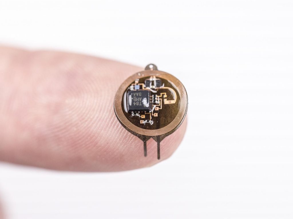
For the first time ever, Northwestern engineers and neurobiologists have wirelessly programmed — and then deprogrammed — mice to socially interact with one another in real time. The advancement is thanks to a first-of-its-kind ultraminiature, wireless, battery-free and fully implantable device that uses light to activate neurons.

This study is the first optogenetics (a method for controlling neurons with light) paper exploring social interactions within groups of animals, which was previously impossible with current technologies.

The research was published May 10 [2021] in the journal Nature Neuroscience.

The thin, flexible, wireless nature of the implant allows the mice to look normal and behave normally in realistic environments, enabling researchers to observe them under natural conditions. Previous research using optogenetics required fiberoptic wires, which restrained mouse movements and caused them to become entangled during social interactions or in complex environments.

“With previous technologies, we were unable to observe multiple animals socially interacting in complex environments because they were tethered,” said Northwestern neurobiologist Yevgenia Kozorovitskiy, who designed the experiment. “The fibers would break or the animals would become entangled. In order to ask more complex questions about animal behavior in realistic environments, we needed this innovative wireless technology. It’s tremendous to get away from the tethers.”

“This paper represents the first time we’ve been able to achieve wireless, battery-free implants for optogenetics with full, independent digital control over multiple devices simultaneously in a given environment,” said Northwestern bioelectronics pioneer John A. Rogers, who led the technology development. “Brain activity in an isolated animal is interesting, but going beyond research on individuals to studies of complex, socially interacting groups is one of the most important and exciting frontiers in neuroscience. We now have the technology to investigate how bonds form and break between individuals in these groups and to examine how social hierarchies arise from these interactions.”

Kozorovitskiy is the Soretta and Henry Shapiro Research Professor of Molecular Biology and associate professor of neurobiology in Northwestern’s Weinberg College of Arts and Sciences. She also is a member of the Chemistry of Life Processes Institute. Rogers is the Louis Simpson and Kimberly Querrey Professor of Materials Science and Engineering, Biomedical Engineering and Neurological Surgery in the McCormick School of Engineering and Northwestern University Feinberg School of Medicine and the director of the Querrey Simpson Institute for Bioelectronics.

Kozorovitskiy and Rogers led the work with Yonggang Huang, the Jan and Marcia Achenbach Professor in Mechanical Engineering at McCormick, and Zhaoqian Xie, a professor of engineering mechanics at Dalian University of Technology in China. The paper’s co-first authors are Yiyuan Yang, Mingzheng Wu and Abraham Vázquez-Guardado — all at Northwestern.

Promise and problems of optogenetics

Because the human brain is a system of nearly 100 billion intertwined neurons, it’s extremely difficult to probe single — or even groups of — neurons. Introduced in animal models around 2005, optogenetics offers control of specific, genetically targeted neurons in order to probe them in unprecedented detail to study their connectivity or neurotransmitter release. Researchers first modify neurons in living mice to express a modified gene from light-sensitive algae. Then they can use external light to specifically control and monitor brain activity. Because of the genetic engineering involved, the method is not yet approved in humans.

“It sounds like sci-fi, but it’s an incredibly useful technique,” Kozorovitskiy said. “Optogenetics could someday soon be used to fix blindness or reverse paralysis.”

Previous optogenetics studies, however, were limited by the available technology to deliver light. Although researchers could easily probe one animal in isolation, it was challenging to simultaneously control neural activity in flexible patterns within groups of animals interacting socially. Fiberoptic wires typically emerged from an animal’s head, connecting to an external light source. Then a software program could be used to turn the light off and on, while monitoring the animal’s behavior.

“As they move around, the fibers tugged in different ways,” Rogers said. “As expected, these effects changed the animal’s patterns of motion. One, therefore, has to wonder: What behavior are you actually studying? Are you studying natural behaviors or behaviors associated with a physical constraint?”

Wireless control in real time

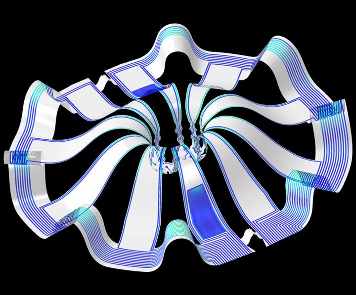
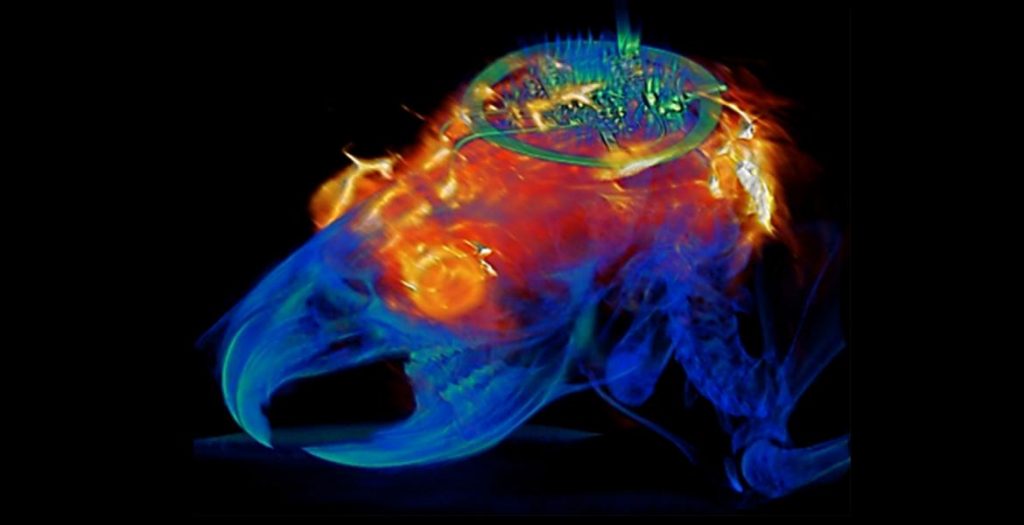
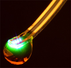
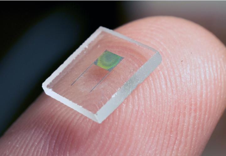
A world-renowned leader in wireless, wearable technology, Rogers and his team developed a tiny, wireless device that gently rests on the skull’s outer surface but beneath the skin and fur of a small animal. The half-millimeter-thick device connects to a fine, flexible filamentary probe with LEDs on the tip, which extend down into the brain through a tiny cranial defect.

The miniature device leverages near-field communication protocols, the same technology used in smartphones for electronic payments. Researchers wirelessly operate the light in real time with a user interface on a computer. An antenna surrounding the animals’ enclosure delivers power to the wireless device, thereby eliminating the need for a bulky, heavy battery.

Activating social connections

To establish proof of principle for Rogers’ technology, Kozorovitskiy and colleagues designed an experiment to explore an optogenetics approach to remote-control social interactions among pairs or groups of mice.

When mice were physically near one another in an enclosed environment, Kozorovitskiy’s team wirelessly synchronously activated a set of neurons in a brain region related to higher order executive function, causing them to increase the frequency and duration of social interactions. Desynchronizing the stimulation promptly decreased social interactions in the same pair of mice. In a group setting, researchers could bias an arbitrarily chosen pair to interact more than others.

“We didn’t actually think this would work,” Kozorovitskiy said. “To our knowledge, this is the first direct evaluation of a major long-standing hypothesis about neural synchrony in social behavior.”

Here’s a citation and a link to the paper,

Wireless multilateral devices for optogenetic studies of individual and social behaviors by Yiyuan Yang, Mingzheng Wu, Amy J. Wegener, Jose G. Grajales-Reyes, Yujun Deng, Taoyi Wang, Raudel Avila, Justin A. Moreno, Samuel Minkowicz, Vasin Dumrongprechachan, Jungyup Lee, Shuangyang Zhang, Alex A. Legaria, Yuhang Ma, Sunita Mehta, Daniel Franklin, Layne Hartman, Wubin Bai, Mengdi Han, Hangbo Zhao, Wei Lu, Yongjoon Yu, Xing Sheng, Anthony Banks, Xinge Yu, Zoe R. Donaldson, Robert W. Gereau IV, Cameron H. Good, Zhaoqian Xie, Yonggang Huang, Yevgenia Kozorovitskiy and John A. Rogers. Nature Neuroscience (2021)
DOI: https://doi.org/10.1038/s41593-021-00849-x Published 10 May 2021

This paper is behind a paywall.

This latest research seems to be the continuation of research featured here in a July 16, 2019 posting: “Controlling neurons with light: no batteries or wires needed.”

Cortical spheroids (like mini-brains) could unlock (larger) brain’s mysteries

A March 19, 2021 Northwestern University news release on EurekAlert announces the creation of a device designed to monitor brain organoids (for anyone unfamiliar with brain organoids there’s more information after the news),

A team of scientists, led by researchers at Northwestern University, Shirley Ryan AbilityLab and the University of Illinois at Chicago (UIC), has developed novel technology promising to increase understanding of how brains develop, and offer answers on repairing brains in the wake of neurotrauma and neurodegenerative diseases.

Their research is the first to combine the most sophisticated 3-D bioelectronic systems with highly advanced 3-D human neural cultures. The goal is to enable precise studies of how human brain circuits develop and repair themselves in vitro. The study is the cover story for the March 19 [March 17, 2021 according to the citation] issue of Science Advances.

The cortical spheroids used in the study, akin to “mini-brains,” were derived from human-induced pluripotent stem cells. Leveraging a 3-D neural interface system that the team developed, scientists were able to create a “mini laboratory in a dish” specifically tailored to study the mini-brains and collect different types of data simultaneously. Scientists incorporated electrodes to record electrical activity. They added tiny heating elements to either keep the brain cultures warm or, in some cases, intentionally overheated the cultures to stress them. They also incorporated tiny probes — such as oxygen sensors and small LED lights — to perform optogenetic experiments. For instance, they introduced genes into the cells that allowed them to control the neural activity using different-colored light pulses.

This platform then enabled scientists to perform complex studies of human tissue without directly involving humans or performing invasive testing. In theory, any person could donate a limited number of their cells (e.g., blood sample, skin biopsy). Scientists can then reprogram these cells to produce a tiny brain spheroid that shares the person’s genetic identity. The authors believe that, by combining this technology with a personalized medicine approach using human stem cell-derived brain cultures, they will be able to glean insights faster and generate better, novel interventions.

“The advances spurred by this research will offer a new frontier in the way we study and understand the brain,” said Shirley Ryan AbilityLab’s Dr. Colin Franz, co-lead author on the paper who led the testing of the cortical spheroids. “Now that the 3-D platform has been developed and validated, we will be able to perform more targeted studies on our patients recovering from neurological injury or battling a neurodegenerative disease.”

Yoonseok Park, postdoctoral fellow at Northwestern University and co-lead author, added, “This is just the beginning of an entirely new class of miniaturized, 3-D bioelectronic systems that we can construct to expand the capacity of the regenerative medicine field. For example, our next generation of device will support the formation of even more complex neural circuits from brain to muscle, and increasingly dynamic tissues like a beating heart.”

Current electrode arrays for tissue cultures are 2-D, flat and unable to match the complex structural designs found throughout nature, such as those found in the human brain. Moreover, even when a system is 3-D, it is extremely challenging to incorporate more than one type of material into a small 3-D structure. With this advance, however, an entire class of 3-D bioelectronics devices has been tailored for the field of regenerative medicine.

“Now, with our small, soft 3-D electronics, the capacity to build devices that mimic the complex biological shapes found in the human body is finally possible, providing a much more holistic understanding of a culture,” said Northwestern’s John Rogers, who led the technology development using technology similar to that found in phones and computers. “We no longer have to compromise function to achieve the optimal form for interfacing with our biology.”

As a next step, scientists will use the devices to better understand neurological disease, test drugs and therapies that have clinical potential, and compare different patient-derived cell models. This understanding will then enable a better grasp of individual differences that may account for the wide variation of outcomes seen in neurological rehabilitation.

“As scientists, our goal is to make laboratory research as clinically relevant as possible,” said Kristen Cotton, research assistant in Dr. Franz’s lab. “This 3-D platform opens the door to new experiments, discovery and scientific advances in regenerative neurorehabilitation medicine that have never been possible.”

Caption: Three dimensional multifunctional neural interfaces for cortical spheroids and engineered assembloids Credit: Northwestern University

As for what brain ogranoids might be, Carl Zimmer in an Aug. 29, 2019 article for the New York Times provides an explanation,

Organoids Are Not Brains. How Are They Making Brain Waves?

Two hundred and fifty miles over Alysson Muotri’s head, a thousand tiny spheres of brain cells were sailing through space.

The clusters, called brain organoids, had been grown a few weeks earlier in the biologist’s lab here at the University of California, San Diego. He and his colleagues altered human skin cells into stem cells, then coaxed them to develop as brain cells do in an embryo.

The organoids grew into balls about the size of a pinhead, each containing hundreds of thousands of cells in a variety of types, each type producing the same chemicals and electrical signals as those cells do in our own brains.

In July, NASA packed the organoids aboard a rocket and sent them to the International Space Station to see how they develop in zero gravity.

Now the organoids were stowed inside a metal box, fed by bags of nutritious broth. “I think they are replicating like crazy at this stage, and so we’re going to have bigger organoids,” Dr. Muotri said in a recent interview in his office overlooking the Pacific.

What, exactly, are they growing into? That’s a question that has scientists and philosophers alike scratching their heads.

On Thursday, Dr. Muotri and his colleagues reported that they  have recorded simple brain waves in these organoids. In mature human brains, such waves are produced by widespread networks of neurons firing in synchrony. Particular wave patterns are linked to particular forms of brain activity, like retrieving memories and dreaming.

As the organoids mature, the researchers also found, the waves change in ways that resemble the changes in the developing brains of premature babies.

“It’s pretty amazing,” said Giorgia Quadrato, a neurobiologist at the University of Southern California who was not involved in the new study. “No one really knew if that was possible.”

But Dr. Quadrato stressed it was important not to read too much into the parallels. What she, Dr. Muotri and other brain organoid experts build are clusters of replicating brain cells, not actual brains.

If you have the time, I recommend reading Zimmer’s article in its entirety. Perhaps not coincidentally, Zimmer has an excerpt titled “Lab-Grown Brain Organoids Aren’t Alive. But They’re Not Not Alive, Either.” published in Slate.com,

From Life’s Edge: The Search For What It Means To Be Alive by Carl Zimmer, published by Dutton, an imprint of Penguin Publishing Group, a division of Penguin Random House, LLC. Copyright © 2021 by Carl Zimmer.

Cleber Trujillo led me to a windowless room banked with refrigerators, incubators, and microscopes. He extended his blue-gloved hands to either side and nearly touched the walls. “This is where we spend half our day,” he said.

In that room Trujillo and a team of graduate students raised a special kind of life. He opened an incubator and picked out a clear plastic box. Raising it above his head, he had me look up at it through its base. Inside the box were six circular wells, each the width of a cookie and filled with what looked like watered-down grape juice. In each well 100 pale globes floated, each the size of a housefly head.

Getting back to the research about monitoring brain organoids, here’s a link to and a citation for the paper about cortical spheroids,

Three-dimensional, multifunctional neural interfaces for cortical spheroids and engineered assembloids by Yoonseok Park, Colin K. Franz, Hanjun Ryu, Haiwen Luan, Kristen Y. Cotton, Jong Uk Kim, Ted S. Chung, Shiwei Zhao, Abraham Vazquez-Guardado, Da Som Yang, Kan Li, Raudel Avila, Jack K. Phillips, Maria J. Quezada, Hokyung Jang, Sung Soo Kwak, Sang Min Won, Kyeongha Kwon, Hyoyoung Jeong, Amay J. Bandodkar, Mengdi Han, Hangbo Zhao, Gabrielle R. Osher, Heling Wang, KunHyuck Lee, Yihui Zhang, Yonggang Huang, John D. Finan and John A. Rogers. Science Advances 17 Mar 2021: Vol. 7, no. 12, eabf9153 DOI: 10.1126/sciadv.abf9153

This paper appears to be open access.

According to a March 22, 2021 posting on the Shirley Riley AbilityLab website, the paper is featured on the front cover of Science Advances (vol. 7 no. 12).

Controlling neurons with light: no batteries or wires needed

Caption: Wireless and battery-free implant with advanced control over targeted neuron groups. Credit: Philipp Gutruf

This January 2, 2019 news item on ScienceDaily describes the object seen in the above and describes the problem it’s designed to solve,

University of Arizona biomedical engineering professor Philipp Gutruf is first author on the paper Fully implantable, optoelectronic systems for battery-free, multimodal operation in neuroscience research, published in Nature Electronics.

Optogenetics is a biological technique that uses light to turn specific neuron groups in the brain on or off. For example, researchers might use optogenetic stimulation to restore movement in case of paralysis or, in the future, to turn off the areas of the brain or spine that cause pain, eliminating the need for — and the increasing dependence on — opioids and other painkillers.

“We’re making these tools to understand how different parts of the brain work,” Gutruf said. “The advantage with optogenetics is that you have cell specificity: You can target specific groups of neurons and investigate their function and relation in the context of the whole brain.”

In optogenetics, researchers load specific neurons with proteins called opsins, which convert light to electrical potentials that make up the function of a neuron. When a researcher shines light on an area of the brain, it activates only the opsin-loaded neurons.

The first iterations of optogenetics involved sending light to the brain through optical fibers, which meant that test subjects were physically tethered to a control station. Researchers went on to develop a battery-free technique using wireless electronics, which meant subjects could move freely.

But these devices still came with their own limitations — they were bulky and often attached visibly outside the skull, they didn’t allow for precise control of the light’s frequency or intensity, and they could only stimulate one area of the brain at a time.

A Dec. 21, 2018 University of Azrizona news release (published Jan. 2, 2019 on EurekAlert), which originated the news item, discusses the work in more detail,

“With this research, we went two to three steps further,” Gutruf said. “We were able to implement digital control over intensity and frequency of the light being emitted, and the devices are very miniaturized, so they can be implanted under the scalp. We can also independently stimulate multiple places in the brain of the same subject, which also wasn’t possible before.”

The ability to control the light’s intensity is critical because it allows researchers to control exactly how much of the brain the light is affecting — the brighter the light, the farther it will reach. In addition, controlling the light’s intensity means controlling the heat generated by the light sources, and avoiding the accidental activation of neurons that are activated by heat.

The wireless, battery-free implants are powered by external oscillating magnetic fields, and, despite their advanced capabilities, are not significantly larger or heavier than past versions. In addition, a new antenna design has eliminated a problem faced by past versions of optogenetic devices, in which the strength of the signal being transmitted to the device varied depending on the angle of the brain: A subject would turn its head and the signal would weaken.

“This system has two antennas in one enclosure, which we switch the signal back and forth very rapidly so we can power the implant at any orientation,” Gutruf said. “In the future, this technique could provide battery-free implants that provide uninterrupted stimulation without the need to remove or replace the device, resulting in less invasive procedures than current pacemaker or stimulation techniques.”

Devices are implanted with a simple surgical procedure similar to surgeries in which humans are fitted with neurostimulators, or “brain pacemakers.” They cause no adverse effects to subjects, and their functionality doesn’t degrade in the body over time. This could have implications for medical devices like pacemakers, which currently need to be replaced every five to 15 years.

The paper also demonstrated that animals implanted with these devices can be safely imaged with computer tomography, or CT, and magnetic resonance imaging, or MRI, which allow for advanced insights into clinically relevant parameters such as the state of bone and tissue and the placement of the device.

This image of a combined MRI (magnetic resonance image) and CT (computer tomography) scan bookends, more or less, the picture of the device which headed this piece,

Combined image analysis with MRI and CT results superimposed on a 3D rendering of the animal implanted with the programmable bilateral multi µ-ILED device. Courtesy: University of Arizona

Here’s a link to and a citation for the paper,

Fully implantable optoelectronic systems for battery-free, multimodal operation in neuroscience research by Philipp Gutruf, Vaishnavi Krishnamurthi, Abraham Vázquez-Guardado, Zhaoqian Xie, Anthony Banks, Chun-Ju Su, Yeshou Xu, Chad R. Haney, Emily A. Waters, Irawati Kandela, Siddharth R. Krishnan, Tyler Ray, John P. Leshock, Yonggang Huang, Debashis Chanda, & John A. Rogers. Nature Electronics volume 1, pages652–660 (2018) DOI: https://doi.org/10.1038/s41928-018-0175-0 Published 13 December 2018

This paper is behind a paywall.

On the verge of controlling neurons by wireless?

Scientists have controlled a mouse’s neurons with a wireless device (and unleashed some paranoid fantasies? well, mine if no one else’s) according to a July 16, 2015 news item on Nanowerk (Note: A link has been removed),

A study showed that scientists can wirelessly determine the path a mouse walks with a press of a button. Researchers at the Washington University School of Medicine, St. Louis, and University of Illinois, Urbana-Champaign, created a remote controlled, next-generation tissue implant that allows neuroscientists to inject drugs and shine lights on neurons deep inside the brains of mice. The revolutionary device is described online in the journal Cell (“Wireless Optofluidic Systems for Programmable In Vivo Pharmacology and Optogenetics”). Its development was partially funded by the [US] National Institutes of Health [NIH].

The researchers have made an image/illustration of the probe available,

Mind Bending Probe Scientists used soft materials to create a brain implant a tenth the width of a human hair that can wirelessly control neurons with lights and drugs. Courtesy of Jeong lab, University of Colorado Boulder.

A July 16, 2015 US NIH National Institute of Neurological Disorders and Stroke news release, which originated the news item, describes the study and notes that instructions for building the implant are included in the published study,

“It unplugs a world of possibilities for scientists to learn how brain circuits work in a more natural setting.” said Michael R. Bruchas, Ph.D., associate professor of anesthesiology and neurobiology at Washington University School of Medicine and a senior author of the study.

The Bruchas lab studies circuits that control a variety of disorders including stress, depression, addiction, and pain. Typically, scientists who study these circuits have to choose between injecting drugs through bulky metal tubes and delivering lights through fiber optic cables. Both options require surgery that can damage parts of the brain and introduce experimental conditions that hinder animals’ natural movements.

To address these issues, Jae-Woong Jeong, Ph.D., a bioengineer formerly at the University of Illinois at Urbana-Champaign, worked with Jordan G. McCall, Ph.D., a graduate student in the Bruchas lab, to construct a remote controlled, optofluidic implant. The device is made out of soft materials that are a tenth the diameter of a human hair and can simultaneously deliver drugs and lights.

“We used powerful nano-manufacturing strategies to fabricate an implant that lets us penetrate deep inside the brain with minimal damage,” said John A. Rogers, Ph.D., professor of materials science and engineering, University of Illinois at Urbana-Champaign and a senior author. “Ultra-miniaturized devices like this have tremendous potential for science and medicine.”

With a thickness of 80 micrometers and a width of 500 micrometers, the optofluidic implant is thinner than the metal tubes, or cannulas, scientists typically use to inject drugs. When the scientists compared the implant with a typical cannula they found that the implant damaged and displaced much less brain tissue.

The scientists tested the device’s drug delivery potential by surgically placing it into the brains of mice. In some experiments, they showed that they could precisely map circuits by using the implant to inject viruses that label cells with genetic dyes. In other experiments, they made mice walk in circles by injecting a drug that mimics morphine into the ventral tegmental area (VTA), a region that controls motivation and addiction.

The researchers also tested the device’s combined light and drug delivery potential when they made mice that have light-sensitive VTA neurons stay on one side of a cage by commanding the implant to shine laser pulses on the cells. The mice lost the preference when the scientists directed the device to simultaneously inject a drug that blocks neuronal communication. In all of the experiments, the mice were about three feet away from the command antenna.

“This is the kind of revolutionary tool development that neuroscientists need to map out brain circuit activity,” said James Gnadt, Ph.D., program director at the NIH’s National Institute of Neurological Disorders and Stroke (NINDS).  “It’s in line with the goals of the NIH’s BRAIN Initiative.”

The researchers fabricated the implant using semi-conductor computer chip manufacturing techniques. It has room for up to four drugs and has four microscale inorganic light-emitting diodes. They installed an expandable material at the bottom of the drug reservoirs to control delivery. When the temperature on an electric heater beneath the reservoir rose then the bottom rapidly expanded and pushed the drug out into the brain.

“We tried at least 30 different prototypes before one finally worked,” said Dr. McCall.

“This was truly an interdisciplinary effort,” said Dr. Jeong, who is now an assistant professor of electrical, computer, and energy engineering at University of Colorado Boulder. “We tried to engineer the implant to meet some of neurosciences greatest unmet needs.”

In the study, the scientists provide detailed instructions for manufacturing the implant.

“A tool is only good if it’s used,” said Dr. Bruchas. “We believe an open, crowdsourcing approach to neuroscience is a great way to understand normal and healthy brain circuitry.”

Here’s a link to and a citation for the paper,

Wireless Optofluidic Systems for Programmable In Vivo Pharmacology and Optogenetics by Jae-Woong Jeong, Jordan G. McCall, Gunchul Shin, Yihui Zhang, Ream Al-Hasani, Minku Kim, Shuo Li, Joo Yong Sim, Kyung-In Jang, Yan Shi, Daniel Y. Hong, Yuhao Liu, Gavin P. Schmitz, Li Xia, Zhubin He, Paul Gamble, Wilson Z. Ray, Yonggang Huang, Michael R. Bruchas, and John A. Rogers.  Cell, July 16, 2015. DOI: 10.1016/j.cell.2015.06.058

This paper is behind a paywall.

I last wrote about wireless activation of neurons in a May 28, 2014 posting which featured research at the University of Massachusetts Medical School.

Silk inks containing enzymes, antibiotics, antibodies, nanoparticles, and growth factors

There’s an almost euphoric tone to a June 16, 2015 Tufts University news release (also on EurekAlert) about research which has resulted in the ability to print silk-based inks,

Silk inks containing enzymes, antibiotics, antibodies, nanoparticles and growth factors could turn inkjet printing into a new, more effective tool for therapeutics, regenerative medicine and biosensing, according to new research led by Tufts University  biomedical engineers and published June 16 [2015] in the journal Advanced Materials online in advance of print.

Until now, heat used in the inkjet printing process made using silk a challenge (as it does for cellulose nanomaterials used in 3D printers, noted in my June 17, 2015 posting), from the Tufts news release,

Inkjet printing is one of the most immediate and accessible forms of printing technology currently available, according to the researchers, and ink-jet printing of biomolecules has been previously proposed by scientists. However, the heat-sensitive nature of these unstable compounds means printed materials rapidly lose functionality, limiting their use.

Enter purified silk protein, or fibroin, which offers intrinsic strength and protective properties that make it well-suited for a range of biomedical and optoelectronic applications. This natural polymer is an ideal “cocoon” that can stabilize compounds such as enzymes, antibodies and growth factors while lending itself to many different mechanically robust formats, said Fiorenzo Omenetto, Ph.D., senior author on the paper and associate dean for research and Frank C. Doble Professor of Engineering at Tufts School of Engineering.

“We thought that if we were able to develop an inkjet-printable silk solution, we would have a universal building block to generate multiple functional printed formats that could lead to a wide variety of applications in which inks remain active over time,” he said.

By using this simple approach and starting with the same base material, the research team created and tested a “custom library” of inkjet-printable, functional silk inks doped with a variety of components:

  • Bacterial-sensing polydiacetylenes (PDAs) printed on surgical gloves; the word “contaminated” printed on the glove changed from blue to red after exposure to E. coli
  • Proteins that stimulate bone growth (BMP-2) printed on a plastic dish to test topographical control of directed tissue growth
  • Sodium ampicillin printed on a bacterial culture to test the effectiveness of a topographical distribution of the antibiotic
  • Gold nanoparticles printed on paper, for possible application in photonics and biology (e.g., color engineering, surface plasmon resonance based sensing and bio-imaging)
  • Enzymes printed on paper to test the ability of the ink to entrain small functional biomolecules

The researchers, who included collaborators from the University of Illinois at Urbana-Champaign, foresee wide potential for future investigation and application of this technology.

For example, Omenetto envisions more work on the bio-sensing gloves, which he says could selectively react to different pathological agents. The ability to print antibiotics in topographical patterns could address the need for “smart” bandages, where therapeutics are incorporated and delivered to match a complex injury.

The published research was restricted to one ink cartridge, but the scientists believe it could extend to multi-cartridge printing combining complex functions.

Omenetto and Kaplan are pioneers in the use of silk as an alternative to plastics. Omenetto’s 2011 TED Talk called silk a “new old material” that could have a profound impact in many technical fields.

Here’s a link to and citation for the paper,

Inkjet Printing of Regenerated Silk Fibroin: From Printable Forms to Printable Functions by Hu Tao, Benedetto marelli, Miaomiao Yang, Bo An, Serdar Onses, John A. Rogers, David L. Kaplan, & Fiorenzo G. Omenetto. Advanced Materials DOI: 10.1002/adma.201501425 First published: 16 June 2015

This article is behind a paywall.

Microbattery from the University of Illinois

Caption: This is an image of the holographically patterned microbattery. Credit: University of Illinois

Caption: This is an image of the holographically patterned microbattery.
Credit: University of Illinois

Hard to believe that’s a battery but the researchers at the University of Illinois  assure us this is so according to a May 11, 2015 news item on Nanowerk,

By combining 3D holographic lithography and 2D photolithography, researchers from the University of Illinois at Urbana-Champaign have demonstrated a high-performance 3D microbattery suitable for large-scale on-chip integration with microelectronic devices.

“This 3D microbattery has exceptional performance and scalability, and we think it will be of importance for many applications,” explained Paul Braun, a professor of materials science and engineering at Illinois. “Micro-scale devices typically utilize power supplied off-chip because of difficulties in miniaturizing energy storage technologies. A miniaturized high-energy and high-power on-chip battery would be highly desirable for applications including autonomous microscale actuators, distributed wireless sensors and transmitters, monitors, and portable and implantable medical devices.”

A May 11, 2015 University of Illinois news release on EurkeAlert, which originated the news item, provides some insight into and detail about the research,

“Due to the complexity of 3D electrodes, it is generally difficult to realize such batteries, let alone the possibility of on-chip integration and scaling. In this project, we developed an effective method to make high-performance 3D lithium-ion microbatteries using processes that are highly compatible with the fabrication of microelectronics,” stated Hailong Ning, a graduate student in the Department of Materials Science and Engineering and first author of the article, “Holographic Patterning of High Performance on-chip 3D Lithium-ion Microbatteries,” appearing in Proceedings of the National Academy of Sciences.

“We utilized 3D holographic lithography to define the interior structure of electrodes and 2D photolithography to create the desired electrode shape.” Ning added. “This work merges important concepts in fabrication, characterization, and modeling, showing that the energy and power of the microbattery are strongly related to the structural parameters of the electrodes such as size, shape, surface area, porosity, and tortuosity. A significant strength of this new method is that these parameters can be easily controlled during lithography steps, which offers unique flexibility for designing next-generation on-chip energy storage devices.”

Enabled by a 3D holographic patterning technique–where multiple optical beams interfere inside the photoresist creating a desirable 3D structure–the battery possesses well-defined, periodically structured porous electrodes, that facilitates the fast transports of electrons and ions inside the battery, offering supercapacitor-like power.

“Although accurate control on the interfering optical beams is required to construct 3D holographic lithography, recent advances have significantly simplified the required optics, enabling creation of structures via a single incident beam and standard photoresist processing. This makes it highly scalable and compatible with microfabrication,” stated John Rogers, a professor of materials science and engineering, who has worked with Braun and his team to develop the technology.

“Micro-engineered battery architectures, combined with high energy material such as tin, offer exciting new battery features including high energy capacity and good cycle lives, which provide the ability to power practical devices,” stated William King, a professor of mechanical science and engineering, who is a co-author of this work.

Here’s a link to and a citation for the paper,

Holographic patterning of high-performance on-chip 3D lithium-ion microbatteries by Hailong Ning, James H. Pikul, Runyu Zhang, Xuejiao Li, Sheng Xu, Junjie Wang, John A. Rogers, William P. King, and Paul V. Braun. PNAS doi: 10.1073/pnas.1423889112

This paper is behind a paywall.

E-tattoo without the nanotech

John Rogers and his team at the University of Illinois and a colleague’s (Yonggang Huang) team at Northwestern University have devised an ‘electronic tattoo’ (a soft, stick-on patch) made up from materials that anyone can purchase off-the-shelf. Rogers is known for his work with nanomaterials (my Aug. 10, 2012 posting titled ‘Surgery with fingertip control‘ mentioned a silicon nanomembrane that can be fitted onto the fingertips for possible use in surgical procedures) and with electronics (my Aug. 12, 2011 posting titled: ‘Electronic tattoos‘ mentioned his earlier attempts at developing e-tattoos).

This latest effort from Rogers and his multi-university team is mentioned in an April 4, 2014 article by Mark Wilson for Fast Company,

About a year ago, University of Illinois researcher John Rogers revealed a pretty amazing creation: a circuit that, rather than living on an inflexible board, could stick to and move with someone’s skin just like an ink stamp. But like any early research, it was mostly a proof-of-concept, and it would require relatively expensive, custom-printed electronics to work.

Today, Rogers, in conjunction with Northwestern University’s Yonggang Huang, has published details on version 2.0 in Science, revealing that this once-esoteric project has more immediate, mass market appeal.

… It means that you could create a wearable electronic that’s one-part special sticky circuit board, every other part whatever-the-hell-you-manufactured-in-China. This flexible circuit could accommodate a stock battery, an accelerometer, a Wi-Fi chip, and a Bluetooth circuitry, for instance, all living on your skin rather than inside your iPhone. And as an added bonus, it would be relatively cheap.

A University of Illinois April ?, 2014 news release describes Rogers, his multi-university team, and their current (pun intended) e-tattoo,

Engineers at the University of Illinois at Urbana-Champaign and Northwestern University have demonstrated thin, soft stick-on patches that stretch and move with the skin and incorporate commercial, off-the-shelf chip-based electronics for sophisticated wireless health monitoring.

The patches stick to the skin like a temporary tattoo and incorporate a unique microfluidic construction with wires folded like origami to allow the patch to bend and flex without being constrained by the rigid electronics components. The patches could be used for everyday health tracking – wirelessly sending updates to your cellphone or computer – and could revolutionize clinical monitoring such as EKG and EEG testing – no bulky wires, pads or tape needed.

“We designed this device to monitor human health 24/7, but without interfering with a person’s daily activity,” said Yonggang Huang, the Northwestern University professor who co-led the work with Illinois professor John A. Rogers. “It is as soft as human skin and can move with your body, but at the same time it has many different monitoring functions. What is very important about this device is it is wirelessly powered and can send high-quality data about the human body to a computer, in real time.”

The researchers did a side-by-side comparison with traditional EKG and EEG monitors and found the wireless patch performed equally to conventional sensors, while being significantly more comfortable for patients. Such a distinction is crucial for long-term monitoring, situations such as stress tests or sleep studies when the outcome depends on the patient’s ability to move and behave naturally, or for patients with fragile skin such as premature newborns.

Rogers’ group at Illinois previously demonstrated skin electronics made of very tiny, ultrathin, specially designed and printed components. While those also offer high-performance monitoring, the ability to incorporate readily available chip-based components provides many important, complementary capabilities in engineering design, at very low cost.

“Our original epidermal devices exploited specialized device geometries – super thin, structured in certain ways,” Rogers said. “But chip-scale devices, batteries, capacitors and other components must be re-formulated for these platforms. There’s a lot of value in complementing this specialized strategy with our new concepts in microfluidics and origami interconnects to enable compatibility with commercial off-the-shelf parts for accelerated development, reduced costs and expanded options in device types.”

The multi-university team turned to soft microfluidic designs to address the challenge of integrating relatively big, bulky chips with the soft, elastic base of the patch. The patch is constructed of a thin elastic envelope filled with fluid. The chip components are suspended on tiny raised support points, bonding them to the underlying patch but allowing the patch to stretch and move.

One of the biggest engineering feats of the patch is the design of the tiny, squiggly wires connecting the electronics components – radios, power inductors, sensors and more. The serpentine-shaped wires are folded like origami, so that no matter which way the patch bends, twists or stretches, the wires can unfold in any direction to accommodate the motion. Since the wires stretch, the chips don’t have to.

Skin-mounted devices could give those interested in fitness tracking a more complete and accurate picture of their activity level.

“When you measure motion on a wristwatch type device, your body is not very accurately or reliably coupled to the device,” said Rogers, a Swanlund Professor of Materials Science and Engineering at the U. of I. “Relative motion causes a lot of background noise. If you have these skin-mounted devices and an ability to locate them on multiple parts of the body, you can get a much deeper and richer set of information than would be possible with devices that are not well coupled with the skin. And that’s just the beginning of the rich range of accurate measurements relevant to physiological health that are possible when you are softly and intimately integrated onto the skin.”

The researchers hope that their sophisticated, integrated sensing systems could not only monitor health but also could help identify problems before the patient may be aware. For example, according to Rogers, data analysis could detect motions associated with Parkinson’s disease at its onset.

“The application of stretchable electronics to medicine has a lot of potential,” Huang said. “If we can continuously monitor our health with a comfortable, small device that attaches to our skin, it could be possible to catch health conditions before experiencing pain, discomfort and illness.”

Here’s a link to and a citation for the paper,

Soft Microfluidic Assemblies of Sensors, Circuits, and Radios for the Skin by Sheng Xu, Yihui Zhang, Lin Jia, Kyle E. Mathewson, Kyung-In Jang, Jeonghyun Kim, Haoran Fu, Xian Huang, Pranav Chava, Renhan Wang, Sanat Bhole, Lizhe Wang, Yoon Joo Na, Yue Guan, Matthew Flavin, Zheshen Han, Yonggang Huang, & John A. Rogers. Science 4 April 2014: Vol. 344 no. 6179 pp. 70-74 DOI: 10.1126/science.1250169

This paper is behind a paywall.

American Assocation for the Advancement of Science (AAAS) meeting in Chicago, Illinois (13 – 17 February 2014)

The 2014 annual meeting of the American Association for the Advancement of Science (AAAS) will take place Feb. 13 – 17, 2014 in Chicago (one of my favourite places), Illinois. It’s always interesting to take a look at the programme and here’s a few of the items I found interesting,

Thursday, Feb. 13, 2014  the AAAS has arranged a number of talks about ‘communicating science and, as usual, bloggers, etc. are confined to presenting under the rubric of social media:

9:00 AM-10:30 AM

Seminar: Communicating Science

11:00 AM-12:30 PM

Seminar: Communicating Science

Engaging with Social Media

To be more specific, here’s the list of presenters for the ‘Journalist’ talk (Note: I have removed links),

Moderator:
Cornelia Dean, The New York Times and Brown University
Speakers:
Carl Zimmer, Independent Science Journalist [Note: Zimmer writes for the NY Times and other prestigious print publications, as well as, being a blogger]

Robert Lee Hotz, The Wall Street Journal

David Baron, Public Radio International

Paula Apsell, NOVA [science program on the US PBS {Public Broadcasting Service} network)

[emphases mine]

Meanwhile, we have this for social media,

Moderator:
Dominique Brossard, University of Wisconsin
Speakers:
Kim Cobb, University of Georgia
Navigating the Science-Social Media Space: Pitfalls and Opportunities
Danielle N. Lee, Cornell University
Raising STEM Awareness Among Under-Served and Under-Represented Audiences
Maggie Koerth-Baker, BoingBoing.net
What’s the Point of Social Media?

It’s nice to see Danielle N. Lee as one of the presenters. Her blog, The Urban Scientist is on the Scientific American blog network (she also featured as a whistle blower and more in the 2013 science blogging scandals [my first post on the topic was Oct. 18, 2013 towards the end of the scandals and I mused on the scandals and discussed  gender in an end-of-year Dec. 31, 2013 posting ) and there’s of course, someone representing BoingBoing, an online publisher,which was conceptualized as a magazine and has now evolved into a group blog.

My basic thesis is that blogs and such are emerging as part of the science media landscape and the types of sessions which isolate bloggers, etc.  do not acknowledge that fact. Yes, it’s true that Zimmer blogs but I can guarantee that the discussion will revolve exclusively around his high profile publishers such as the NY Times and how the participants can get their stories in front of mainstream media journalists and as for the social media session that’s going to focus on how scientists can directly approach their publics.

Moving on, there’s a nanotechnology aspect to the following presentation, although you’d never guess it from the title,

 Preserving Our Cultural Heritage: Science in the Service of Art
Friday, 14 February 2014: 10:00 AM-11:30 AM
Acapulco (Hyatt Regency Chicago)
In 2009 a group of chemists and materials scientists from a wide range of institutions came together for a workshop on “Chemistry and Materials Research at the Interface Between Science and Art,” co-sponsored by the Andrew W. Mellon Foundation and the National Science Foundation. One of the workshop conclusions was that scientists in academia need to be encouraged to collaborate with their peers in cultural heritage institutions, to both increase scientist knowledge of this heritage and also to develop the necessary tools and apply the science to be able to preserve it. The session covers different collaborations that are ongoing in this area, relating to different mediums of art and different technologies that can be applied. The session will also include recent results and successes in this process, both in better understanding of materials as well as in developments for their conservation. The discussion will also address what is needed for collaborations like this to continue to flourish and grow.

One doesn’t get to the ‘nano’ part until looking at the speakers’ list (Note: Links have been removed),

Organizer:
Nicholas Bigelow, University of Rochester
Co-Organizer:
Leonor Sierra, University of Rochester
Speakers:
Nicholas Bigelow, University of Rochester
21st Century Tools for 19th Century Nanotechnology ‘[emphasis mine]
Richard Van Duyne, Northwestern University
Detecting Organic Dyestuffs in Art with SERS
Anikó Bezur, Yale University
Aiming for a Perfect Match: Pairing Collections-Based Scientific Research with Academia

The 19th Century nanotechnology referred to in the title of Biglow’s talk is the daggeureotype (a type of 19th century photographic process) which gained a lot of attention in the last few years when a display of irreplaceable pieces started showing signs of visible (25 pieces) and catastrophic (five pieces) deterioration. There’s more about this fascinating story in my Jan. 10, 2013 posting.

Saturday, Feb.15, 2014, Alan Alda will be at the meeting as a plenary speaker,

Alan Alda: Getting Beyond a Blind Date with Science
Plenary Lecture
Saturday, 15 February 2014: 5:00 PM-6:00 PM
Imperial Ballroom (Fairmont Chicago)
Alan Alda is an actor, writer, director, and visiting professor at the Alan Alda Center for Communicating Science at Stony Brook University, where he helps current and future scientists learn to communicate more clearly and vividly with the public. In collaboration with theater arts faculty at Stony Brook, he is pioneering the use of improvisational theater exercises to help scientists connect more directly with people outside their field. Alda is best known for his award-winning work in movies, theater, and television, but he also has a distinguished record in the public communication of science. For 13 years he hosted the PBS series Scientific American Frontiers, which he has called “the best thing I ever did in front of a camera.” After interviewing hundreds of scientists around the world, he became convinced that many researchers have wonderful stories but need to learn how to tell them better. That realization inspired the creation of Stony Brook’s multidisciplinary Alan Alda Center for Communicating Science in 2009.

The last two sessions I’m highlighting are on standard nanotechnology topics. On Sunday, Feb. 16, 2014, there’s

Nanoelectronics for Renewable Energy: How Nanoscale Innovations Address Global Needs
Sunday, 16 February 2014: 1:30 PM-4:30 PM
Regency B (Hyatt Regency Chicago)
Sometimes it’s possible to get a handle on the world’s biggest problems by thinking creatively on a very small scale—and advances in the rapidly maturing field of nanoelectronics prove it. Innovations that hold promise for broader and faster adoption of renewable energy technologies loom large against a backdrop of population growth, rapid industrialization in developing countries, and initiatives to decrease reliance on both fossil fuels and nuclear power. In this symposium, researchers from the U.S. and Europe will review the latest progress in nanoelectronics for renewable energy across a series of interrelated programs. For instance, new manufacturing approaches such as nanoimprinting, nanotransfer, and spray-on fabrication of organic semiconductors not only point the way toward low-cost production of large-scale electronics such as solar panels, they also enable and inspire novel nanoelectronic device designs. These device-level innovations range from ultrasensitive molecular sensors to nanomagnet logic circuits, and they are of particular interest in solar energy applications. Many lines of research appear to be converging on nanostructure-based solar cells that will be vastly more efficient in capturing sunlight (or even heat) and converting it to electrical power. In addition to outlining these promising paths toward higher-efficiency, lower-cost photovoltaics, the symposium will highlight some of the remaining hurdles, including needed advances in fundamental science.
Organizer:
Patrick Regan, Technical University Munich
Co-organizers:
William Gilroy, University of Notre Dame
and Hillary Sanctuary, Swiss Federal Institute of Technology (EPFL)

On Monday, Feb. 17, 2014,  nanotechnology features in the final plenary session,

John A. Rogers: Stretchy Electronics That Dissolve in Your Body
Plenary Lecture
Monday, 17 February 2014: 8:30 AM-9:30 AM
Imperial Ballroom (Fairmont Chicago)
Dr. John Rogers’ research includes fundamental and applied aspects of nano- and molecular scale fabrication. He also studies materials and patterning techniques for unusual electronic and photonic devices, with an emphasis on bio-integrated and bio-inspired systems. He received a Ph.D. in physical chemistry from Massachusetts Institute of Technology in 2005. He has published more than 350 papers and is an inventor on over 80 patents and patent applications, many of which are licensed or in active use by large companies and startups that he co-founded. He previously worked for Bell Laboratories as director of its research program in condensed matter physics. He has received recognition including a MacArthur Fellowship from the John D. and Catherine T. MacArthur Foundation, the Lemelson-MIT Prize, the National Security Science and Engineering Faculty Fellowship from the U.S. Department of Defense, the George Smith Award from IEEE, the Robert Henry Thurston Award from American Society of Mechanical Engineers, the Mid-Career Researcher Award from Materials Research Society, the Leo Hendrick Baekeland Award from the American Chemical Society, and the Daniel Drucker Eminent Faculty Award from the University of Illinois.
Speaker:
John Rogers, Ph. D., University of Illinois, Urbana-Champaign

You can find out more about registration and public events for the AAAS 2014 annual meeting here.

Organizer:
Nicholas Bigelow, University of Rochester
Co-Organizer:
Leonor Sierra, University of Rochester
Speakers:
Nicholas Bigelow, University of Rochester
21st Century Tools for 19th Century Nanotechnology

Richard Van Duyne, Northwestern University
Detecting Organic Dyestuffs in Art with SERS

Anikó Bezur, Yale University
Aiming for a Perfect Match: Pairing Collections-Based Scientific Research with Academia

Seeing things from a bug’s perspective—a new type of digital camera

The new digital cameras exploit large arrays of tiny focusing lenses and miniaturized detectors in hemispherical layouts, just like eyes found in arthropods

The new digital cameras exploit large arrays of tiny focusing lenses and miniaturized detectors in hemispherical layouts, just like eyes found in arthropods

A May 1, 2013 news item on Nanowerk provides some details about a new ‘bug-eyed’ digital camera,

An interdisciplinary team of researchers has created the first digital cameras with designs that mimic those of ocular systems found in dragonflies, bees, praying mantises and other insects. This class of technology offers exceptionally wide-angle fields of view, with low aberrations, high acuity to motion, and nearly infinite depth of field.

Taking cues from Mother Nature, the cameras exploit large arrays of tiny focusing lenses and miniaturized detectors in hemispherical layouts, just like eyes found in arthropods. The devices combine soft, rubbery optics with high performance silicon electronics and detectors, using ideas first established in research on skin and brain monitoring systems by John A. Rogers, a Swanlund Chair Professor at the University of Illinois at Urbana-Champaign, and his collaborators.

The May 1, 2013 University of Illinois news release by John Kubetz, which originated the news item, describes the special properties of an insect eye and how the camera mimics those properties,

Eyes in arthropods use compound designs, in which arrays of smaller eyes act together to provide image perception. Each small eye, known as an ommatidium, consists of a corneal lens, a crystalline cone, and a light sensitive organ at the base. The entire system is configured to provide exceptional properties in imaging, many of which lie beyond the reach of existing man-made cameras.

The researchers developed new ideas in materials and fabrication strategies allowing construction of artificial ommatidia in large, interconnected arrays in hemispherical layouts. Building such systems represents a daunting task, as all established camera technologies rely on bulk glass lenses and detectors constructed on the planar surfaces of silicon wafers which cannot be bent or flexed, much less formed into a hemispherical shape.

“A critical feature of our fly’s eye cameras is that they incorporate integrated microlenses, photodetectors, and electronics on hemispherically curved surfaces,” said Jianliang Xiao, an assistant professor of mechanical engineering at University of Colorado Boulder and coauthor of the study. “To realize this outcome, we used soft, rubbery optics bonded to detectors/electronics in mesh layouts that can be stretched and deformed, reversibly and without damage.”

On a more technical note, from the news release,

The fabrication starts with electronics, detectors and lens arrays formed on flat surfaces using advanced techniques adapted from the semiconductor industry, said Xiao [Jianliang Xiao, an assistant professor of mechanical engineering at University of Colorado Boulder and coauthor of the study], who began working on the project as a postdoctoral researcher in Rogers’ lab at Illinois. The lens sheet—made from a polymer material similar to a contact lens—and the electronics/detectors are then aligned and bonded together. Pneumatic pressure deforms the resulting system into the desired hemispherical shape, in a process much like blowing up a balloon, but with precision engineering control.

The individual electronic detectors and microlenses are coupled together to avoid any relative motion during this deformation process. Here, the spaces between these artificial ommatidia can stretch to allow transformation in geometry from planar to hemispherical. The electrical interconnections are thin, and narrow, in filamentary serpentine shapes; they deform as tiny springs during the stretching process.

According to the researchers, each microlens produces a small image of an object with a form dictated by the parameters of the lens and the viewing angle. An individual detector responds only if a portion of the image formed by the associated microlens overlaps the active area. The detectors stimulated in this way produce a sampled image of the object that can then be reconstructed using models of the optics.

Katherine Bourzac in her May 1, 2013 article for Nature magazine provides some additional insight and a perspective (intentional wordplay) from a researcher who has an idea of how he might like to integrate this new type of camera into his own work,

Insect eyes are made up of hundreds or even thousands of light-sensing structures called ommatidia. Each contains a lens and a cone that funnels light to a photosensitive organ. The long, thin ommatidia are bunched together to form the hemispherical eye, with each ommatidium pointing in a slightly different direction. This structure gives bugs a wide field of view, with objects in the periphery just as clear as those in the centre of the visual field, and high motion sensitivity. It also allows a large depth of field — objects are in focus whether they’re nearby or at a distance.

“The whole thing [the new digital camera] is stretchy and thin, and we blow it up like a balloon” so that it curves like a compound eye, says Rogers. The current prototype produces black-and-white images only, but Rogers says a colour version could be made with the same design.

With the basic designs in place, Rogers says, his team can now increase the resolution of the camera by incorporating more ommatidia. “We’d like to do a dragonfly, with 20,000 ommatidia,” he says, which will require some miniaturization of the components.

Alexander Borst, who builds miniature flying robots at the Max Planck Institute of Neurobiology in Martinsried, Germany, says that he is eager to integrate the camera into his machines. Insects’ wide field of vision helps them to monitor and stabilize their position during flight; robots with artificial compound eyes might be better fliers, he says.

For interested parties, here’s a link to and a citation for the research paper,

Digital cameras with designs inspired by the arthropod eye by Young Min Song, Yizhu Xie, Viktor Malyarchuk, Jianliang Xiao, Inhwa Jung, Ki-Joong Choi, Zhuangjian Liu, Hyunsung Park, Chaofeng Lu, Rak-Hwan Kim, Rui Li, Kenneth B. Crozier, Yonggang Huang, & John A. Rogers.
Nature 497, 95–99 (02 May 2013) doi:10.1038/nature12083 Published online 01 May 2013

This article is behind a paywall.

I last mentioned John A. Rogers and the University of Illinois in a Feb. 28, 2013 posting about a bendable, stretchable lithium-ion battery.

Bend it, twist it, any way you want to—a foldable lithium-ion battery

Feb. 26, 2013 news item on ScienceDaily features an extraordinary lithium-ion battery,

Northwestern University’s Yonggang Huang and the University of Illinois’ John A. Rogers are the first to demonstrate a stretchable lithium-ion battery — a flexible device capable of powering their innovative stretchable electronics.

No longer needing to be connected by a cord to an electrical outlet, the stretchable electronic devices now could be used anywhere, including inside the human body. The implantable electronics could monitor anything from brain waves to heart activity, succeeding where flat, rigid batteries would fail.

Huang and Rogers have demonstrated a battery that continues to work — powering a commercial light-emitting diode (LED) — even when stretched, folded, twisted and mounted on a human elbow. The battery can work for eight to nine hours before it needs recharging, which can be done wirelessly.

The researchers at Northwestern have produced a video where they demonstrate the battery’s ‘stretchability’,

The Northwestern University Feb. 26, 2013 news release by Megan Fellman, which originated the news item, offers this detail,

“We start with a lot of battery components side by side in a very small space, and we connect them with tightly packed, long wavy lines,” said Huang, a corresponding author of the paper. “These wires provide the flexibility. When we stretch the battery, the wavy interconnecting lines unfurl, much like yarn unspooling. And we can stretch the device a great deal and still have a working battery.”

The power and voltage of the stretchable battery are similar to a conventional lithium-ion battery of the same size, but the flexible battery can stretch up to 300 percent of its original size and still function.

Huang and Rogers have been working together for the last six years on stretchable electronics, and designing a cordless power supply has been a major challenge. Now they have solved the problem with their clever “space filling technique,” which delivers a small, high-powered battery.

For their stretchable electronic circuits, the two developed “pop-up” technology that allows circuits to bend, stretch and twist. They created an array of tiny circuit elements connected by metal wire “pop-up bridges.” When the array is stretched, the wires — not the rigid circuits — pop up.

This approach works for circuits but not for a stretchable battery. A lot of space is needed in between components for the “pop-up” interconnect to work. Circuits can be spaced out enough in an array, but battery components must be packed tightly to produce a powerful but small battery. There is not enough space between battery components for the “pop-up” technology to work.

Huang’s design solution is to use metal wire interconnects that are long, wavy lines, filling the small space between battery components. (The power travels through the interconnects.)

The unique mechanism is a “spring within a spring”: The line connecting the components is a large “S” shape and within that “S” are many smaller “S’s.” When the battery is stretched, the large “S” first stretches out and disappears, leaving a line of small squiggles. The stretching continues, with the small squiggles disappearing as the interconnect between electrodes becomes taut.

“We call this ordered unraveling,” Huang said. “And this is how we can produce a battery that stretches up to 300 percent of its original size.”

The stretching process is reversible, and the battery can be recharged wirelessly. The battery’s design allows for the integration of stretchable, inductive coils to enable charging through an external source but without the need for a physical connection.

Huang, Rogers and their teams found the battery capable of 20 cycles of recharging with little loss in capacity. The system they report in the paper consists of a square array of 100 electrode disks, electrically connected in parallel.

I’d like to see this battery actually powering a device even though the stretching is quite alluring in its way. For those who are interested here’s a citation and a link to the research paper,

Stretchable batteries with self-similar serpentine interconnects and integrated wireless recharging systems by Sheng Xu, Yihui Zhang, Jiung Cho, Juhwan Lee, Xian Huang, Lin Jia, Jonathan A. Fan, Yewang Su, Jessica Su, Huigang Zhang, Huanyu Cheng, Bingwei Lu,           Cunjiang Yu, Chi Chuang, Tae-il Kim, Taeseup Song, Kazuyo Shigeta, Sen Kang, Canan Dagdeviren, Ivan Petrov  et al.   Nature Communications 4, Article number: 1543 doi: 10.1038/ncomms2553  Published 26 February 2013

The article is behind a paywall.