Tag Archives: Kazunori Kataoka

CRISPR-CAS9 and gold

As so often happens in the sciences, now that the initial euphoria has expended itself problems (and solutions) with CRISPR ((clustered regularly interspaced short palindromic repeats))-CAAS9 are being disclosed to those of us who are not experts. From an Oct. 3, 2017 article by Bob Yirka for phys.org,

A team of researchers from the University of California and the University of Tokyo has found a way to use the CRISPR gene editing technique that does not rely on a virus for delivery. In their paper published in the journal Nature Biomedical Engineering, the group describes the new technique, how well it works and improvements that need to be made to make it a viable gene editing tool.

CRISPR-Cas9 has been in the news a lot lately because it allows researchers to directly edit genes—either disabling unwanted parts or replacing them altogether. But despite many success stories, the technique still suffers from a major deficit that prevents it from being used as a true medical tool—it sometimes makes mistakes. Those mistakes can cause small or big problems for a host depending on what goes wrong. Prior research has suggested that the majority of mistakes are due to delivery problems, which means that a replacement for the virus part of the technique is required. In this new effort, the researchers report that they have discovered just a such a replacement, and it worked so well that it was able to repair a gene mutation in a Duchenne muscular dystrophy mouse model. The team has named the new technique CRISPR-Gold, because a gold nanoparticle was used to deliver the gene editing molecules instead of a virus.

An Oct. 2, 2017 article by Abby Olena for The Scientist lays out the CRISPR-CAS9 problems the scientists are trying to solve (Note: Links have been removed),

While promising, applications of CRISPR-Cas9 gene editing have so far been limited by the challenges of delivery—namely, how to get all the CRISPR parts to every cell that needs them. In a study published today (October 2) in Nature Biomedical Engineering, researchers have successfully repaired a mutation in the gene for dystrophin in a mouse model of Duchenne muscular dystrophy by injecting a vehicle they call CRISPR-Gold, which contains the Cas9 protein, guide RNA, and donor DNA, all wrapped around a tiny gold ball.

The authors have made “great progress in the gene editing area,” says Tufts University biomedical engineer Qiaobing Xu, who did not participate in the work but penned an accompanying commentary. Because their approach is nonviral, Xu explains, it will minimize the potential off-target effects that result from constant Cas9 activity, which occurs when users deliver the Cas9 template with a viral vector.

Duchenne muscular dystrophy is a degenerative disease of the muscles caused by a lack of the protein dystrophin. In about a third of patients, the gene for dystrophin has small deletions or single base mutations that render it nonfunctional, which makes this gene an excellent candidate for gene editing. Researchers have previously used viral delivery of CRISPR-Cas9 components to delete the mutated exon and achieve clinical improvements in mouse models of the disease.

“In this paper, we were actually able to correct [the gene for] dystrophin back to the wild-type sequence” via homology-directed repair (HDR), coauthor Niren Murthy, a drug delivery researcher at the University of California, Berkeley, tells The Scientist. “The other way of treating this is to do something called exon skipping, which is where you delete some of the exons and you can get dystrophin to be produced, but it’s not [as functional as] the wild-type protein.”

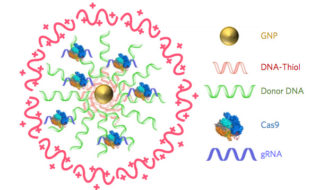
The research team created CRISPR-Gold by covering a central gold nanoparticle with DNA that they modified so it would stick to the particle. This gold-conjugated DNA bound the donor DNA needed for HDR, which the Cas9 protein and guide RNA bound to in turn. They coated the entire complex with a polymer that seems to trigger endocytosis and then facilitate escape of the Cas9 protein, guide RNA, and template DNA from endosomes within cells.

In order to do HDR, “you have to provide the cell [with] the Cas9 enzyme, guide RNA by which you target Cas9 to a particular part of the genome, and a big chunk of DNA, which will be used as a template to edit the mutant sequence to wild-type,” explains coauthor Irina Conboy, who studies tissue repair at the University of California, Berkeley. “They all have to be present at the same time and at the same place, so in our system you have a nanoparticle which simultaneously delivers all of those three key components in their active state.”

Olena’s article carries on to describe how the team created CRISPR-Gold and more.

Additional technical details are available in an Oct. 3, 2017 University of California at Berkeley news release by Brett Israel (also on EurekAlert), which originated the news item (Note: A link has been removed) ,

Scientists at the University of California, Berkeley, have engineered a new way to deliver CRISPR-Cas9 gene-editing technology inside cells and have demonstrated in mice that the technology can repair the mutation that causes Duchenne muscular dystrophy, a severe muscle-wasting disease. A new study shows that a single injection of CRISPR-Gold, as the new delivery system is called, into mice with Duchenne muscular dystrophy led to an 18-times-higher correction rate and a two-fold increase in a strength and agility test compared to control groups.

Diagram of CRISPR-Gold

CRISPR–Gold is composed of 15 nanometer gold nanoparticles that are conjugated to thiol-modified oligonucleotides (DNA-Thiol), which are hybridized with single-stranded donor DNA and subsequently complexed with Cas9 and encapsulated by a polymer that disrupts the endosome of the cell.

Since 2012, when study co-author Jennifer Doudna, a professor of molecular and cell biology and of chemistry at UC Berkeley, and colleague Emmanuelle Charpentier, of the Max Planck Institute for Infection Biology, repurposed the Cas9 protein to create a cheap, precise and easy-to-use gene editor, researchers have hoped that therapies based on CRISPR-Cas9 would one day revolutionize the treatment of genetic diseases. Yet developing treatments for genetic diseases remains a big challenge in medicine. This is because most genetic diseases can be cured only if the disease-causing gene mutation is corrected back to the normal sequence, and this is impossible to do with conventional therapeutics.

CRISPR/Cas9, however, can correct gene mutations by cutting the mutated DNA and triggering homology-directed DNA repair. However, strategies for safely delivering the necessary components (Cas9, guide RNA that directs Cas9 to a specific gene, and donor DNA) into cells need to be developed before the potential of CRISPR-Cas9-based therapeutics can be realized. A common technique to deliver CRISPR-Cas9 into cells employs viruses, but that technique has a number of complications. CRISPR-Gold does not need viruses.

In the new study, research lead by the laboratories of Berkeley bioengineering professors Niren Murthy and Irina Conboy demonstrated that their novel approach, called CRISPR-Gold because gold nanoparticles are a key component, can deliver Cas9 – the protein that binds and cuts DNA – along with guide RNA and donor DNA into the cells of a living organism to fix a gene mutation.

“CRISPR-Gold is the first example of a delivery vehicle that can deliver all of the CRISPR components needed to correct gene mutations, without the use of viruses,” Murthy said.

The study was published October 2 [2017] in the journal Nature Biomedical Engineering.

CRISPR-Gold repairs DNA mutations through a process called homology-directed repair. Scientists have struggled to develop homology-directed repair-based therapeutics because they require activity at the same place and time as Cas9 protein, an RNA guide that recognizes the mutation and donor DNA to correct the mutation.

To overcome these challenges, the Berkeley scientists invented a delivery vessel that binds all of these components together, and then releases them when the vessel is inside a wide variety of cell types, triggering homology directed repair. CRISPR-Gold’s gold nanoparticles coat the donor DNA and also bind Cas9. When injected into mice, their cells recognize a marker in CRISPR-Gold and then import the delivery vessel. Then, through a series of cellular mechanisms, CRISPR-Gold is released into the cells’ cytoplasm and breaks apart, rapidly releasing Cas9 and donor DNA.

Schematic of CRISPR-Gold's method of action

CRISPR-Gold’s method of action (Click to enlarge).

A single injection of CRISPR-Gold into muscle tissue of mice that model Duchenne muscular dystrophy restored 5.4 percent of the dystrophin gene, which causes the disease, to the wild- type, or normal, sequence. This correction rate was approximately 18 times higher than in mice treated with Cas9 and donor DNA by themselves, which experienced only a 0.3 percent correction rate.

Importantly, the study authors note, CRISPR-Gold faithfully restored the normal sequence of dystrophin, which is a significant improvement over previously published approaches that only removed the faulty part of the gene, making it shorter and converting one disease into another, milder disease.

CRISPR-Gold was also able to reduce tissue fibrosis – the hallmark of diseases where muscles do not function properly – and enhanced strength and agility in mice with Duchenne muscular dystrophy. CRISPR-Gold-treated mice showed a two-fold increase in hanging time in a common test for mouse strength and agility, compared to mice injected with a control.

“These experiments suggest that it will be possible to develop non-viral CRISPR therapeutics that can safely correct gene mutations, via the process of homology-directed repair, by simply developing nanoparticles that can simultaneously encapsulate all of the CRISPR components,” Murthy said.

CRISPR-Cas9

CRISPR in action: A model of the Cas9 protein cutting a double-stranded piece of DNA

The study found that CRISPR-Gold’s approach to Cas9 protein delivery is safer than viral delivery of CRISPR, which, in addition to toxicity, amplifies the side effects of Cas9 through continuous expression of this DNA-cutting enzyme. When the research team tested CRISPR-Gold’s gene-editing capability in mice, they found that CRISPR-Gold efficiently corrected the DNA mutation that causes Duchenne muscular dystrophy, with minimal collateral DNA damage.

The researchers quantified CRISPR-Gold’s off-target DNA damage and found damage levels similar to the that of a typical DNA sequencing error in a typical cell that was not exposed to CRISPR (0.005 – 0.2 percent). To test for possible immunogenicity, the blood stream cytokine profiles of mice were analyzed at 24 hours and two weeks after the CRISPR-Gold injection. CRISPR-Gold did not cause an acute up-regulation of inflammatory cytokines in plasma, after multiple injections, or weight loss, suggesting that CRISPR-Gold can be used multiple times safely, and that it has a high therapeutic window for gene editing in muscle tissue.

“CRISPR-Gold and, more broadly, CRISPR-nanoparticles open a new way for safer, accurately controlled delivery of gene-editing tools,” Conboy said. “Ultimately, these techniques could be developed into a new medicine for Duchenne muscular dystrophy and a number of other genetic diseases.”

A clinical trial will be needed to discern whether CRISPR-Gold is an effective treatment for genetic diseases in humans. Study co-authors Kunwoo Lee and Hyo Min Park have formed a start-up company, GenEdit (Murthy has an ownership stake in GenEdit), which is focused on translating the CRISPR-Gold technology into humans. The labs of Murthy and Conboy are also working on the next generation of particles that can deliver CRISPR into tissues from the blood stream and would preferentially target adult stem cells, which are considered the best targets for gene correction because stem and progenitor cells are capable of gene editing, self-renewal and differentiation.

“Genetic diseases cause devastating levels of mortality and morbidity, and new strategies for treating them are greatly needed,” Murthy said. “CRISPR-Gold was able to correct disease-causing gene mutations in vivo, via the non-viral delivery of Cas9 protein, guide RNA and donor DNA, and therefore has the potential to develop into a therapeutic for treating genetic diseases.”

The study was funded by the National Institutes of Health, the W.M. Keck Foundation, the Moore Foundation, the Li Ka Shing Foundation, Calico, Packer, Roger’s and SENS, and the Center of Innovation (COI) Program of the Japan Science and Technology Agency.

Here’s a link to and a citation for the paper,

Nanoparticle delivery of Cas9 ribonucleoprotein and donor DNA in vivo induces homology-directed DNA repair by Kunwoo Lee, Michael Conboy, Hyo Min Park, Fuguo Jiang, Hyun Jin Kim, Mark A. Dewitt, Vanessa A. Mackley, Kevin Chang, Anirudh Rao, Colin Skinner, Tamanna Shobha, Melod Mehdipour, Hui Liu, Wen-chin Huang, Freeman Lan, Nicolas L. Bray, Song Li, Jacob E. Corn, Kazunori Kataoka, Jennifer A. Doudna, Irina Conboy, & Niren Murthy. Nature Biomedical Engineering (2017) doi:10.1038/s41551-017-0137-2 Published online: 02 October 2017

This paper is behind a paywall.
Nanozymes as an antidote for pesticides

Should you have concerns about exposure to pesticides or chemical warfare agents (timely given events in Syria as per this April 4, 2017 news item on CBC [Canadian Broadcasting News Corporation] online) , scientists at the Lomonosov Moscow State University have developed a possible antidote according to a March 8,, 2017 news item on phys.org,

Members of the Faculty of Chemistry of the Lomonosov Moscow State University have developed novel nanosized agents that could be used as efficient protective and antidote modalities against the impact of neurotoxic organophosphorus compounds such as pesticides and chemical warfare agents. …

A March 7, 2017 Lomonosov Moscow State University press release on EurekAlert, which originated the news item, describes the work in detail,

A group of scientists from the Faculty of Chemistry under the leadership of Prof. Alexander Kabanov has focused their research supported by a “megagrant” on the nanoparticle-based delivery to an organism of enzymes, capable of destroying toxic organophosphorous compounds. Development of first nanosized drugs has started more than 30 years ago and already in the 90-s first nanomedicines for cancer treatment entered the market. First such medicines were based on liposomes – spherical vesicles made of lipid bilayers. The new technology, developed by Kabanov and his colleagues, uses an enzyme, synthesized at the Lomonosov Moscow State University, encapsulated into a biodegradable polymer coat, based on an amino acid (glutamic acid).

Alexander Kabanov, Doctor of Chemistry, Professor at the Eshelman School of Pharmacy of the University of North Carolina (USA) and the Faculty of Chemistry, M. V. Lomonosov Moscow State University, one of the authors of the article explains: “At the end of the 80-s my team (at that time in Moscow) and independently Japanese colleagues led by Prof. Kazunori Kataoka from Tokyo began using polymer micelles for small molecules delivery. Soon the nanomedicine field has “exploded”. Currently hundreds of laboratories across the globe work in this area, applying a wide variety of approaches to creation of such nanosized agents. A medicine on the basis of polymeric micelles, developed by a Korean company Samyang Biopharm, was approved for human use in 2006.”

Professor Kabanov’s team after moving to the USA in 1994 focused on development of polymer micelles, which could include biopolymers due to electrostatic interactions. Initially chemists were interested in usage of micelles for RNA and DNA delivery but later on scientists started actively utilizing this approach for delivery of proteins and, namely, enzymes, to the brain and other organs.

Alexander Kabanov says: “At the time I worked at the University of Nebraska Medical Center, in Omaha (USA) and by 2010 we had a lot of results in this area. That’s why when my colleague from the Chemical Enzymology Department of the Lomonosov Moscow State University, Prof. Natalia Klyachko offered me to apply for a megagrant the research theme of the new laboratory was quite obvious. Specifically, to use our delivery approach, which we’ve called a “nanozyme”, for “improvement” of enzymes, developed by colleagues at the Lomonosov Moscow State University for its further medical application.”

Scientists together with the group of enzymologists from the Lomonosov Moscow State University under the leadership of Elena Efremenko, Doctor of Biological Sciences, have chosen organophosphorus hydrolase as a one of the delivered enzymes. Organophosphorus hydrolase is capable of degrading toxic pesticides and chemical warfare agents with very high rate. However, it has disadvantages: because of its bacterial origin, an immune response is observed as a result of its delivery to an organism of mammals. Moreover, organophosphorus hydrolase is quickly removed from the body. Chemists have solved this problem with the help of a “self-assembly” approach: as a result of inclusion of organophosphorus hydrolase enzyme in a nanozyme particles the immune response becomes weaker and, on the contrary, both the storage stability of the enzyme and its lifetime after delivery to an organism considerably increase. Rat experiments have proved that such nanozyme efficiently protects organisms against lethal doses of highly toxic pesticides and even chemical warfare agents, such as VX nerve gas.

Alexander Kabanov summarizes: “The simplicity of our approach is very important. You could get an organophosphorus hydrolase nanozyme by simple mixing of aqueous solutions of anenzyme and safe biocompatible polymer. This nanozyme is self-assembled due to electrostatic interaction between a protein (enzyme) and polymer”.

According to the scientist’s words the simplicity and technological effectiveness of the approach along with the obtained promising results of animal experiments bring hope that this modality could be successful and in clinical use.

Members of the Faculty of Chemistry of the Lomonosov Moscow State University, along with scientists from the 27th Central Research Institute of the Ministry of Defense of the Russian Federation, the Eshelman School of Pharmacy of the University of North Carolina at Chapel Hill (USA) and the University of Nebraska Medical Center (UNC) have taken part in the Project.

Here’s a link to and a citation for the paper,

A simple and highly effective catalytic nanozyme scavenger for organophosphorus neurotoxins by Elena N. Efremenko, Ilya V. Lyagin, Natalia L. Klyachko, Tatiana Bronich, Natalia V. Zavyalova, Yuhang Jiang, Alexander V. Kabanov. Journal of Controlled Release Volume 247, 10 February 2017, Pages 175–181  http://dx.doi.org/10.1016/j.jconrel.2016.12.037

This paper is behind a paywall.

pH dependent nanoparticle-based contrast agent for MRIs (magnetic resonance images)

This news about a safer and more effective contrast agent for MRIs (magnetic resonance images) developed by Japanese scientists come from a June 6, 2016 article by Heather Zeiger on phys.org. First some explanations,

Magnetic resonance imaging relies on the excitation and subsequent relaxation of protons. In clinical MRI studies, the signal is determined by the relaxation time of the hydrogen protons in water. To get a stronger signal, scientists can use contrast agents to shorten the relaxation time of the protons.

MRI is non-invasive and does not involve radiation, making it a safe diagnostic tool. However, its weak signal makes tumor detection difficult. The ideal contrast agent would select for malignant tumors, making its location and diagnosis much more obvious.

Nanoparticle contrast agents have been of interested because nanoparticles can be functionalized and, as in this study, can contain various metals. Researchers have attempted to functionalize nanoparticles with ligands that attach to chemical factors on the surface of cancer cells. However, cancer cells tend to be compositionally heterogeneous, leading some researchers to look for nanoparticles that respond to differences in pH or redox potential compared to normal cells.

Now for the research,

Researchers from the University of Tokyo, Tokyo Institute of Technology, Kawasaki Institute of Industry Promotion, and the Japan Agency for Quantum and Radiological Science and Technology have developed a contrast agent from calcium phosphate-based nanoparticles that release a manganese ion an acidic environment. …

Peng Mi, Daisuke Kokuryo, Horacio Cabral, Hailiang Wu, Yasuko Terada, Tsuneo Saga, Ichio Aoki, Nobuhiro Nishiyama, and Kazunori Kataoka developed a contrast agent that is comprised of Mn2+– doped CaP nanoparticles with a PEG shell. They reasoned that using CaP nanoparticles, which are known to be pH sensitive, would allow the targeted release of Mn2+ ions in the tumor microenvironment. The tumor microenvironment tends to have a lower pH than the normal regions to rapid cell metabolism in an oxygen-depleted environment. Manganese ions were tested because they are paramagnetic, which makes for a good contrast agent. They also bind to proteins creating a slowly rotating manganese-protein system that results in sharp contrast enhancement.

These results were promising, so Peng Mi, et al. then tested whether the CaPMnPEG contrast agent worked in solid tumors. Because Mn2+ remains confined within the nanoparticle matrix at physiological pH, CaPMnPEG demonstrate a much lower toxicity [emphasis mine] compared to MnCl2. MRI studies showed a tumor-to-normal contrast of 131% after 30 minute, which is much higher than Gd-DTPA [emphasis mine], a clinically approved contrast agent. After an hour, the tumor-to-normal ratio was 160% and remained around 170% for several hours.

Three-dimensional MRI studies of solid tumors showed that without the addition of CaPMnPEG, only blood vessels were visible. However, upon adding CaPMnPEG, the tumor was easily distinguishable. Additionally, there is evidence that excess Mn2+ leaves the plasma after an hour. The contrast signal remained strong for several hours indicating that protein binding rather than Mn2+ concentration is important for signal enhancement.

Finally, tests with metastatic tumors in the liver (C26 colon cancer cells) showed that CaPMnPEG works well in solid organ analysis and is highly sensitive to detecting millimeter-sized micrometastasis [emphasis mine]. Unlike other contrast agents used in the clinic, CaPMnPEG provided a contrast signal that lasted for several hours after injection. After an hour, the signal was enhanced by 25% and after two hours, the signal was enhanced by 39%.

This is exciting stuff. Bravo to the researchers!

Here’s a link to and citation for the paper,

A pH-activatable nanoparticle with signal-amplification capabilities for non-invasive imaging of tumour malignancy by Peng Mi, Daisuke Kokuryo, Horacio Cabral, Hailiang Wu, Yasuko Terada, Tsuneo Saga, Ichio Aoki, Nobuhiro Nishiyama, & Kazunori Kataoka. Nature Nanotechnology (2016) doi:10.1038/nnano.2016.72 Published online 16 May 2016

This paper is behind a paywall.

A city of science in Japan: Kawasaki (Kanagawa)

Happily, I’m getting more nanotechnology (for the most part) information from Japan. Given Japan’s prominence in this field of endeavour I’ve long felt FrogHeart has not adequately represented Japanese contributions. Now that I’m receiving English language translations, I hope to better address the situation.

This morning (March 26, 2015), there were two news releases from Kawasaki INnovation Gateway at SKYFRONT (KING SKYFRONT), Coastal Area International Strategy Office, Kawasaki City, Japan in my mailbox. Before getting on to the news releases, here’s a little about  the city of Kawasaki and about its innovation gateway. From the Kawasaki, Kanagawa entry in Wikipedia (Note: Links have been removed),

Kawasaki (川崎市 Kawasaki-shi?) is a city in Kanagawa Prefecture, Japan, located between Tokyo and Yokohama. It is the 9th most populated city in Japan and one of the main cities forming the Greater Tokyo Area and Keihin Industrial Area.

Kawasaki occupies a belt of land stretching about 30 kilometres (19 mi) along the south bank of the Tama River, which divides it from Tokyo. The eastern end of the belt, centered on JR Kawasaki Station, is flat and largely consists of industrial zones and densely built working-class housing, the Western end mountainous and more suburban. The coastline of Tokyo Bay is occupied by vast heavy industrial complexes built on reclaimed land.

There is a 2014 video about Kawasaki’s innovation gateway, which despite its 14 mins. 39 secs. running time I am embedding here. (Caution: They highlight their animal testing facility at some length.)

Now on to the two news releases. The first concerns research on gold nanoparticles that was published in 2014. From a March 26, 2015 Kawasaki INnovation Gateway news release,

Gold nanoparticles size up to cancer treatment

Incorporating gold nanoparticles helps optimise treatment carrier size and stability to improve delivery of cancer treatment to cells.

Treatments that attack cancer cells through the targeted silencing of cancer genes could be developed using small interfering RNA molecules (siRNA). However delivering the siRNA into the cells intact is a challenge as it is readily degraded by enzymes in the blood and small enough to be eliminated from the blood stream by kidney filtration.  Now Kazunori Kataoka at the University of Tokyo and colleagues at Tokyo Institute of Technology have designed a protective treatment delivery vehicle with optimum stability and size for delivering siRNA to cells.

The researchers formed a polymer complex with a single siRNA molecule. The siRNA-loaded complex was then bonded to a 20 nm gold nanoparticle, which thanks to advances in synthesis techniques can be produced with a reliably low size distribution. The resulting nanoarchitecture had the optimum overall size – small enough to infiltrate cells while large enough to accumulate.

In an assay containing heparin – a biological anti-coagulant with a high negative charge density – the complex was found to release the siRNA due to electrostatic interactions. However when the gold nanoparticle was incorporated the complex remained stable. Instead, release of the siRNA from the complex with the gold nanoparticle could be triggered once inside the cell by the presence of glutathione, which is present in high concentrations in intracellular fluid. The glutathione bonded with the gold nanoparticles and the complex, detaching them from each other and leaving the siRNA prone to release.

The researchers further tested their carrier in a subcutaneous tumour model. The authors concluded that the complex bonded to the gold nanoparticle “enabled the efficient tumor accumulation of siRNA and significant in vivo gene silencing effect in the tumor, demonstrating the potential for siRNA-based cancer therapies.”

The news release provides links to the March 2015 newsletter which highlights this research and to the specific article and video,

March 2015 Issue of Kawasaki SkyFront iNewsletter: http://inewsletter-king-skyfront.jp/en/

Contents

Feature video on Professor Kataoka’s research : http://inewsletter-king-skyfront.jp/en/video_feature/vol_3/feature01/

Research highlights: http://inewsletter-king-skyfront.jp/en/research_highlights/vol_3/research01/

Here’s a link to and a citation for the paper,

Precise Engineering of siRNA Delivery Vehicles to Tumors Using Polyion Complexes and Gold Nanoparticles by Hyun Jin Kim, Hiroyasu Takemoto, Yu Yi, Meng Zheng, Yoshinori Maeda, Hiroyuki Chaya, Kotaro Hayashi, Peng Mi, Frederico Pittella, R. James Christie, Kazuko Toh, Yu Matsumoto, Nobuhiro Nishiyama, Kanjiro Miyata, and Kazunori Kataoka. ACS Nano, 2014, 8 (9), pp 8979–8991 DOI: 10.1021/nn502125h Publication Date (Web): August 18, 2014
Copyright © 2014 American Chemical Society

This article is behind a paywall.

The second March 26, 2015 Kawasaki INnovation Gateway news release concerns a DNA chip and food-borne pathogens,

Rapid and efficient DNA chip technology for testing 14 major types of food borne pathogens

Conventional methods for testing food-borne pathogens is based on the cultivation of pathogens, a process that is complicated and time consuming. So there is demand for alternative methods to test for food-borne pathogens that are simpler, quick and applicable to a wide range of potential applications.

Now Toshiba Ltd and Kawasaki City Institute for Public Health have collaborated in the development of a rapid and efficient automatic abbreviated DNA detection technology that can test for 14 major types of food borne pathogens. The so called ‘DNA chip card’ employs electrochemical DNA chips and overcomes the complicated procedures associated with genetic testing of conventional methods. The ‘DNA chip card’ is expected to find applications in hygiene management in food manufacture, pharmaceuticals, and cosmetics.

Details

The so-called automatic abbreviated DNA detection technology ‘DNA chip card’ was developed by Toshiba Ltd and in a collaboration with Kawasaki City Institute for Public Health, used to simultaneously detect 14 different types of food-borne pathogens in less than 90 minutes. The detection sensitivity depends on the target pathogen and has a range of 1E+01~05 cfu/mL.

Notably, such tests would usually take 4-5 days using conventional methods based on pathogen cultivation. Furthermore, in contrast to conventional DNA protocols that require high levels of skill and expertise, the ‘DNA chip card’ only requires the operator to inject nucleic acid, thereby making the procedure easier to use and without specialized operating skills.

Examples of pathogens associated with food poisoning that were tested with the “DNA chip card”

Enterohemorrhagic Escherichia coli

Salmonella

Campylobacter

Vibrio parahaemolyticus

Shigella

Staphylococcus aureus

Enterotoxigenic Escherichia coli

Enteroaggregative Escherichia coli

Enteropathogenic Escherichia coli

Clostridium perfringens

Bacillus cereus

Yersinia

Listeria

Vibrio cholerae

I think 14 is the highest number of tests I’ve seen for one of these chips. This chip is quite an achievement.

One final bit from the news release about the DNA chip provides a brief description of the gateway and something they call King SkyFront,

About KING SKYFRONT

The Kawasaki INnovation Gateway (KING) SKYFRONT is the flagship science and technology innovation hub of Kawasaki City. KING SKYFRONT is a 40 hectare area located in the Tonomachi area of the Keihin Industrial Region that spans Tokyo and Kanagawa Prefecture and Tokyo International Airport (also often referred to as Haneda Airport).

KING SKYFRONT was launched in 2013 as a base for scholars, industrialists and government administrators to work together to devise real life solutions to global issues in the life sciences and environment.

I find this emphasis on the city interesting. It seems that cities are becoming increasingly important and active where science research and development are concerned. Europe seems to have adopted a biannual event wherein a city is declared a European City of Science in conjunction with the EuroScience Open Forum (ESOF) conferences. The first such city was Dublin in 2012 (I believe the Irish came up with the concept themselves) and was later adopted by Copenhagen for 2014. The latest city to embrace the banner will be Manchester in 2016.