Tag Archives: Korea

Ceria-zirconia nanoparticles for sepsis treatment

South Korean researchers are looking at a new way of dealing with infections (sepsis) according to a July 6, 2017 news item on phys.org,

During sepsis, cells are swamped with reactive oxygen species generated in an aberrant response of the immune system to a local infection. If this fatal inflammatory path could be interfered, new treatment schemes could be developed. Now, Korean scientists report in the journal Angewandte Chemie that zirconia-doped ceria nanoparticles act as effective scavengers of these oxygen radicals, promoting a greatly enhanced surviving rate in sepsis model organisms.

A July 6, 2017 Wiley (Publishers) press release, which originated the news item, provides more detail,

Sepsis proceeds as a vicious cycle of inflammatory reactions of the immune system to a local infection. Fatal consequences can be falling blood pressure and the collapse of organ function. As resistance against antibiotics is growing, scientists turn to the inflammatory pathway as an alternative target for new treatment strategies. Taeghwan Heyon from Seoul National University, Seung-Hoon Lee at Seoul National University Hospital, South Korea, and collaborators explore ceria nanoparticles for their ability to scavenge reactive oxygen species, which play a key role in the inflammatory process. By quickly converting between two oxidation states, the cerium ion can quench typical oxygen radical species like the superoxide anion, the hydroxyl radical anion, or even hydrogen peroxide. But in the living cell, this can only happen if two conditions are met.

The first condition is the size and nature of the particles. Small, two-nanometer-sized particles were coated by a hydrophilic shell of poly(ethylene glycol)-connected phospholipids to make them soluble so that they can enter the cell and remain there. Second, the cerium ion responsible for the quenching (Ce3+) should be accessible on the surface of the nanoparticles, and it must be regenerated after the reactions. Here, the scientists found out that a certain amount of zirconium ions in the structure helped, because “the Zr4+ ions control the Ce3+-to-Ce4+ ratio as well as the rate of conversion between the two oxidation states,” they argued.

The prepared nanoparticles were then tested for their ability to detoxify reactive oxygen species, not only in the test tube, but also in live animal models. The results were clear, as the authors stated: “A single dose of ceria-zirconia nanoparticles successfully attenuated the vicious cycle of inflammatory responses in two sepsis models.” The nanoparticles accumulated in organs where severe immune responses occurred, and they were successful in the eradication of reactive oxygen species, as evidenced with fluorescence microscopy and several other techniques. And importantly, the treated mice and rats had a far higher survival rate.

This work demonstrates that other approaches in sepsis treatment than killing bacteria with antibiotics are possible. Targeting the inflammatory signal pathways in macrophages is a very promising option, and the authors have shown that effective scavenging of reactive oxygen species and stopping inflammation is possible with a suitably designed chemical system like this cerium ion redox system provided by nanoparticles.

Here’s a link to and a citation for the paper,

Ceria–Zirconia Nanoparticles as an Enhanced Multi-Antioxidant for Sepsis Treatment by Min Soh, Dr. Dong-Wan Kang, Dr. Han-Gil Jeong, Dr. Dokyoon Kim, Dr. Do Yeon Kim, Dr. Wookjin Yang, Changyeong Song, Seungmin Baik, In-Young Choi, Seul-Ki Ki, Hyek Jin Kwon, Dr. Taeho Kim, Prof. Dr. Chi Kyung Kim, Prof. Dr. Seung-Hoon Lee, and Prof. Dr. Taeghwan Hyeon. Angewandte Chemie DOI: 10.1002/anie.201704904 Version of Record online: 5 JUL 2017

© 2017 Wiley-VCH Verlag GmbH & Co. KGaA, Weinheim

This paper is behind a paywall.

Graphene-based neural probes

I have two news bits (dated almost one month apart) about the use of graphene in neural probes, one from the European Union and the other from Korea.

European Union (EU)

This work is being announced by the European Commission’s (a subset of the EU) Graphene Flagship (one of two mega-funding projects announced in 2013; 1B Euros each over ten years for the Graphene Flagship and the Human Brain Project).

According to a March 27, 2017 news item on ScienceDaily, researchers have developed a graphene-based neural probe that has been tested on rats,

Measuring brain activity with precision is essential to developing further understanding of diseases such as epilepsy and disorders that affect brain function and motor control. Neural probes with high spatial resolution are needed for both recording and stimulating specific functional areas of the brain. Now, researchers from the Graphene Flagship have developed a new device for recording brain activity in high resolution while maintaining excellent signal to noise ratio (SNR). Based on graphene field-effect transistors, the flexible devices open up new possibilities for the development of functional implants and interfaces.

The research, published in 2D Materials, was a collaborative effort involving Flagship partners Technical University of Munich (TU Munich; Germany), Institut d’Investigacions Biomèdiques August Pi i Sunyer (IDIBAPS; Spain), Spanish National Research Council (CSIC; Spain), The Biomedical Research Networking Center in Bioengineering, Biomaterials and Nanomedicine (CIBER-BBN; Spain) and the Catalan Institute of Nanoscience and Nanotechnology (ICN2; Spain).
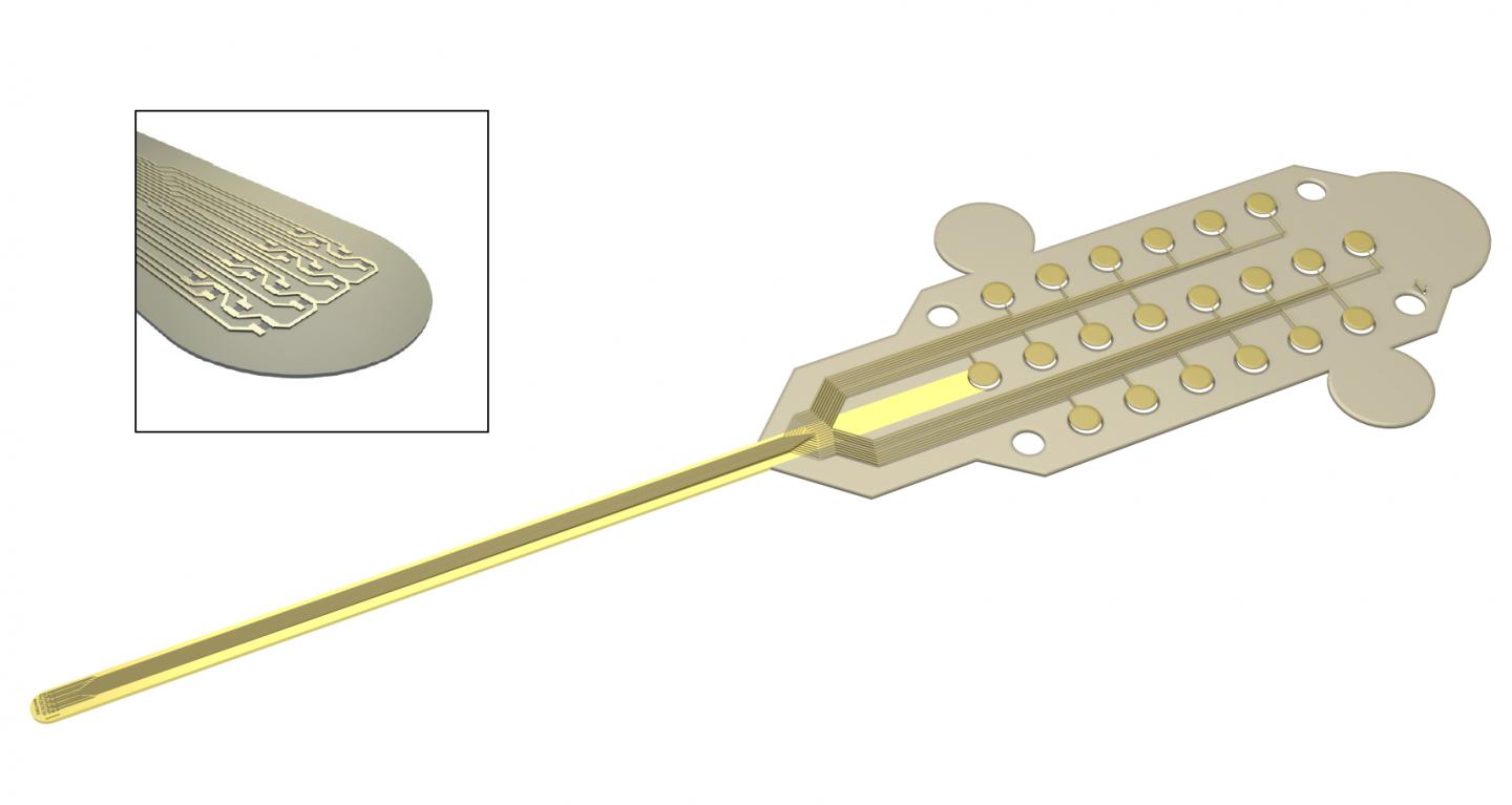
Caption: Graphene transistors integrated in a flexible neural probe enables electrical signals from neurons to be measured with high accuracy and density. Inset: The tip of the probe contains 16 flexible graphene transistors. Credit: ICN2

A March 27, 2017 Graphene Flagship press release on EurekAlert, which originated the news item, describes the work,  in more detail,

The devices were used to record the large signals generated by pre-epileptic activity in rats, as well as the smaller levels of brain activity during sleep and in response to visual light stimulation. These types of activities lead to much smaller electrical signals, and are at the level of typical brain activity. Neural activity is detected through the highly localised electric fields generated when neurons fire, so densely packed, ultra-small measuring devices is important for accurate brain readings.

The neural probes are placed directly on the surface of the brain, so safety is of paramount importance for the development of graphene-based neural implant devices. Importantly, the researchers determined that the graphene-based probes are non-toxic, and did not induce any significant inflammation.

Devices implanted in the brain as neural prosthesis for therapeutic brain stimulation technologies and interfaces for sensory and motor devices, such as artificial limbs, are an important goal for improving quality of life for patients. This work represents a first step towards the use of graphene in research as well as clinical neural devices, showing that graphene-based technologies can deliver the high resolution and high SNR needed for these applications.

First author Benno Blaschke (TU Munich) said “Graphene is one of the few materials that allows recording in a transistor configuration and simultaneously complies with all other requirements for neural probes such as flexibility, biocompability and chemical stability. Although graphene is ideally suited for flexible electronics, it was a great challenge to transfer our fabrication process from rigid substrates to flexible ones. The next step is to optimize the wafer-scale fabrication process and improve device flexibility and stability.”

Jose Antonio Garrido (ICN2), led the research. He said “Mechanical compliance is an important requirement for safe neural probes and interfaces. Currently, the focus is on ultra-soft materials that can adapt conformally to the brain surface. Graphene neural interfaces have shown already great potential, but we have to improve on the yield and homogeneity of the device production in order to advance towards a real technology. Once we have demonstrated the proof of concept in animal studies, the next goal will be to work towards the first human clinical trial with graphene devices during intraoperative mapping of the brain. This means addressing all regulatory issues associated to medical devices such as safety, biocompatibility, etc.”
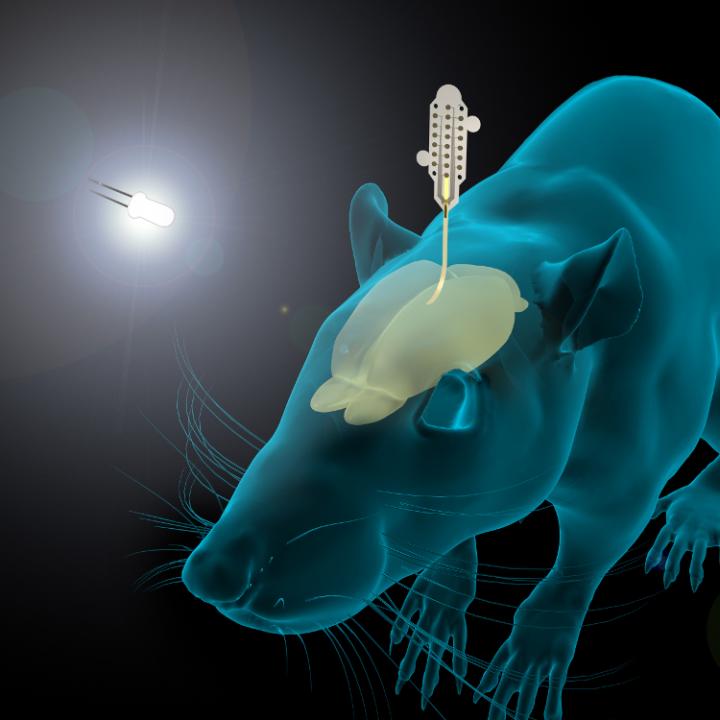
Caption: The graphene-based neural probes were used to detect rats’ responses to visual stimulation, as well as neural signals during sleep. Both types of signals are small, and typically difficult to measure. Credit: ICN2

Here’s a link to and a citation for the paper,

Mapping brain activity with flexible graphene micro-transistors by Benno M Blaschke, Núria Tort-Colet, Anton Guimerà-Brunet, Julia Weinert, Lionel Rousseau, Axel Heimann, Simon Drieschner, Oliver Kempski, Rosa Villa, Maria V Sanchez-Vives. 2D Materials, Volume 4, Number 2 DOI https://doi.org/10.1088/2053-1583/aa5eff Published 24 February 2017

© 2017 IOP Publishing Ltd

This paper is behind a paywall.

Korea

While this research from Korea was published more recently, the probe itself has not been subjected to in vivo (animal testing). From an April 19, 2017 news item on ScienceDaily,

Electrodes placed in the brain record neural activity, and can help treat neural diseases like Parkinson’s and epilepsy. Interest is also growing in developing better brain-machine interfaces, in which electrodes can help control prosthetic limbs. Progress in these fields is hindered by limitations in electrodes, which are relatively stiff and can damage soft brain tissue.

Designing smaller, gentler electrodes that still pick up brain signals is a challenge because brain signals are so weak. Typically, the smaller the electrode, the harder it is to detect a signal. However, a team from the Daegu Gyeongbuk Institute of Science & Technology [DGIST} in Korea developed new probes that are small, flexible and read brain signals clearly.

This is a pretty interesting way to illustrate the research,
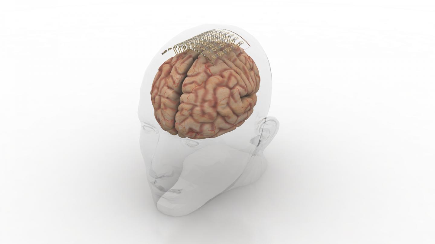
Caption: Graphene and gold make a better brain probe. Credit: DGIST

An April 19, 2017 DGIST press release (also on EurekAlert), which originated the news item, expands on the theme (Note: A link has been removed),

The probe consists of an electrode, which records the brain signal. The signal travels down an interconnection line to a connector, which transfers the signal to machines measuring and analysing the signals.

The electrode starts with a thin gold base. Attached to the base are tiny zinc oxide nanowires, which are coated in a thin layer of gold, and then a layer of conducting polymer called PEDOT. These combined materials increase the probe’s effective surface area, conducting properties, and strength of the electrode, while still maintaining flexibility and compatibility with soft tissue.

Packing several long, thin nanowires together onto one probe enables the scientists to make a smaller electrode that retains the same effective surface area of a larger, flat electrode. This means the electrode can shrink, but not reduce signal detection. The interconnection line is made of a mix of graphene and gold. Graphene is flexible and gold is an excellent conductor. The researchers tested the probe and found it read rat brain signals very clearly, much better than a standard flat, gold electrode.

“Our graphene and nanowires-based flexible electrode array can be useful for monitoring and recording the functions of the nervous system, or to deliver electrical signals to the brain,” the researchers conclude in their paper recently published in the journal ACS Applied Materials and Interfaces.

The probe requires further clinical tests before widespread commercialization. The researchers are also interested in developing a wireless version to make it more convenient for a variety of applications.

Here’s a link to and a citation for the paper,

Enhancement of Interface Characteristics of Neural Probe Based on Graphene, ZnO Nanowires, and Conducting Polymer PEDOT by Mingyu Ryu, Jae Hoon Yang, Yumi Ahn, Minkyung Sim, Kyung Hwa Lee, Kyungsoo Kim, Taeju Lee, Seung-Jun Yoo, So Yeun Kim, Cheil Moon, Minkyu Je, Ji-Woong Choi, Youngu Lee, and Jae Eun Jang. ACS Appl. Mater. Interfaces, 2017, 9 (12), pp 10577–10586 DOI: 10.1021/acsami.7b02975 Publication Date (Web): March 7, 2017

Copyright © 2017 American Chemical Society

This paper is behind a paywall.

Nanozymes as an antidote for pesticides

Should you have concerns about exposure to pesticides or chemical warfare agents (timely given events in Syria as per this April 4, 2017 news item on CBC [Canadian Broadcasting News Corporation] online) , scientists at the Lomonosov Moscow State University have developed a possible antidote according to a March 8,, 2017 news item on phys.org,

Members of the Faculty of Chemistry of the Lomonosov Moscow State University have developed novel nanosized agents that could be used as efficient protective and antidote modalities against the impact of neurotoxic organophosphorus compounds such as pesticides and chemical warfare agents. …

A March 7, 2017 Lomonosov Moscow State University press release on EurekAlert, which originated the news item, describes the work in detail,

A group of scientists from the Faculty of Chemistry under the leadership of Prof. Alexander Kabanov has focused their research supported by a “megagrant” on the nanoparticle-based delivery to an organism of enzymes, capable of destroying toxic organophosphorous compounds. Development of first nanosized drugs has started more than 30 years ago and already in the 90-s first nanomedicines for cancer treatment entered the market. First such medicines were based on liposomes – spherical vesicles made of lipid bilayers. The new technology, developed by Kabanov and his colleagues, uses an enzyme, synthesized at the Lomonosov Moscow State University, encapsulated into a biodegradable polymer coat, based on an amino acid (glutamic acid).

Alexander Kabanov, Doctor of Chemistry, Professor at the Eshelman School of Pharmacy of the University of North Carolina (USA) and the Faculty of Chemistry, M. V. Lomonosov Moscow State University, one of the authors of the article explains: “At the end of the 80-s my team (at that time in Moscow) and independently Japanese colleagues led by Prof. Kazunori Kataoka from Tokyo began using polymer micelles for small molecules delivery. Soon the nanomedicine field has “exploded”. Currently hundreds of laboratories across the globe work in this area, applying a wide variety of approaches to creation of such nanosized agents. A medicine on the basis of polymeric micelles, developed by a Korean company Samyang Biopharm, was approved for human use in 2006.”

Professor Kabanov’s team after moving to the USA in 1994 focused on development of polymer micelles, which could include biopolymers due to electrostatic interactions. Initially chemists were interested in usage of micelles for RNA and DNA delivery but later on scientists started actively utilizing this approach for delivery of proteins and, namely, enzymes, to the brain and other organs.

Alexander Kabanov says: “At the time I worked at the University of Nebraska Medical Center, in Omaha (USA) and by 2010 we had a lot of results in this area. That’s why when my colleague from the Chemical Enzymology Department of the Lomonosov Moscow State University, Prof. Natalia Klyachko offered me to apply for a megagrant the research theme of the new laboratory was quite obvious. Specifically, to use our delivery approach, which we’ve called a “nanozyme”, for “improvement” of enzymes, developed by colleagues at the Lomonosov Moscow State University for its further medical application.”

Scientists together with the group of enzymologists from the Lomonosov Moscow State University under the leadership of Elena Efremenko, Doctor of Biological Sciences, have chosen organophosphorus hydrolase as a one of the delivered enzymes. Organophosphorus hydrolase is capable of degrading toxic pesticides and chemical warfare agents with very high rate. However, it has disadvantages: because of its bacterial origin, an immune response is observed as a result of its delivery to an organism of mammals. Moreover, organophosphorus hydrolase is quickly removed from the body. Chemists have solved this problem with the help of a “self-assembly” approach: as a result of inclusion of organophosphorus hydrolase enzyme in a nanozyme particles the immune response becomes weaker and, on the contrary, both the storage stability of the enzyme and its lifetime after delivery to an organism considerably increase. Rat experiments have proved that such nanozyme efficiently protects organisms against lethal doses of highly toxic pesticides and even chemical warfare agents, such as VX nerve gas.

Alexander Kabanov summarizes: “The simplicity of our approach is very important. You could get an organophosphorus hydrolase nanozyme by simple mixing of aqueous solutions of anenzyme and safe biocompatible polymer. This nanozyme is self-assembled due to electrostatic interaction between a protein (enzyme) and polymer”.

According to the scientist’s words the simplicity and technological effectiveness of the approach along with the obtained promising results of animal experiments bring hope that this modality could be successful and in clinical use.

Members of the Faculty of Chemistry of the Lomonosov Moscow State University, along with scientists from the 27th Central Research Institute of the Ministry of Defense of the Russian Federation, the Eshelman School of Pharmacy of the University of North Carolina at Chapel Hill (USA) and the University of Nebraska Medical Center (UNC) have taken part in the Project.

Here’s a link to and a citation for the paper,

A simple and highly effective catalytic nanozyme scavenger for organophosphorus neurotoxins by Elena N. Efremenko, Ilya V. Lyagin, Natalia L. Klyachko, Tatiana Bronich, Natalia V. Zavyalova, Yuhang Jiang, Alexander V. Kabanov. Journal of Controlled Release Volume 247, 10 February 2017, Pages 175–181  http://dx.doi.org/10.1016/j.jconrel.2016.12.037

This paper is behind a paywall.

Nanotechnology-enabled acupuncture needles

An Oct. 17, 2016 news item on phys.org makes an announcement about nanotechnology-enabled acupuncture needles (Note: The writing style is a little unusual for this kind of announcement),

A Daegu Gyeongbuk Institute of Science and Technology [Korea] research team led by Professor Su-Il In, who developed acupuncture needles combined with nanotechnology, was recognized as the world’s first application of this technology. This development is expected to open new directions in the oriental medicine research field.

Professor Su-Il In’s research team from the Department of Energy Systems Engineering succeeded in developing porous acupuncture needles (hereafter PANs) that offer enhanced therapeutic properties by applying nanotechnology on the acupuncture needles for the first time in the world.

The findings of this experiment, which was conducted in collaboration with DGIST’s research team and the Addiction Control Research Center at Daegu Haany University, have attracted the attention of the relevant academic field in light of the fact that the experiment combined nanotechnology with acupuncture needles.

An Oct. 17, 2016 DGIST press release on EurekAlert, which originated the news item, provides more technical detail,

Professor In’s research team developed PANs with fine pores ranging in sizes from nanometers (nm= one billionth of a meter) to micrometers (? = one millionth of a meter) on the surface of the needles using a nano-electrochemical method.

PANs are formed by anodization, and are characterized by a widened surface of the needles through the following process: anion (F-) contained in the electrolyte bored into the surface of the metal needles (positive) and created fine and uniform pores.

PANs are expected to be as effective as conventional large and long needles by minimizing the sense of pain during acupuncture treatment while expanding the surface area of the needle 20 times greater than conventional acupuncture ones.

Through electrophysiological experiments with rats, In’s research team proved that PANs excel in transferring signals from a spinal dorsal horn by the in vivo stimulation of Shenmen (HT7) points, and in particular, demonstrated that the efficacy of PANs is superior to conventional acupuncture needles in treating alcohol and cocaine addiction in animal experiments.

Applications for international patents for the fabrication technology of PANs developed by DGIST have already been submitted in countries such as the US, China, and Europe. In addition, in the domestic oriental medicine field, the fact that the efficacy of acupuncture needles has been improved through their structural transformation by applying nanotechnology has been recognized and evaluated as the first such instance in the thousand-year history of eastern medicine.

Professor Su-Il In from DGIST’s Department of Energy Systems Engineering said, “The development of nanotechnology has taken science and technology to the next level in various fields such as solar cells, quantum computers, display development, and the like. Based on this experiment’s achievement of combining nanotechnology and oriental medicine, I will continue to conduct research in order to be at the forefront of the scientific population of oriental medicine.”

Director Jae-ha Yang from Daegu Haany University said, “In western medicine, nanotechnology is widely used from diagnosis to treatment; but in eastern medicine, particularly in acupuncture therapy, it is rare to utilize nano science. The findings of this study are expected to open new directions in the field of eastern medicine where nano science is rarely explored and utilized.”

Here’s a link to and a citation for the paper,

Hierarchical Micro/Nano-Porous Acupuncture Needles Offering Enhanced Therapeutic Properties by Su-ll In, Young S. Gwak, Hye Rim Kim, Abdul Razzaq, Kyeong-Seok Lee, Hee Young Kim, SuChan Chang, Bong Hyo Lee, Craig A. Grimes, & Chae Ha Yang. Scientific Reports 6, Article number: 34061 (2016) doi:10.1038/srep34061 Published online: 07 October 2016

This paper is open access.

Glucose-sensing contact lens invented by US and Korean researchers

Blood tests for glucose levels may one day be a feature of the past according to an Oct. 3, 2016 news item on ScienceDaily,

Blood testing is the standard option for checking glucose levels, but a new technology could allow non-invasive testing via a contact lens that samples glucose levels in tears.

“There’s no noninvasive method to do this,” said Wei-Chuan Shih, a researcher with the University of Houston [UH] who worked with colleagues at UH and in Korea to develop the project, described in the high-impact journal Advanced Materials. “It always requires a blood draw. This is unfortunately the state of the art.”

A Sept. 27, 2016 UH news release (also on EurekAlert) by Jeannie Kever, which originated the news item, describes the proposed technology,

… glucose is a good target for optical sensing, and especially for what is known as surface-enhanced Raman scattering spectroscopy [also known as surface-enhanced Raman scattering or surface-enhanced Raman spectroscopy, and SERS], said Shih, an associate professor of electrical and computer engineering whose lab, the NanoBioPhotonics Group, works on optical biosensing enabled by nanoplasmonics.

This is an alternative approach, in contrast to a Raman spectroscopy-based noninvasive glucose sensor Shih developed as a Ph.D. student at the Massachusetts Institute of Technology. He holds two patents for technologies related to directly probing skin tissue using laser light to extract information about glucose concentrations.

The paper describes the development of a tiny device, built from multiple layers of gold nanowires stacked on top of a gold film and produced using solvent-assisted nanotransfer printing, which optimized the use of surface-enhanced Raman scattering to take advantage of the technique’s ability to detect small molecular samples.

Surface-enhanced Raman scattering – named for Indian physicist C.V. Raman [Raman scattering; SERS history begins in 1973 according to its Wikipedia entry], who discovered the effect in 1928 – uses information about how light interacts with a material to determine properties of the molecules that make up the material.

The device enhances the sensing properties of the technique by creating “hot spots,” or narrow gaps within the nanostructure which intensified the Raman signal, the researchers said.

Researchers created the glucose sensing contact lens to demonstrate the versatility of the technology. The contact lens concept isn’t unheard of – Google has submitted a patent for a multi-sensor contact lens, which the company says can also detect glucose levels in tears – but the researchers say this technology would also have a number of other applications.

“It should be noted that glucose is present not only in the blood but also in tears, and thus accurate monitoring of the glucose level in human tears by employing a contact-lens-type sensor can be an alternative approach for noninvasive glucose monitoring,” the researchers wrote.

“Everyone knows tears have a lot to mine,” Shih said. “The question is, whether you have a detector that is capable of mining it, and how significant is it for real diagnostics.”

In addition to Shih, authors on the paper include Yeon Sik Jung, Jae Won Jeong and Kwang-Min Baek, all with the Korea Advanced Institute of Science and Technology; Seung Yong Lee of the Korea Institute of Science and Technology, and Md Masud Parvez Arnob of UH.

Although non-invasive glucose sensing is just one potential application of the technology, Shih said it provided a good way to prove the technology. “It’s one of the grand challenges to be solved,” he said. “It’s a needle in a haystack challenge.”

Scientists know that glucose is present in tears, but Shih said how tear glucose levels correlate with blood glucose levels hasn’t been established. The more important finding, he said, is that the structure is an effective mechanism for using surface-enhanced Raman scattering spectroscopy.

Although traditional nanofabrication techniques rely on a hard substrate – usually glass or a silicon wafer – Shih said researchers wanted a flexible nanostructure, which would be more suited to wearable electronics. The layered nanoarray was produced on a hard substrate but lifted off and printed onto a soft contact, he said.

Here’s a link to and a citation for the paper,

Wafer Scale Phase-Engineered 1T- and 2H-MoSe2/Mo Core–Shell 3D-Hierarchical Nanostructures toward Efficient Electrocatalytic Hydrogen Evolution Reaction by Yindong Qu, Henry Medina, Sheng-Wen Wang, Yi-Chung Wang, Chia-Wei Chen, Teng-Yu Su, Arumugam Manikandan, Kuangye Wang, Yu-Chuan Shih, Je-Wei Chang, Hao-Chung Kuo, Chi-Yung Lee, Shih-Yuan Lu, Guozhen Shen, Zhiming M. Wang, and Yu-Lun Chueh. Advanced Materials DOI: 10.1002/adma.201602697 Version of Record online: 26 SEP 2016

© 2016 WILEY-VCH Verlag GmbH & Co. KGaA, Weinheim

This paper is behind a paywall.

Placenta-on-a-chip for research into causes for preterm birth

Preterm birth (premature baby) research has received a boost with this latest work from the University of Pennsylvania. A July 21, 2016 news item on phys.org tells us more,

Researchers at the University of Pennsylvania have developed the first placenta-on-a-chip that can fully model the transport of nutrients across the placental barrier.

A July 21, 2016 University of Pennsylvania news release, which originated the news item, provides more detail about the chip and the research (Note: Links have been removed),

The flash-drive-sized device contains two layers of human cells that model the interface between mother and fetus. Microfluidic channels on either side of those layers allow researchers to study how molecules are transported through, or are blocked by, that interface.

Like other organs-on-chips, such as ones developed to simulate lungs, intestines and eyes, the placenta-on-a-chip provides a unique capability to mimic and study the function of that human organ in ways that have not been possible using traditional tools.

Research on the team’s placenta-on-a-chip is part of a nationwide effort sponsored by the March of Dimes to identify causes of preterm birth and ways to prevent it. Prematurely born babies may experience lifelong, debilitating consequences, but the underlying mechanisms of this condition are not well understood due in part to the difficulties of experimenting with intact, living human placentae.

The research was led by Dan Huh, the Wilf Family Term Assistant Professor of Bioengineering in Penn’s School of Engineering and Applied Science, and Cassidy Blundell, a graduate student in the Huh lab. They collaborated with Samuel Parry, the Franklin Payne Professor of Obstetrics and Gynecology; Christos Coutifaris, the Nancy and Richard Wolfson Professor of Obstetrics and Gynecology in Penn’s Perelman School of Medicine; and Emily Su, assistant professor of obstetrics and gynecology in the Anschutz Medical School of the University of Colorado Denver.

The researchers’ placenta-on-a-chip is a clear silicone device with two parallel microfluidic channels separated by a porous membrane. On one side of those pores, trophoblast cells, which are found at the placental interface with maternal blood, are grown. On the other side are endothelial cells, found on the interior of fetal blood vessels. The layers of those two cell types mimic the placental barrier, the gatekeeper between the maternal and fetal circulatory systems.

“That barrier,” Blundell said, “mediates all transport between mother and fetus during pregnancy. Nutrients, but also foreign agents like viruses, need to be either transported by that barrier or stopped.”

“One of the most important function of the placental barrier is transport,” Huh said, “so it’s essential for us to mimic that functionality.”

In 2013, Huh and his collaborators at Seoul National University conducted a preliminary study to create a microfluidic device for culturing trophoblast cells and fetal endothelial cells. This model, however, lacked the ability to form physiological placental tissue and accurately simulate transport function of the placental barrier.

In their new study, the Penn researchers have demonstrated that the two layers of cells continue to grow and develop while inside the chip, undergoing a process known as “syncytialization.”

“The placental cells change over the course of pregnancy,” Huh said. “During pregnancy, the placental trophoblast cells actually fuse with one another to form an interesting tissue called syncytium. The barrier also becomes thinner as the pregnancy progresses, and with our new model we’re able to reproduce this change.

“This process is very important because it affects placental transport and was a critical aspect not represented in our previous model.”

The Penn team validated the new model by showing glucose transfer rates across this syncytialized barrier matched those measured in perfusion studies of donated human placentae.

While useful in providing this type of baseline, donated placental tissue can be problematic for doing many of the types of studies necessary for fully understanding the structure and function of the placenta, especially as it pertains to diseases and disorders.

“The placenta is arguably the least understood organ in the human body,” Huh said, “and much remains to be learned about how transport between mother and fetus works at the tissue, cellular and molecular levels. An isolated whole organ is an not ideal platform for these types of mechanistic studies.”

“Beyond the scarcity of samples,” Blundell said, “there’s a limited lifespan of how long the tissue remains viable, for only a few hours after delivery, and the system that is used to perfuse the tissue and perform transport studies is complex.”

While the placenta-on-a-chip is still in the early stages of testing, researchers at Penn and beyond are already planning to use it in studies on preterm birth.

“This effort,” Parry said, “was part of the much larger Prematurity Research Center here at Penn, one of five centers around the country funded by the March of Dimes to study the causes of preterm birth. The rate of preterm birth is about 10 to 11 percent of all pregnancies. That rate has not been decreasing, and interventions to prevent preterm birth have been largely unsuccessful.”

As part of a $10 million grant from the March of Dimes that established the Center, Parry and his colleagues research metabolic changes that may be associated with preterm birth using in vitro placental cell lines and ex vivo placental tissue. The grant also supported their work with the Huh lab to develop new tools that could model preterm birth-associated placental dysfunction and inform such research efforts.

“Since publishing this paper,” Samuel Parry said, “we’ve reached out to the principal investigators at the other four March of Dimes sites and offered to provide them this model to use in their experiments.”

“Eventually,” Huh said, “we hope to leverage the unique capabilities of our model to demonstrate the potential of organ-on-a-chip technology as a new strategy to innovate basic and translational research in reproductive biology and medicine.”

Here’s a link to and a citation for the paper,

A microphysiological model of the human placental barrier by Cassidy Blundell, Emily R. Tess, Ariana S. R. Schanzer, Christos Coutifaris, Emily J. Su, Samuel Parry. and Dongeun Huh. Lab Chip, 2016, Advance Article DOI: 10.1039/C6LC00259E First published online 20 May 2016

I believe this paper is behind a paywall.

One final note, I thought this was a really well written news release.

Not exactly ‘Prey’: self-organizing materials that can mimic swarm behaviour

Prey, a 2002 novel by Michael Crichton, focused on nanotechnology and other emerging technologies and how their development could lead to unleashing swarms of nanobots with agendas of their own. Crichton’s swarms had collective artificial intelligence, and could massive themselves together to take on different macroscale shapes to achieve their own ends. This latest development has nowhere near that potential—not yet and probably never. From a July 21, 2016 news item on ScienceDaily,

A new study by an international team of researchers, affiliated with Ulsan National Institute of Science and Technology (UNIST) [Korea] has announced that they have succeeded in demonstarting control over the interactions occurring among microscopic spheres, which cause them to self-propel into swarms, chains, and clusters.

The research published in the current online edition of Nature Materials takes lessons from cooperation in nature, including that observed in honey bee swarms and bacterial clusters. In the study, the team has successfully demonstrated the self-organizing pattern formation in active materials at microscale by modifying only one parameter.

A July 21, 2016 UNIST press release, which originated the news item, expands on the theme,

This breakthrough comes from a research, conducted by Dr. Steve Granick (School of Natural Science, UNIST) of IBS Center for Soft and Living Matter in collaboration with Dr. Erik Luijten from Northwestern University. Ming Han, a PhD student in Luijten’s laboratory, and Jing Yan, a former graduate student at the University of Illinois, served as co-first authors of the paper.

Researchers expect that such active particles could open a new class of technologies with applications in medicine, chemistry, and engineering as well as advance scientists’ fundamental understanding of collective, dynamic behavior in systems.

According to the research team, the significance of team work was stressed by both Dr. Luijten and Dr. Granick as this current breakthrough is part of a longtime partnership using a new class of soft-matter particles known as Janus colloids, which Dr. Granick had earlier created in his laboratory. The theoretical computer simulations were completed by the team, led by Dr. Luijten and Dr. Granick used these colloids to experimentally test the collective, dynamic behavior in the laboratory.

The micron-sized spheres, typically suspended in solution, were named after the Roman god with two faces as they have attractive interactions on one side and negative charges on the other side.

The electrostatic interactions between the two sides of the self-propelled spheres could be manipulated by subjecting the colloids to an electric field. Some experienced stronger repulsions between their forward-facing sides, while others went through the opposite. Along with them, another set remained completely neutral. This imbalance caused the self-propelled particles to swim and self-organize into one of the following patterns, which are swarms, chains, clusters and isotropic gases.

To avoid head-to-head collisions, head-repulsive particles swam side-by-side, forming into swarms. Depending on the electric-field frequency, tail-repulsive particles positioned their tails apart, thus encouraging them to face each other to form jammed clusters of high local density. Also, swimmers with equal-and-opposite charges attracted one another into connected chains.

Dr. Granick states, “This truly is a joint work of the technological know-how by the Korean IBS and the University of Illinois, as well as the computer simulations technology by Northwestern University.” He expects that this breakthrough has probable application in sensing, drug delivery, or even microrobotics.

With this discovery, a drug could be placed within particles, for instance, that cluster into the delivery spot. Moreover, alterations in the environment could be perceived if the system unexpectedly switches from swarming to forming chains.

Here’s a link to and a citation for the paper,

Reconfiguring active particles by electrostatic imbalance by Jing Yan, Ming Han, Jie Zhang, Cong Xu, Erik Luijten, & Steve Granick. Nature Materials (2016)  doi:10.1038/nmat4696 Published online 11 July 2016

This paper is behind a paywall.

Making perovskite solar cells more stable and more humidity tolerant

Living in what’s considered a humid environment the news of solar cells that are humidity-resistant caught my attention. From a July 18, 2016 news item on phys.org,

Widely known as one of the cleanest and most renewable energy sources, solar energy is a fast growing alternative to fossil fuels. Among the various types of solar materials, organometal halide perovskite in particular has attracted researchers’ attention thanks to its superior optical and electronic properties. With a dramatic increase in the power conversion efficiency (PCE) from 3% in 2009 to as high as over 22% today [according to my July 13, 2016 posting that efficiency could now be as high as 31%], perovskite solar cells are considered as a promising next-generation energy device; only except that perovskite is weak to water and quickly loses its stability and performance in a damp, humid environment.

A team of Korean researchers led by Taiho Park at Pohang University of Science and Technology (POSTECH), Korea, has found a new method to improve not only the efficiency, but stability and humidity tolerance of perovskite solar cells. Park and his students, Guan-Woo Kim and Gyeongho Kang, designed a hydrophobic conducting polymer that has high hole mobility without the need of additives, which tend to easily absorb moisture in the air. …

A July 18, 2016 Pohang University of Science and Technology (POSTECH) press release on EurekAlert, which originated the news item, provides more information about the work,

Perovskite solar cells in general consist of a transparent electrode, an electron transport layer, perovskite, a hole transport layer, and a metal electrode. The hole transport layer is important because it not only transports holes to the electrode but also prevents perovskite from being directly exposed to air. Spiro-MeOTAD, a conventionally used hole-transport material, needs additives due to its intrinsically low hole mobility. However, Bis(trifluoromethane)sulfonimide lithium salt (LiTFSI), one of the common additives, is prone to suck in moisture in the air. Moreover, Spiro-MeOTAD forms a slightly hydrophilic layer that easily dissolves in water, and thus it cannot work as a moisture barrier itself.

Park’s team focused on an idea of an additive-free (dopant-free) polymeric hole transport layer. They designed and synthesized a hydrophobic conducting polymer by combining benzodithiophene (BDT) and benzothiadiazole (BT). As the new polymer has a face-on orientation, which helps vertical charge transport of holes, the researchers were able to achieve high hole mobility without any additives.

Park and colleagues confirmed that the perovskite solar cells with the new polymer showed high efficiency of 17.3% and dramatically improved stability — the cells retained the high efficiency for over 1400 hours, almost two months, under 75 percent humidity.

“We believe that our findings will bring perovskite one step closer to use and accelerate the commercialization of perovskite solar cells,” commented Taiho Park, a professor with the Department of Chemical Engineering at POSTECH.

Here’s a link to and a citation for the paper,

Dopant-free polymeric hole transport materials for highly efficient and stable perovskite solar cells by Guan-Woo Kim, Gyeongho Kang, Jinseck Kim, Gang-Young Lee, Hong Il Kim, Limok Pyeon, Jaechol Lee, and Taiho Park. Energy Environ. Sci., 2016,9, 2326-2333 DOI: 10.1039/C6EE00709K First published online 28 Apr 2016

I wonder if the press release was originally written in April 2016? That would explain the difference in efficiency I noted earlier in the press release. Getting back to the paper, it is open access with three different means of accessing the material from the publisher, the Royal Society of Chemistry.

Inspiration from the sea for titanium implants (mussels) and adhesive panels for flexible sensors (octopuses/octopi/octopodes)

I have two sea-inspired news bits both of which concern adhesion.

Mussels and titanium implants

A July 8, 2016 news item on ScienceDaily features some mussel-inspired research from Japan into how to make better titanium implants,

Titanium is used medically in applications such as artificial joints and dental implants. While it is strong and is not harmful to tissues, the metal lacks some of the beneficial biological properties of natural tissues such as bones and natural teeth. Now, based on insights from mussels–which are able to attach themselves very tightly to even metallic surfaces due to special proteins found in their byssal threads–scientists from RIKEN have successfully attached a biologically active molecule to a titanium surface, paving the way for implants that can be more biologically beneficial.

A July 11, 2016 RIKEN press release (also on EurekAlert but dated July 8, 2016), which originated the news item, provides more information,

The work began from earlier discoveries that mussels can attach to smooth surfaces so effectively thanks to a protein, L-DOPA, which is known to be able to bind very strongly to smooth surfaces such as rocks, ceramics, or metals (…). Interestingly, the same protein functions in humans as a precursor to dopamine, and is used as a treatment for Parkinson’s disease.

According to Chen Zhang of the RIKEN Nano Medical Engineering Laboratory, the first author of the paper published in Angewandte Chemie, “We thought it would be interesting to try to use various techniques to attach a biologically active protein—in our case we chose insulin-like growth factor-1, a promoter of cell proliferation—to a titanium surface like those used in implants” (…).

Using a combination of recombinant DNA technology and treatment with tyrosinase, they were able to create a hybrid protein that contained active parts of both the growth factor and L-DOPA. Tests showed that the proteins were able to fold normally, and further experiments in cell cultures demonstrated that the IGF-1 was still functioning normally. Thanks to the incorporation of the L-DOPA, the team was able to confirm that the proteins bound strongly to the titanium surface, and remained attached even when the metal was washed with phosphate-buffered saline, a water-based solution. Zhang says, “This is similar to the powerful properties of mussel adhesive, which can remain fixed to metallic materials even underwater.”

According to Yoshihiro Ito, Team Leader of the Emergent Bioengineering Research Team of the RIKEN Center for Emergent Matter Science, “We are very excited by this finding, because the modification process is a universal one that could be used with other proteins. It could allow us to prepare new cell-growth enhancing materials, with potential applications in cell culture systems and regenerative medicine. And it is particularly interesting that this is an example of biomimetics, where nature can teach us new ways to do things. The mussel has given us insights that could be used to allow us to live healthier lives.”

The work was done by RIKEN researchers in collaboration with Professor Peibiao Zhang of the Changchun Institute of Applied Chemistry, Chinese Academy of Sciences, and Professor Yi Wang of the School of Pharmaceutical Sciences, Jilin University. The work was partially supported by the Japan Society for the Promotion of Science KAKENHI (Grant Number 15H01810 and 22220009), CAS-JSPS joint fund (GJHZ1519), and RIKEN MOST joint project.

Here’s a link to and a citation for the paper,

A Bioorthogonal Approach for the Preparation of a Titanium-Binding Insulin-like Growth-Factor-1 Derivative by using Tyrosinase by Chen Zhang, Hideyuki Miyatake, Yu Wang, Takehiko Inaba, Yi Wang, Peibiao Zhang, and Prof. Yoshihiro Ito. Angewandte Chemie International Edition DOI: 10.1002/anie.201603155 Version of Record online: 6 JUL 2016

© 2016 WILEY-VCH Verlag GmbH & Co. KGaA, Weinheim

This paper is behind a paywall.

Octopuses/octopi/octopodes and adhesive panels

Before launching into the science part of this news bit, here’s some grammar (from the Octopus Wikipedia entry; Note: Links have been removed),

The standard pluralized form of “octopus” in the English language is “octopuses” /ˈɒktəpʊsɪz/,[10] although the Ancient Greek plural “octopodes” /ɒkˈtɒpədiːz/, has also been used historically.[9] The alternative plural “octopi” — which misguidedly assumes it is a Latin “-us”-word — is considered grammatically incorrect.[11][12][13][14] It is nevertheless used enough to make it notable, and was formally acknowledged by the descriptivist Merriam-Webster 11th Collegiate Dictionary and Webster’s New World College Dictionary. The Oxford English Dictionary (2008 Draft Revision)[15] lists “octopuses”, “octopi”, and “octopodes”, in that order, labelling “octopodes” as rare and noting that “octopi” derives from the apprehension that octōpus comes from Latin.[16] In contrast, New Oxford American Dictionary (3rd Edition 2010) lists “octopuses” as the only acceptable pluralization, with a usage note indicating “octopodes” as being still occasionally used but “octopi” as being incorrect.[17]

Now the news. A July 12, 2016 news item on Nanowerk highlights some research into adhesives and octopuses,

With increased study of bio-adhesives, a significant effort has been made in search for novel adhesives that will combine reversibility, repeated usage, stronger bonds and faster bonding time, non-toxic, and more importantly be effective in wet and other extreme conditions.

A team of Korean scientists-made up of scientists from Korea Institute of Science and Technology (KIST) and UNIST has recently found a way to make building flexible pressure sensors easier–by mimicking the suction cups on octopus’s tentacles.

A July 5, 2016 UNIST (Ulsan National Institute of Science and Technology) press release, which originated the news item, provides more information,

According to the research team, “Although flexible pressure sensors might give future prosthetics and robots a better sense of touch, building them requires a lot of laborious transferring of nano- and microribbons of inorganic semiconductor materials onto polymer sheets.”

In search of an easier way to process this transfer printing, Prof. Hyunhyub Ko (School of Energy and Chemical Engineering, UNIST) and his colleagues turned to the octopus suction cups for inspiration.

An octopus uses its tentacles to move to a new location and uses suction cups underneath each tentacle to grab onto something. Each suction cup contains a cavity whose pressure is controlled by surrounding muscles. These can be made thinner or thicker on demand, increasing or decreasing air pressure inside the cup, allowing for sucking and releasing as desired.

By mimicking muscle actuation to control cavity-pressure-induced adhesion of octopus suckers, Prof. Ko and his team engineered octopus-inspired smart adhesive pads. They used the rubbery material polydimethylsiloxane (PDMS) to create an array of microscale suckers, which included pores that are coated with a thermally responsive polymer to create sucker-like walls.

The team discovered that the best way to replicate organic nature of muscle contractions would be through applied heat. Indeed, at room temperature, the walls of each pit sit in an ‘open’ state, but when the mat is heated to 32°C, the walls contract, creating suction, therby allowing the entire mate to adhere to a material (mimicking the suction function of an octopus). The adhesive strength also spiked from .32 kilopascals to 94 kilopascals at high temperature.

The team reports that the mat worked as envisioned—they made some indium gallium arsenide transistors that sat on a flexible substrate and also used it to move some nanomaterials to a different type of flexible material.

Prof. Ko and his team expect that their smart adhesive pads can be used as the substrate for wearable health sensors, such as Band-Aids or sensors that stick to the skin at normal body temperatures but fall off when rinsed under cold water.

Here’s a link to and a citation for the paper,

Octopus-Inspired Smart Adhesive Pads for Transfer Printing of Semiconducting Nanomembranes by Hochan Lee, Doo-Seung Um, Youngsu Lee, Seongdong Lim, Hyung-jun Kim,  and Hyunhyub Ko. Advanced Materials DOI: 10.1002/adma.201601407 Version of Record online: 20 JUN 2016

© 2016 WILEY-VCH Verlag GmbH & Co. KGaA, Weinheim

This paper is behind a paywall.

Korea Advanced Institute of Science and Technology (KAIST) at summer 2016 World Economic Forum in China

From the Ideas Lab at the 2016 World Economic Forum at Davos to offering expertise at the 2016 World Economic Forum in Tanjin, China that is taking place from June 26 – 28, 2016.

Here’s more from a June 24, 2016 KAIST news release on EurekAlert,

Scientific and technological breakthroughs are more important than ever as a key agent to drive social, economic, and political changes and advancements in today’s world. The World Economic Forum (WEF), an international organization that provides one of the broadest engagement platforms to address issues of major concern to the global community, will discuss the effects of these breakthroughs at its 10th Annual Meeting of the New Champions, a.k.a., the Summer Davos Forum, in Tianjin, China, June 26-28, 2016.

Three professors from the Korea Advanced Institute of Science and Technology (KAIST) will join the Annual Meeting and offer their expertise in the fields of biotechnology, artificial intelligence, and robotics to explore the conference theme, “The Fourth Industrial Revolution and Its Transformational Impact.” The Fourth Industrial Revolution, a term coined by WEF founder, Klaus Schwab, is characterized by a range of new technologies that fuse the physical, digital, and biological worlds, such as the Internet of Things, cloud computing, and automation.

Distinguished Professor Sang Yup Lee of the Chemical and Biomolecular Engineering Department will speak at the Experts Reception to be held on June 25, 2016 on the topic of “The Summer Davos Forum and Science and Technology in Asia.” On June 27, 2016, he will participate in two separate discussion sessions.

In the first session entitled “What If Drugs Are Printed from the Internet?” Professor Lee will discuss the future of medicine being impacted by advancements in biotechnology and 3D printing technology with Nita A. Farahany, a Duke University professor, under the moderation of Clare Matterson, the Director of Strategy at Wellcome Trust in the United Kingdom. The discussants will note recent developments made in the way patients receive their medicine, for example, downloading drugs directly from the internet and the production of yeast strains to make opioids for pain treatment through systems metabolic engineering, and predicting how these emerging technologies will transform the landscape of the pharmaceutical industry in the years to come.

In the second session, “Lessons for Life,” Professor Lee will talk about how to nurture life-long learning and creativity to support personal and professional growth necessary in an era of the new industrial revolution.

During the Annual Meeting, Professors Jong-Hwan Kim of the Electrical Engineering School and David Hyunchul Shim of the Aerospace Department will host, together with researchers from Carnegie Mellon University and AnthroTronix, an engineering research and development company, a technological exhibition on robotics. Professor Kim, the founder of the internally renowned Robot World Cup, will showcase his humanoid micro-robots that play soccer, displaying their various cutting-edge technologies such as imaging processing, artificial intelligence, walking, and balancing. Professor Shim will present a human-like robotic piloting system, PIBOT, which autonomously operates a simulated flight program, grabbing control sticks and guiding an airplane from take offs to landings.

In addition, the two professors will join Professor Lee, who is also a moderator, to host a KAIST-led session on June 26, 2016, entitled “Science in Depth: From Deep Learning to Autonomous Machines.” Professors Kim and Shim will explore new opportunities and challenges in their fields from machine learning to autonomous robotics including unmanned vehicles and drones.

Since 2011, KAIST has been participating in the World Economic Forum’s two flagship conferences, the January and June Davos Forums, to introduce outstanding talents, share their latest research achievements, and interact with global leaders.

KAIST President Steve Kang said, “It is important for KAIST to be involved in global talks that identify issues critical to humanity and seek answers to solve them, where our skills and knowledge in science and technology could play a meaningful role. The Annual Meeting in China will become another venue to accomplish this.”

I mentioned KAIST and the Ideas Lab at the 2016 Davos meeting in this Nov. 20, 2015 posting and was able to clear up my (and possible other people’s) confusion as to what the Fourth Industrial revolution might be in my Dec. 3, 2015 posting.