Tag Archives: Linac Coherent Light Source (LCLS)

Nanoparticle snapshots with femtosecond photography

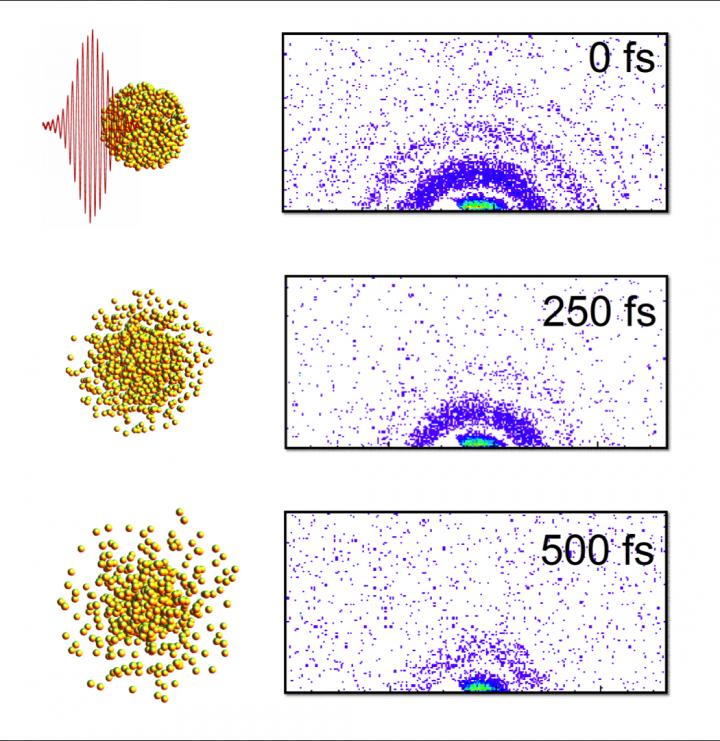
Caption: Here are "stills" from an X-ray "movie" of an exploding nanoparticle. The nanoparticle is superheated with an intense optical pulse and subsequently explodes (left). A series of ultrafast x-ray diffraction images (right) maps the process and contains information how the explosion starts with surface softening and proceeds from the outside in. Credit: Christoph Bostedt

Caption: Here are “stills” from an X-ray “movie” of an exploding nanoparticle. The nanoparticle is superheated with an intense optical pulse and subsequently explodes (left). A series of ultrafast x-ray diffraction images (right) maps the process and contains information how the explosion starts with surface softening and proceeds from the outside in. Credit: Christoph Bostedt

A Feb. 10, 2016 news item on Nanotechnology Now provides more information about the ‘snapshots,

Just as a photographer needs a camera with a split-second shutter speed to capture rapid motion, scientists looking at the behavior of tiny materials need special instruments with the capacity to see changes that happen in the blink of an eye.

An international team of researchers led by X-ray scientist Christoph Bostedt of the U.S. Department of Energy’s (DOE) Argonne National Laboratory and Tais Gorkhover of DOE’s SLAC National Accelerator Laboratory used two special lasers to observe the dynamics of a small sample of xenon as it was heated to a plasma.

A Feb. 10, 2016 Argonne National Laboratory news release (also on EurekAlert) by Jared Sagoff, which originated the news item, provides more technical details,

Bostedt and Gorkhover were able to use the Linac Coherent Light Source (LCLS) at SLAC to make observations of the sample in time steps of approximately a hundred femtoseconds – a femtosecond being one millionth of a billionth of a second [emphasis mine]. The exposure time of the individual images was so short that the quickly moving particles in the gas phase appeared frozen. “The advantage of a machine like the LCLS is that it gives us the equivalent of high-speed flash photography as opposed to a pinhole camera,” Bostedt said. The LCLS is a DOE Office of Science User Facility.

The researchers used an optical laser to heat the sample cluster and an X-ray laser to probe the dynamics of the cluster as it changed over time. As the laser heated the cluster, the photons freed electrons initially bound to the atoms; however, these electrons still remained loosely bound to the cluster.

By imaging exploding nanoparticles, the team was able to make measurements of how they change over time in extreme environments. “Ultimately, we want to understand how the energy from the light affects the system,” Gorkhover said.

“There are really no other techniques that give us this good a resolution in both time and space simultaneously,” she added. “Other methods require us to take averages over many different ‘exposures,’ which can obscure relevant details. Additionally, techniques like electron microscopy involve a substrate material that can interfere with the behavior of the sample.”

According to Bostedt, the research could also impact the study of aerosols in the environment or in combustion, as the dual-laser “pump and probe” model could be adapted to study materials in the gas phase. “Although our material goes from solid to plasma very quickly, there are other types of materials you could study with this or a similar technique,” he said.

I marvel at how very brief the time intervals are at the femtoscale and for that matter, the other subatomic scales.

Here’s a link to and a citation for the paper,

Femtosecond and nanometre visualization of structural dynamics in superheated nanoparticles by Tais Gorkhover, Sebastian Schorb, Ryan Coffee, Marcus Adolph, Lutz Foucar, Daniela Rupp, Andrew Aquila, John D. Bozek, Sascha W. Epp, Benjamin Erk, Lars Gumprecht, Lotte Holmegaard, Andreas Hartmann, Robert Hartmann, Günter Hauser, Peter Holl, Andre Hömke, Per Johnsson, Nils Kimmel, Kai-Uwe Kühnel, Marc Messerschmidt, Christian Reich, Arnaud Rouzée, Benedikt Rudek, Carlo Schmidt et al. Nature Photonics 10, 93–97 (2016) doi:10.1038/nphoton.2015.264 Published online 25 January 2016

This paper is behind a paywall.

Stress makes quantum dots ‘breathe’

A March 19, 2015 news item on ScienceDaily describes some new research on quantum dots,

Researchers at the Department of Energy’s SLAC National Accelerator Laboratory watched nanoscale semiconductor crystals expand and shrink in response to powerful pulses of laser light. This ultrafast “breathing” provides new insight about how such tiny structures change shape as they start to melt — information that can help guide researchers in tailoring their use for a range of applications.

In the experiment using SLAC’s Linac Coherent Light Source (LCLS) X-ray laser, a DOE Office of Science User Facility, researchers first exposed the nanocrystals to a burst of laser light, followed closely by an ultrabright X-ray pulse that recorded the resulting structural changes in atomic-scale detail at the onset of melting.

“This is the first time we could measure the details of how these ultrasmall materials react when strained to their limits,” said Aaron Lindenberg, an assistant professor at SLAC and Stanford who led the experiment. The results were published March 12 [2015] in Nature Communications.

A March 18, 2015 SLAC news release, which originated the news item, provides a general description of quantum dots,

The crystals studied at SLAC are known as “quantum dots” because they display unique traits at the nanoscale that defy the classical physics governing their properties at larger scales. The crystals can be tuned by changing their size and shape to emit specific colors of light, for example.

So scientists have worked to incorporate them in solar panels to make them more efficient and in computer displays to improve resolution while consuming less battery power. These materials have also been studied for potential use in batteries and fuel cells and for targeted drug delivery.

Scientists have also discovered that these and other nanomaterials, which may contain just tens or hundreds of atoms, can be far more damage-resistant than larger bits of the same materials because they exhibit a more perfect crystal structure at the tiniest scales. This property could prove useful in battery components, for example, as smaller particles may be able to withstand more charging cycles than larger ones before degrading.

The news release then goes on to describe the latest research showing the dots ‘breathe’ (Note: A link has been removed),

In the LCLS experiment, researchers studied spheres and nanowires made of cadmium sulfide and cadmium selenide that were just 3 to 5 nanometers, or billionths of a meter, across. The nanowires were up to 25 nanometers long. By comparison, amino acids – the building blocks of proteins – are about 1 nanometer in length, and individual atoms are measured in tenths of nanometers.

By examining the nanocrystals from many different angles with X-ray pulses, researchers reconstructed how they change shape when hit with an optical laser pulse. They were surprised to see the spheres and nanowires expand in width by about 1 percent and then quickly contract within femtoseconds, or quadrillionths of a second. They also found that the nanowires don’t expand in length, and showed that the way the crystals respond to strain was coupled to how their structure melts.

In an earlier, separate study, another team of researchers had used LCLS to explore the response of larger gold particles on longer timescales.

“In the future, we want to extend these experiments to more complex and technologically relevant nanostructures, and also to enable X-ray exploration of nanoscale devices while they are operating,” Lindenberg said. “Knowing how materials change under strain can be used together with simulations to design new materials with novel properties.”

Participating researchers were from SLAC, Stanford and two of their joint institutes, the Stanford Institute for Materials and Energy Sciences (SIMES) and Stanford PULSE Institute; University of California, Berkeley; University of Duisburg-Essen in Germany; and Argonne National Laboratory. The work was supported by the DOE Office of Science and the German Research Council.

Here’s a link to and a citation for the paper,

Visualization of nanocrystal breathing modes at extreme strains by Erzsi Szilagyi, Joshua S. Wittenberg, Timothy A. Miller, Katie Lutker, Florian Quirin, Henrik Lemke, Diling Zhu, Matthieu Chollet, Joseph Robinson, Haidan Wen, Klaus Sokolowski-Tinten, & Aaron M. Lindenberg. Nature Communications 6, Article number: 6577 doi:10.1038/ncomms7577 Published 12 March 2015

This paper is behind a paywall but there is a free preview available through ReadCube Access.