Tag Archives: lung cancer

Cooking up a lung one way or the other

I have two stories about lungs and they are entirely different with the older one being a bioengineering story from the US and the more recent one being an artificial tissue story from the University of Toronto and the University of Ottawa (both in Canada).

Lab grown lungs

The Canadian Broadcasting Corporation’s Quirks and Quarks radio programme posted a December 29, 2018 news item (with embedded radio files) about bioengineered lunjgs,

There are two major components to building an organ: the structure and the right cells on that structure. A team led by Dr. Joan Nichols, a Professor of Internal Medicine, Microbiology and Immunology at the University of Texas Medical Branch in Galveston, were able to tackle both parts of the problem

In their experiment they used a donor organ for the structure. They took a lung from an unrelated pig, and stripped it of its cells, leaving a scaffold of collagen, a tough, flexible protein.  This provided a pre-made appropriate structure, though in future they think it may be possible to use 3-D printing technology to get the same result.

They then added cultured cells from the animal who would be receiving the transplant – so the lung was made of the animal’s own cells. Cultured lung and blood vessel cells were placed on the scaffold and it was  placed in a tank for 30 days with a cocktail of nutrients to help the cells stick to the scaffold and proliferate. The result was a kind of baby lung.

They then transplanted the bio-engineered, though immature, lung into the recipient animal where they hoped it would continue to develop and mature – growing to become a healthy, functioning organ.

The recipients of the bio-engineered lungs were four pigs adult pigs, which appeared to tolerate the transplants well. In order to study the development of the bio-engineered lungs, they euthanized the animals at different times: 10 hours, two weeks, one month and two months after transplantation.

They found that as early as two weeks, the bio-engineered lung had integrated into the recipient animals’ body, building a strong network of blood vessels essential for the lung to survive. There was no evidence of pulmonary edema, the build of fluid in the lungs, which is usually a sign of the blood vessels not working efficiently.  There was no sign of rejection of the transplanted organs, and the pigs were healthy up to the point where they were euthanized.

One lingering concern is how well the bio-engineered lungs delivered oxygen. The four pigs who received the trasplant [sic] had one original functioning lung, so they didn’t depend on their new bio-engineered lung for breathing. The scientists were not sure that the bio-engineered lung was mature enough to handle the full load of oxygen on its own.

You can hear Bob McDonald’s (host of Quirks & Quarks, a Canadian Broadcasting Corporation science radio programme) interview lead scientist, Dr. Joan Nichols if you go to here. (Note: I find he overmodulates his voice but some may find he has a ‘friendly’ voice.)

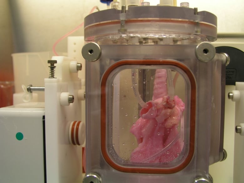
This is an image of the lung scaffold produced by the team,

Lung scaffold in the bioreactor chamber on Day 1 of the experiment, before the cells from the study pig were added. (Credit: Joan Nichols) [downloaded from https://www.cbc.ca/radio/quirks/dec-29-2018-water-on-mars-lab-grown-lungs-and-more-the-biggest-science-stories-of-2018-1.4940811/lab-grown-lungs-are-transplanted-in-pigs-today-they-may-help-humans-tomorrow-1.4940822]

Here’s more technical detail in an August 1, 2018i University of Texas Medical Branch (UTMB) news release (also on EurekAlert), which originally announced the research,

A research team at the University of Texas Medical Branch at Galveston have bioengineered lungs and transplanted them into adult pigs with no medical complication.

In 2014, Joan Nichols and Joaquin Cortiella from The University of Texas Medical Branch at Galveston were the first research team to successfully bioengineer human lungs in a lab. In a paper now available in Science Translational Medicine, they provide details of how their work has progressed from 2014 to the point no complications have occurred in the pigs as part of standard preclinical testing.

“The number of people who have developed severe lung injuries has increased worldwide, while the number of available transplantable organs have decreased,” said Cortiella, professor of pediatric anesthesia. “Our ultimate goal is to eventually provide new options for the many people awaiting a transplant,” said Nichols, professor of internal medicine and associate director of the Galveston National Laboratory at UTMB.

To produce a bioengineered lung, a support scaffold is needed that meets the structural needs of a lung. A support scaffold was created using a lung from an unrelated animal that was treated using a special mixture of sugar and detergent to eliminate all cells and blood in the lung, leaving only the scaffolding proteins or skeleton of the lung behind. This is a lung-shaped scaffold made totally from lung proteins.

The cells used to produce each bioengineered lung came from a single lung removed from each of the study animals. This was the source of the cells used to produce a tissue-matched bioengineered lung for each animal in the study. The lung scaffold was placed into a tank filled with a carefully blended cocktail of nutrients and the animals’ own cells were added to the scaffold following a carefully designed protocol or recipe. The bioengineered lungs were grown in a bioreactor for 30 days prior to transplantation. Animal recipients were survived for 10 hours, two weeks, one month and two months after transplantation, allowing the research team to examine development of the lung tissue following transplantation and how the bioengineered lung would integrate with the body.

All of the pigs that received a bioengineered lung stayed healthy. As early as two weeks post-transplant, the bioengineered lung had established the strong network of blood vessels needed for the lung to survive.

“We saw no signs of pulmonary edema, which is usually a sign of the vasculature not being mature enough,” said Nichols and Cortiella. “The bioengineered lungs continued to develop post-transplant without any infusions of growth factors, the body provided all of the building blocks that the new lungs needed.”

Nichols said that the focus of the study was to learn how well the bioengineered lung adapted and continued to mature within a large, living body. They didn’t evaluate how much the bioengineered lung provided oxygenation to the animal.

“We do know that the animals had 100 percent oxygen saturation, as they had one normal functioning lung,” said Cortiella. “Even after two months, the bioengineered lung was not yet mature enough for us to stop the animal from breathing on the normal lung and switch to just the bioengineered lung.”

For this reason, future studies will look at long-term survival and maturation of the tissues as well as gas exchange capability.

The researchers said that with enough funding, they could grow lungs to transplant into people in compassionate use circumstances within five to 10 years.

“It has taken a lot of heart and 15 years of research to get us this far, our team has done something incredible with a ridiculously small budget and an amazingly dedicated group of people,” Nichols and Cortiella said.

Here’s a citation and another link for the paper,

Production and transplantation of bioengineered lung into a large-animal model by Joan E. Nichols, Saverio La Francesca, Jean A. Niles, Stephanie P. Vega, Lissenya B. Argueta, Luba Frank, David C. Christiani, Richard B. Pyles, Blanca E. Himes, Ruyang Zhang, Su Li, Jason Sakamoto, Jessica Rhudy, Greg Hendricks, Filippo Begarani, Xuewu Liu, Igor Patrikeev, Rahul Pal, Emiliya Usheva, Grace Vargas, Aaron Miller, Lee Woodson, Adam Wacher, Maria Grimaldo, Daniil Weaver, Ron Mlcak, and Joaquin Cortiella. Science Translational Medicine 01 Aug 2018: Vol. 10, Issue 452, eaao3926 DOI: 10.1126/scitranslmed.aao3926

This paper is behind a paywall.

Artificial lung cancer tissue

The research teams at the University of Toronto and the University of Ottawa worked on creating artificial lung tissue but other applications are possible too. First, there’s the announcement in a February 25, 2019 news item on phys.org,

A 3-D hydrogel created by researchers in U of T Engineering Professor Molly Shoichet’s lab is helping University of Ottawa researchers to quickly screen hundreds of potential drugs for their ability to fight highly invasive cancers.

Cell invasion is a critical hallmark of metastatic cancers, such as certain types of lung and brain cancer. Fighting these cancers requires therapies that can both kill cancer cells as well as prevent cell invasion of healthy tissue. Today, most cancer drugs are only screened for their ability to kill cancer cells.

“In highly invasive diseases, there is a crucial need to screen for both of these functions,” says Shoichet. “We now have a way to do this.”

A February 25, 2019 University of Toronto news release (also on EurekAlert), which originated the news item, offers more detail ,

In their latest research, the team used hydrogels to mimic the environment of lung cancer, selectively allowing cancer cells, and not healthy cells, to invade. In their latest research, the team used hydrogels to mimic the environment of lung cancer, selectively allowing cancer cells, and not healthy cells, to invade. This emulated environment enabled their collaborators in Professor Bill Stanford’s lab at University of Ottawa to screen for both cancer-cell growth and invasion. The study, led by Roger Y. Tam, a research associate in Shochet’s lab, was recently published in Advanced Materials.

“We can conduct this in a 384-well plate, which is no bigger than your hand. And with image-analysis software, we can automate this method to enable quick, targeted screenings for hundreds of potential cancer treatments,” says Shoichet.

One example is the researchers’ drug screening for lymphangioleiomyomatosis (LAM), a rare lung disease affecting women. Shoichet and her team were inspired by the work of Green Eggs and LAM, a Toronto-based organization raising awareness of the disease.

Using their hydrogels, they were able to automate and screen more than 800 drugs, thereby uncovering treatments that could target disease growth and invasion.

In the ongoing collaboration, the researchers plan to next screen multiple drugs at different doses to gain greater insight into new treatment methods for LAM. The strategies and insights they gain could also help identify new drugs for other invasive cancers.

Shoichet, who was recently named a Distinguished Woman in Chemistry or Chemical Engineering, also plans to patent the hydrogel technology.

“This has, and continues to be, a great collaboration that is advancing knowledge at the intersection of engineering and biology,” says Shoichet.

I note that Shoichet (pronounced ShoyKet) is getting ready to patent this work. I do have a question about this and it’s not up to Shoichet to answer as she didn’t create the system. Will the taxpayers who funded her work receive any financial benefits should the hydrogel prove to be successful or will we be paying double, both supporting her research and paying for the hydrogel through our healthcare costs?

Getting back to the research, here’s a link to and a citation for the paper,

Rationally Designed 3D Hydrogels Model Invasive Lung Diseases Enabling High‐Content Drug Screening by Roger Y. Tam, Julien Yockell‐Lelièvre, Laura J. Smith, Lisa M. Julian, Alexander E. G. Baker, Chandarong Choey, Mohamed S. Hasim, Jim Dimitroulakos, William L. Stanford, Molly S. Shoichet. Advanced Materials Volume 31, Issue 7 February 15, 2019 1806214 First published online: 27 December 2018 DOI: https://doi.org/10.1002/adma.201806214

This paper is behind a paywall.

Explaining the link between air pollution and heart disease?

An April 26, 2017 news item on Nanowerk announces research that may explain the link between heart disease and air pollution (Note: A link has been removed),

Tiny particles in air pollution have been associated with cardiovascular disease, which can lead to premature death. But how particles inhaled into the lungs can affect blood vessels and the heart has remained a mystery.

Now, scientists have found evidence in human and animal studies that inhaled nanoparticles can travel from the lungs into the bloodstream, potentially explaining the link between air pollution and cardiovascular disease. Their results appear in the journal ACS Nano (“Inhaled Nanoparticles Accumulate at Sites of Vascular Disease”).

An April 26, 2017 American Chemical Society news release on EurekAlert, which originated the news item,  expands on the theme,

The World Health Organization estimates that in 2012, about 72 percent of premature deaths related to outdoor air pollution were due to ischemic heart disease and strokes. Pulmonary disease, respiratory infections and lung cancer were linked to the other 28 percent. Many scientists have suspected that fine particles travel from the lungs into the bloodstream, but evidence supporting this assumption in humans has been challenging to collect. So Mark Miller and colleagues at the University of Edinburgh in the United Kingdom and the National Institute for Public Health and the Environment in the Netherlands used a selection of specialized techniques to track the fate of inhaled gold nanoparticles.

In the new study, 14 healthy volunteers, 12 surgical patients and several mouse models inhaled gold nanoparticles, which have been safely used in medical imaging and drug delivery. Soon after exposure, the nanoparticles were detected in blood and urine. Importantly, the nanoparticles appeared to preferentially accumulate at inflamed vascular sites, including carotid plaques in patients at risk of a stroke. The findings suggest that nanoparticles can travel from the lungs into the bloodstream and reach susceptible areas of the cardiovascular system where they could possibly increase the likelihood of a heart attack or stroke, the researchers say.

Here’s a link to and a citation for the paper,

Inhaled Nanoparticles Accumulate at Sites of Vascular Disease by Mark R. Miller, Jennifer B. Raftis, Jeremy P. Langrish, Steven G. McLean, Pawitrabhorn Samutrtai, Shea P. Connell, Simon Wilson, Alex T. Vesey, Paul H. B. Fokkens, A. John F. Boere, Petra Krystek, Colin J. Campbell, Patrick W. F. Hadoke, Ken Donaldson, Flemming R. Cassee, David E. Newby, Rodger Duffin, and Nicholas L. Mills. ACS Nano, Article ASAP DOI: 10.1021/acsnano.6b08551 Publication Date (Web): April 26, 2017

Copyright © 2017 American Chemical Society

This paper is behind a paywall.

Soy and cellulose come together for a bionano air filter

A Jan. 18, 2017 news item on Nanowerk describes research into an environmentally friendly air filter from Washington State University,

Washington State University researchers have developed a soy-based air filter that can capture toxic chemicals, such as carbon monoxide and formaldehyde, which current air filters can’t.

The research could lead to better air purifiers, particularly in regions of the world that suffer from very poor air quality. …

Working with researchers from the University of Science and Technology Beijing, the WSU team, including Weihong (Katie) Zhong, professor in the School of Mechanical and Materials Engineering, and graduate student Hamid Souzandeh, used a pure soy protein along with bacterial cellulose for an all-natural, biodegradable, inexpensive air filter.

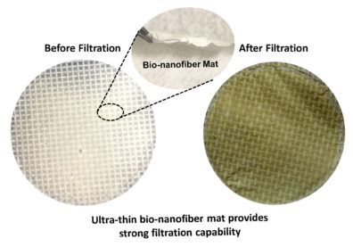
Here’s an image the researchers have made available,

Bionano air filter before and after filtration. Courtesy: Washington State University

A Jan. 12, 2017 Washington State University news release by Tilda Hilding, which originated the news item, expands on the theme,

Poor air quality causes health problems worldwide and is a factor in diseases such as asthma, heart disease and lung cancer. Commercial air purifiers aim for removing the small particles that are present in soot, smoke or car exhaust because these damaging particles are inhaled directly into the lungs.

With many sources of pollution in some parts of the world, however, air pollution also can contain a mix of hazardous gaseous molecules, such as carbon monoxide, formaldehyde, sulfur dioxide and other volatile organic compounds.

Typical air filters, which are usually made of micron-sized fibers of synthetic plastics, physically filter the small particles but aren’t able to chemically capture gaseous molecules. Furthermore, they’re most often made of glass and petroleum products, which leads to secondary pollution, Zhong said.

Soy captures nearly all pollutants

The WSU and Chinese team developed a new kind of air filtering material that uses natural, purified soy protein and bacterial cellulose – an organic compound produced by bacteria. The soy protein and cellulose are cost effective and already used in numerous applications, such as adhesives, plastic products, tissue regeneration materials and wound dressings.

Soy contains a large number of functional chemical groups – it includes 18 types of amino groups. Each of the chemical groups has the potential to capture passing pollution at the molecular level. The researchers used an acrylic acid treatment to disentangle the very rigid soy protein, so that the chemical groups can be more exposed to the pollutants.

The resulting filter was able to remove nearly all of the small particles as well as chemical pollutants, said Zhong.

Filters are economical, biodegradable

Especially in very polluted environments, people might be breathing an unknown mix of pollutants that could prove challenging to purify. But, with its large number of functional groups, the soy protein is able to attract a wide variety of polluting molecules.

“We can take advantage from those chemical groups to grab the toxics in the air,” Zhong said.

The materials are also cost-effective and biodegradable. Soybeans are among the most abundant plants in the world, she added.

Zhong occasionally visits her native China and has personally experienced the heavy pollution in Beijing as sunny skies turn to gray smog within a few days.

“Air pollution is a very serious health issue,” she said. “If we can improve indoor air quality, it would help a lot of people.”

Patents filed on filters, paper towels

In addition to the soy-based filters, the researchers have also developed gelatin- and cellulose-based air filters. They are also applying the filter material on top of low-cost and disposable paper towel to reinforce it and to improve its performance. They have filed patents on the technology and are interested in commercialization opportunities.

Here’s a link to and a citation for the paper,

Soy protein isolate/bacterial cellulose composite membranes for high efficiency particulate air filtration by Xiaobing Liu, Hamid Souzandeh, Yudong Zheng, Yajie Xie, Wei-Hong Zhong, Cai Wang. Composites Science and Technology Volume 138, 18 January 2017, Pages 124–133         http://dx.doi.org/10.1016/j.compscitech.2016.11.022

This paper is behind a paywall.

Sniffing out disease (Na-Nose)

The ‘artificial nose’ is not a newcomer to this blog. The most recent post prior to this is a March 15, 2016 piece about Disney using an artificial nose for art conservation. Today’s (Jan. 9, 2016) piece concerns itself with work from Israel and ‘sniffing out’ disease, according to a Dec. 30, 2016 news item in Sputnik News,

A team from the Israel Institute of Technology has developed a device that from a single breath can identify diseases such as multiple forms of cancer, Parkinson’s disease, and multiple sclerosis. While the machine is still in the experimental stages, it has a high degree of promise for use in non-invasive diagnoses of serious illnesses.

The international team demonstrated that a medical theory first proposed by the Greek physician Hippocrates some 2400 years ago is true, certain diseases leave a “breathprint” on the exhalations of those afflicted. The researchers created a prototype for a machine that can pick up on those diseases using the outgoing breath of a patient. The machine, called the Na-Nose, tests breath samples for the presence of trace amounts of chemicals that are indicative of 17 different illnesses.

A Dec. 22, 2016 Technion Israel Institute of Technology press release offers more detail about the work,

An international team of 56 researchers in five countries has confirmed a hypothesis first proposed by the ancient Greeks – that different diseases are characterized by different “chemical signatures” identifiable in breath samples. …

Diagnostic techniques based on breath samples have been demonstrated in the past, but until now, there has not been scientific proof of the hypothesis that different and unrelated diseases are characterized by distinct chemical breath signatures. And technologies developed to date for this type of diagnosis have been limited to detecting a small number of clinical disorders, without differentiation between unrelated diseases.

The study of more than 1,400 patients included 17 different and unrelated diseases: lung cancer, colorectal cancer, head and neck cancer, ovarian cancer, bladder cancer, prostate cancer, kidney cancer, stomach cancer, Crohn’s disease, ulcerative colitis, irritable bowel syndrome, Parkinson’s disease (two types), multiple sclerosis, pulmonary hypertension, preeclampsia and chronic kidney disease. Samples were collected between January 2011 and June 2014 from in 14 departments at 9 medical centers in 5 countries: Israel, France, the USA, Latvia and China.

The researchers tested the chemical composition of the breath samples using an accepted analytical method (mass spectrometry), which enabled accurate quantitative detection of the chemical compounds they contained. 13 chemical components were identified, in different compositions, in all 17 of the diseases.

According to Prof. Haick, “each of these diseases is characterized by a unique fingerprint, meaning a different composition of these 13 chemical components.  Just as each of us has a unique fingerprint that distinguishes us from others, each disease has a chemical signature that distinguishes it from other diseases and from a normal state of health. These odor signatures are what enables us to identify the diseases using the technology that we developed.”

With a new technology called “artificially intelligent nanoarray,” developed by Prof. Haick, the researchers were able to corroborate the clinical efficacy of the diagnostic technology. The array enables fast and inexpensive diagnosis and classification of diseases, based on “smelling” the patient’s breath, and using artificial intelligence to analyze the data obtained from the sensors. Some of the sensors are based on layers of gold nanoscale particles and others contain a random network of carbon nanotubes coated with an organic layer for sensing and identification purposes.

The study also assessed the efficiency of the artificially intelligent nanoarray in detecting and classifying various diseases using breath signatures. To verify the reliability of the system, the team also examined the effect of various factors (such as gender, age, smoking habits and geographic location) on the sample composition, and found their effect to be negligible, and without impairment on the array’s sensitivity.

“Each of the sensors responds to a wide range of exhalation components,” explain Prof. Haick and his previous Ph.D student, Dr. Morad Nakhleh, “and integration of the information provides detailed data about the unique breath signatures characteristic of the various diseases. Our system has detected and classified various diseases with an average accuracy of 86%.

This is a new and promising direction for diagnosis and classification of diseases, which is characterized not only by considerable accuracy but also by low cost, low electricity consumption, miniaturization, comfort and the possibility of repeating the test easily.”

“Breath is an excellent raw material for diagnosis,” said Prof. Haick. “It is available without the need for invasive and unpleasant procedures, it’s not dangerous, and you can sample it again and again if necessary.”

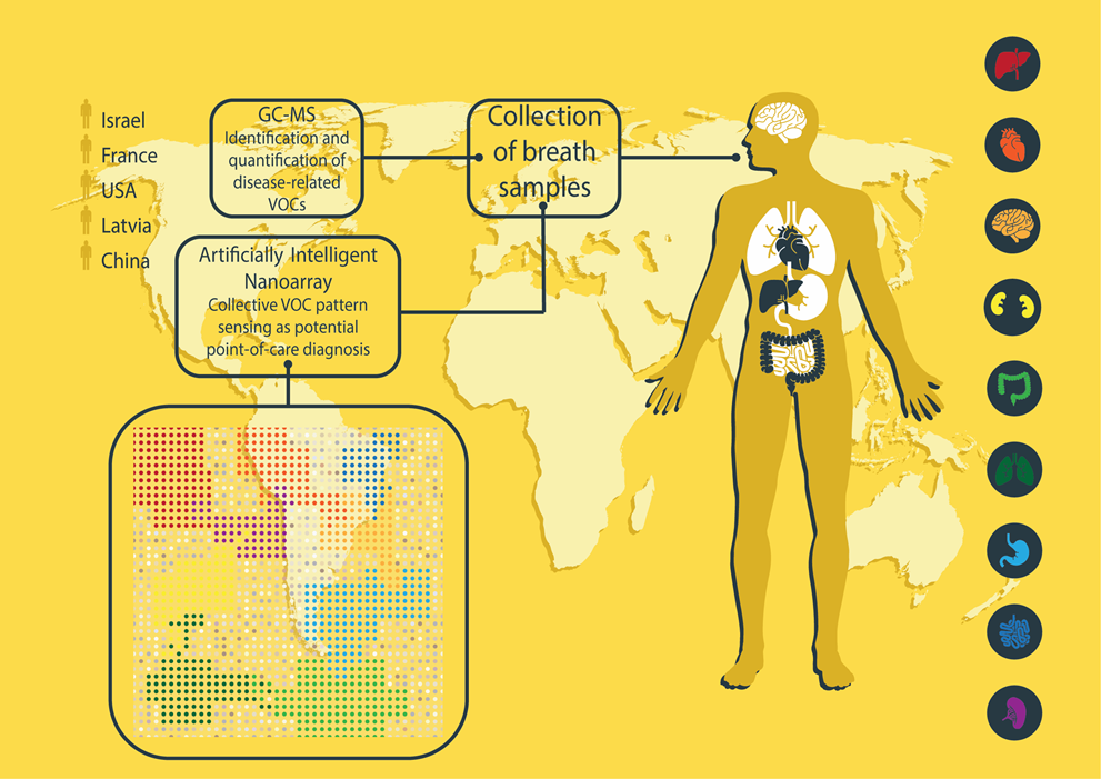
Here’s a schematic of the study, which the researchers have made available,

Diagram: A schematic view of the study. Two breath samples were taken from each subject, one was sent for chemical mapping using mass spectrometry, and the other was analyzed in the new system, which produced a clinical diagnosis based on the chemical fingerprint of the breath sample. Courtesy: Tech;nion

There is also a video, which covers much of the same ground as the press release but also includes information about the possible use of the Na-Nose technology in the European Union’s SniffPhone project,

Here’s a link to and a citation for the paper,

Diagnosis and Classification of 17 Diseases from 1404 Subjects via Pattern Analysis of Exhaled Molecules by Morad K. Nakhleh, Haitham Amal, Raneen Jeries, Yoav Y. Broza, Manal Aboud, Alaa Gharra, Hodaya Ivgi, Salam Khatib, Shifaa Badarneh, Lior Har-Shai, Lea Glass-Marmor, Izabella Lejbkowicz, Ariel Miller, Samih Badarny, Raz Winer, John Finberg, Sylvia Cohen-Kaminsky, Frédéric Perros, David Montani, Barbara Girerd, Gilles Garcia, Gérald Simonneau, Farid Nakhoul, Shira Baram, Raed Salim, Marwan Hakim, Maayan Gruber, Ohad Ronen, Tal Marshak, Ilana Doweck, Ofer Nativ, Zaher Bahouth, Da-you Shi, Wei Zhang, Qing-ling Hua, Yue-yin Pan, Li Tao, Hu Liu, Amir Karban, Eduard Koifman, Tova Rainis, Roberts Skapars, Armands Sivins, Guntis Ancans, Inta Liepniece-Karele, Ilze Kikuste, Ieva Lasina, Ivars Tolmanis, Douglas Johnson, Stuart Z. Millstone, Jennifer Fulton, John W. Wells, Larry H. Wilf, Marc Humbert, Marcis Leja, Nir Peled, and Hossam Haick. ACS Nano, Article ASAP DOI: 10.1021/acsnano.6b04930 Publication Date (Web): December 21, 2016

Copyright © 2017 American Chemical Society

This paper appears to be open access.

As for SniffPhone, they’re hoping that Na-Nose or something like it will allow them to modify smartphones in a way that will allow diseases to be detected.

I can’t help wondering who will own the data if your smartphone detects a disease. If you think that’s an idle question, here’s an excerpt from Sue Halpern’s Dec. 22, 2016 review of two books (“Weapons of Math Destruction: How Big Data Increases Inequality and Threatens Democracy” by Cathy O’Neil and “Virtual Competition: The Promise and Perils of the Algorithm-Driven Economy” by Ariel Ezrachi and Maurice E. Stucke) for the New York Times Review of Books,

We give our data away. We give it away in drips and drops, not thinking that data brokers will collect it and sell it, let alone that it will be used against us. There are now private, unregulated DNA databases culled, in part, from DNA samples people supply to genealogical websites in pursuit of their ancestry. These samples are available online to be compared with crime scene DNA without a warrant or court order. (Police are also amassing their own DNA databases by swabbing cheeks during routine stops.) In the estimation of the Electronic Frontier Foundation, this will make it more likely that people will be implicated in crimes they did not commit.

Or consider the data from fitness trackers, like Fitbit. As reported in The Intercept:

During a 2013 FTC panel on “Connected Health and Fitness,” University of Colorado law professor Scott Peppet said, “I can paint an incredibly detailed and rich picture of who you are based on your Fitbit data,” adding, “That data is so high quality that I can do things like price insurance premiums or I could probably evaluate your credit score incredibly accurately.”

Halpern’s piece is well worth reading in its entirety.