Tag Archives: Macquarie University

Ancient Namibian gemstone could be key to new light-based quantum computers

Researchers in Scotland, the US, Australia, and Denmark have a found a solution to a problem with creating light-based computers according to an April 15, 2022 news item on phys.org,

A special form of light made using an ancient Namibian gemstone could be the key to new light-based quantum computers, which could solve long-held scientific mysteries, according to new research led by the University of St Andrews.

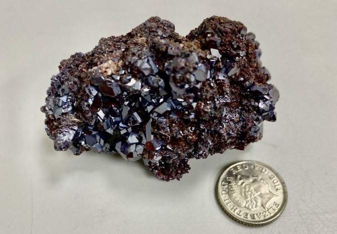
The research, conducted in collaboration with scientists at Harvard University in the US, Macquarie University in Australia and Aarhus University in Denmark and published in Nature Materials, used a naturally mined cuprous oxide (Cu2O) gemstone from Namibia to produce Rydberg polaritons, the largest hybrid particles of light and matter ever created.

Cuprous oxide – the mined crystal from Namibia used for making Rydberg polaritons. Courtesy: University of St. Andrews

An April 15, 2022 University of St. Andrews press release, which originated the news item, describes Rydberg polaritons and explains why they could be the key to light-based quantum computing,

Rydberg polaritons switch continually from light to matter and back again. In Rydberg polaritons, light and matter are like two sides of a coin, and the matter side is what makes polaritons interact with each other.

This interaction is crucial because this is what allows the creation of quantum simulators, a special type of quantum computer, where information is stored in quantum bits. These quantum bits [qubits], unlike the binary bits in classical computers that can only be 0 or 1, can take any value between 0 and 1. They can therefore store much more information and perform several processes simultaneously.

This capability could allow quantum simulators to solve important mysteries of physics, chemistry and biology, for example, how to make high-temperature superconductors for highspeed trains, how cheaper fertilisers could be made potentially solving global hunger, or how proteins fold making it easier to produce more effective drugs.

Project lead Dr Hamid Ohadi, of the School of Physics and Astronomy at the University of St Andrews, said: “Making a quantum simulator with light is the holy grail of science. We have taken a huge leap towards this by creating Rydberg polaritons, the key ingredient of it.”

To create Rydberg polaritons, the researchers trapped light between two highly reflective mirrors. A cuprous oxide crystal from a stone mined in Namibia was then thinned and polished to a 30-micrometer thick slab (thinner than a strand of human hair) and sandwiched between the two mirrors to make Rydberg polaritons 100 times larger than ever demonstrated before.

One of the leading authors Dr Sai Kiran Rajendran, of the School of Physics and Astronomy at the University of St Andrews, said: “Purchasing the stone on eBay was easy. The challenge was to make Rydberg polaritons that exist in an extremely narrow colour range.”

The team is currently further refining these methods in order to explore the possibility of making quantum circuits, which are the next ingredient for quantum simulators.

The research was funded by UK Engineering and Physical Sciences Research Council (EPSRC).

Here’s a link to and a citation for the paper,

Rydberg exciton–polaritons in a Cu2O microcavity by Konstantinos Orfanakis, Sai Kiran Rajendran, Valentin Walther, Thomas Volz, Thomas Pohl & Hamid Ohadi. Nature Materials (2022) DOI: DOIhttps://doi.org/10.1038/s41563-022-01230-4 Published: 14 April 2022

This paper is behind a paywall.

Finding killer bacteria with quantum dots and a smartphone

An August 5, 2019 news item on Nanowerk announces a new technology for detecting killer bacteria (Note: A link has been removed),

A combination of off-the-shelf quantum dots and a smartphone camera soon could allow doctors to identify antibiotic-resistant bacteria in just 40 minutes, potentially saving patient lives.

Staphylococcus aureus (golden staph), is a common form of bacterium that causes serious and sometimes fatal conditions such as pneumonia and heart valve infections. Of particular concern is a strain that does not respond to methicillin, the antibiotic of first resort, and is known as methicillin-resistant S. aureus, or MRSA.

Recent reports estimate that 700 000 deaths globally could be attributed to antimicrobial resistance, such as methicillin-resistance. Rapid identification of MRSA is essential for effective treatment, but current methods make it a challenging process, even within well-equipped hospitals.

Soon, however, that may change, using nothing except existing technology.

Researchers from Macquarie University and the University of New South Wales, both in Australia, have demonstrated a proof-of-concept device that uses bacterial DNA to identify the presence of Staphylococcus aureus positively in a patient sample – and to determine if it will respond to frontline antibiotics.

An August 12,2019 Macquarie University press release (also on EurekAlert but published August 4, 2019), which originated the news item, delves into the work,

In a paper published in the international peer-reviewed journal Sensors and Actuators B: Chemical the Macquarie University team of Dr Vinoth Kumar Rajendran, Professor Peter Bergquist and Associate Professor Anwar Sunna with Dr Padmavathy Bakthavathsalam (UNSW) reveal a new way to confirm the presence of the bacterium, using a mobile phone and some ultra-tiny semiconductor particles known as quantum dots.

“Our team is using Synthetic Biology and NanoBiotechnology to address biomedical challenges. Rapid and simple ways of identifying the cause of infections and starting appropriate treatments are critical for treating patients effectively,” says Associate Professor Anwar Sunna, head of the Sunna Lab at Macquarie University.

“This is true in routine clinical situations, but also in the emerging field of personalised medicine.”

The researchers’ approach identifies the specific strain of golden staph by using a method called convective polymerase chain reaction (or cPCR). This is a derivative of a widely -employed technique in which a small segment of DNA is copied thousands of times, creating multiple samples suitable for testing.

Vinoth Kumar and colleagues then subject the DNA copies to a process known as lateral flow immunoassay – a paper-based diagnostic tool used to confirm the presence or absence of a target biomarker. The researchers use probes fitted with quantum dots to detect two unique genes, that confirms the presence of methicillin resistance in golden staph

A chemical added at the PCR stage to the DNA tested makes the sample fluoresce when the genes are detected by the quantum dots – a reaction that can be captured easily using the camera on a mobile phone.

The result is a simple and rapid method of detecting the presence of the bacterium, while simultaneously ruling first-line treatment in or out.

Although currently at proof-of-concept stage, the researchers say their system which is powered by a simple battery is suitable for rapid detection in different settings.

“We can see this being used easily not only in hospitals, but also in GP clinics and at patient bedsides,” says lead author, Macquarie’s Vinoth Kumar Rajendran.

Here’s a link to and a citation for the paper,

Smartphone detection of antibiotic resistance using convective PCR and a lateral flow assay by Vinoth Kumar Rajendran, Padmavathy Bakthavathsalam, Peter L.Bergquist, Anwar Sunna. Sensors and Actuators B: Chemical Volume 298, 1 November 2019,126849 DOI: https://doi.org/10.1016/j.snb.2019.126849 Available online 23 July 2019

This paper is behind a paywall.

Luminescent upconversion nanoparticles could make imaging more efficient

Researchers at the University of Adelaide (Australia) have found a way to embed luminiscent nanoparticles in glass, according to a June 8, 2016 news item on Nanotechnology,

This new “hybrid glass” successfully combines the properties of these special luminescent (or light-emitting) nanoparticles with the well-known aspects of glass, such as transparency and the ability to be processed into various shapes including very fine optical fibres.

The research, in collaboration with Macquarie University and University of Melbourne, has been published online in the journal Advanced Optical Materials.

A June 7, 2016 University of Adelaide press release (also on EurekAlert), which originated the news item, offers more detail,

“These novel luminescent nanoparticles, called upconversion nanoparticles, have become promising candidates for a whole variety of ultra-high tech applications such as biological sensing, biomedical imaging and 3D volumetric displays,” says lead author Dr Tim Zhao, from the University of Adelaide’s School of Physical Sciences and Institute for Photonics and Advanced Sensing (IPAS).

“Integrating these nanoparticles into glass, which is usually inert, opens up exciting possibilities for new hybrid materials and devices that can take advantage of the properties of nanoparticles in ways we haven’t been able to do before. For example, neuroscientists currently use dye injected into the brain and lasers to be able to guide a glass pipette to the site they are interested in. If fluorescent nanoparticles were embedded in the glass pipettes, the unique luminescence of the hybrid glass could act like a torch to guide the pipette directly to the individual neurons of interest.”

Although this method was developed with upconversion nanoparticles, the researchers believe their new ‘direct-doping’ approach can be generalised to other nanoparticles with interesting photonic, electronic and magnetic properties. There will be many applications – depending on the properties of the nanoparticle.

“If we infuse glass with a nanoparticle that is sensitive to radiation and then draw that hybrid glass into a fibre, we could have a remote sensor suitable for nuclear facilities,” says Dr Zhao.

To date, the method used to integrate upconversion nanoparticles into glass has relied on the in-situ growth of the nanoparticles within the glass.

“We’ve seen remarkable progress in this area but the control over the nanoparticles and the glass compositions has been limited, restricting the development of many proposed applications,” says project leader Professor Heike Ebendorff-Heideprem, Deputy Director of IPAS.

“With our new direct doping method, which involves synthesizing the nanoparticles and glass separately and then combining them using the right conditions, we’ve been able to keep the nanoparticles intact and well dispersed throughout the glass. The nanoparticles remain functional and the glass transparency is still very close to its original quality. We are heading towards a whole new world of hybrid glass and devices for light-based technologies.”

Here’s a link to and a citation for the paper,

Upconversion Nanocrystal-Doped Glass: A New Paradigm for Photonic Materials by Jiangbo Zhao, Xianlin Zheng, Erik P. Schartner, Paul Ionescu, Run Zhang, Tich-Lam Nguyen, Dayong Jin, and Heike Ebendorff-Heidepriem. Advanced Optical Materials DOI: 10.1002/adom.201600296 Version of Record online: 30 MAY 2016

© 2016 WILEY-VCH Verlag GmbH & Co. KGaA, Weinheim

This paper is behind a paywall.

Nanodiamonds and a broken heart

Nanowerk’s March 3, 2013 news item highlights some research with biomedical applications taking place in Australia,

Researchers at Macquarie University have been perfecting a technique that may help see nanodiamonds used in biomedical applications. PhD student Jana Say has been working on processing the raw diamonds so that they might be used as a tag for biological molecules.

“We are working with nanodiamonds to process them so that they are stable enough to be used as a probe for single-molecule interactions. As diamonds are made from carbon, they are non-toxic which makes them advantageous for many biological applications over other nanoparticles,” says Say.

The Macquarie University Feb. 13, 2013 news release, which originated the Nanowerk news item, provides more detail,

The processing technique has already contributed to the success of research projects that have used the diamonds. Working with an international team, Say’s diamonds were able to be optically trapped and manipulated in three-dimensions – this first time this has been achieved.

The diamonds themselves are incredibly small, 5000 times smaller than a human hair, and so the real strength in Say’s technique is her ability to consistently produce stable samples.

“The real challenge is reliably producing the same sample. It’s a very repetitive and involved process to prepare and characterise these diamonds,” she says.

Say, under the supervision of Dr Louise Brown of the Department of Chemistry and Biomolecular Sciecnes, has plans to continue to develop these diamonds and collaborate with other researchers to explore their full potential.

“Jana’s work is incredibly important,” says Dr Brown. “These diamonds were recently used in a project which won a Macquarie University research excellence award for demonstrating that nanodiamonds can be isolated and made to emit light. With this work, we continue to make real breakthroughs in this area and are contributing to the long term goals in ultrasensitive imaging and sensing technologies.”

For anyone who’s interested, here’s a citation and link to the research paper,

Three-dimensional optical manipulation of a single electron spin by Michael Geiselmann, Mathieu L. Juan, Jan Renger, Jana M. Say, Louise J. Brown,  F. Javier García de Abajo, Frank Koppens & Romain Quidant. Nature Nanotechnology (2013) doi:10.1038/nnano.2012.259 Published online 10 February 2013

The article is behind a paywall. One final comment, I very much enjoyed the fanciful title for the news release about this work, Diamonds may mend a broken heart: Researchers perfect nanodiamonds for use in biomedical applications.