Tag Archives: medical diagnostics

Graphene and smart textiles

Here’s one of the more recent efforts to create fibres that are electronic and capable of being woven into a smart textile. (Details about a previous effort can be found at the end of this post.) Now for this one, from a Dec. 3, 2018 news item on ScienceDaily,

The quest to create affordable, durable and mass-produced ‘smart textiles’ has been given fresh impetus through the use of the wonder material Graphene.

An international team of scientists, led by Professor Monica Craciun from the University of Exeter Engineering department, has pioneered a new technique to create fully electronic fibres that can be incorporated into the production of everyday clothing.

A Dec. 3, 2018 University of Exeter press release (also on EurekAlert), provides more detail about the problems associated with wearable electronics and the solution being offered (Note: A link has been removed),

Currently, wearable electronics are achieved by essentially gluing devices to fabrics, which can mean they are too rigid and susceptible to malfunctioning.

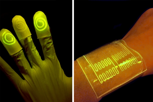
The new research instead integrates the electronic devices into the fabric of the material, by coating electronic fibres with light-weight, durable components that will allow images to be shown directly on the fabric.

The research team believe that the discovery could revolutionise the creation of wearable electronic devices for use in a range of every day applications, as well as health monitoring, such as heart rates and blood pressure, and medical diagnostics.

The international collaborative research, which includes experts from the Centre for Graphene Science at the University of Exeter, the Universities of Aveiro and Lisbon in Portugal, and CenTexBel in Belgium, is published in the scientific journal Flexible Electronics.

Professor Craciun, co-author of the research said: “For truly wearable electronic devices to be achieved, it is vital that the components are able to be incorporated within the material, and not simply added to it.

Dr Elias Torres Alonso, Research Scientist at Graphenea and former PhD student in Professor Craciun’s team at Exeter added “This new research opens up the gateway for smart textiles to play a pivotal role in so many fields in the not-too-distant future.  By weaving the graphene fibres into the fabric, we have created a new technique to all the full integration of electronics into textiles. The only limits from now are really within our own imagination.”

At just one atom thick, graphene is the thinnest substance capable of conducting electricity. It is very flexible and is one of the strongest known materials. The race has been on for scientists and engineers to adapt graphene for the use in wearable electronic devices in recent years.

This new research used existing polypropylene fibres – typically used in a host of commercial applications in the textile industry – to attach the new, graphene-based electronic fibres to create touch-sensor and light-emitting devices.

The new technique means that the fabrics can incorporate truly wearable displays without the need for electrodes, wires of additional materials.

Professor Saverio Russo, co-author and from the University of Exeter Physics department, added: “The incorporation of electronic devices on fabrics is something that scientists have tried to produce for a number of years, and is a truly game-changing advancement for modern technology.”

Dr Ana Neves, co-author and also from Exeter’s Engineering department added “The key to this new technique is that the textile fibres are flexible, comfortable and light, while being durable enough to cope with the demands of modern life.”

In 2015, an international team of scientists, including Professor Craciun, Professor Russo and Dr Ana Neves from the University of Exeter, have pioneered a new technique to embed transparent, flexible graphene electrodes into fibres commonly associated with the textile industry.

Here’s a link to and a citation for the paper,

Graphene electronic fibres with touch-sensing and light-emitting functionalities for smart textiles by Elias Torres Alonso, Daniela P. Rodrigues, Mukond Khetani, Dong-Wook Shin, Adolfo De Sanctis, Hugo Joulie, Isabel de Schrijver, Anna Baldycheva, Helena Alves, Ana I. S. Neves, Saverio Russo & Monica F. Craciun. Flexible Electronicsvolume 2, Article number: 25 (2018) DOI: https://doi.org/10.1038/s41528-018-0040-2 Published 25 September 2018

This paper is open access.

I have an earlier post about an effort to weave electronics into textiles for soldiers, from an April 5, 2012 posting,

I gather that today’s soldier (aka, warfighter)  is carrying as many batteries as weapons. Apparently, the average soldier carries a couple of kilos worth of batteries and cables to keep their various pieces of equipment operational. The UK’s Centre for Defence Enterprise (part of the Ministry of Defence) has announced that this situation is about to change as a consequence of a recently funded research project with a company called Intelligent Textiles. From Bob Yirka’s April 3, 2012 news item for physorg.com,

To get rid of the cables, a company called Intelligent Textiles has come up with a type of yarn that can conduct electricity, which can be woven directly into the fabric of the uniform. And because they allow the uniform itself to become one large conductive unit, the need for multiple batteries can be eliminated as well.

I dug down to find more information about this UK initiative and the Intelligent Textiles company but the trail seems to end in 2015. Still, I did find a Canadian connection (for those who don’t know I’m a Canuck) and more about Intelligent Textile’s work with the British military in this Sept. 21, 2015 article by Barry Collins for alphr.com (Note: Links have been removed),

A two-person firm operating from a small workshop in Staines-upon-Thames, Intelligent Textiles has recently landed a multimillion-pound deal with the US Department of Defense, and is working with the Ministry of Defence (MoD) to bring its potentially life-saving technology to British soldiers. Not bad for a company that only a few years ago was selling novelty cushions.

Intelligent Textiles was born in 2002, almost by accident. Asha Peta Thompson, an arts student at Central Saint Martins, had been using textiles to teach children with special needs. That work led to a research grant from Brunel University, where she was part of a team tasked with creating a “talking jacket” for the disabled. The garment was designed to help cerebral palsy sufferers to communicate, by pressing a button on the jacket to say “my name is Peter”, for example, instead of having a Stephen Hawking-like communicator in front of them.

Another member of that Brunel team was engineering lecturer Dr Stan Swallow, who was providing the electronics expertise for the project. Pretty soon, the pair realised the prototype waistcoat they were working on wasn’t going to work: it was cumbersome, stuffed with wires, and difficult to manufacture. “That’s when we had the idea that we could weave tiny mechanical switches into the surface of the fabric,” said Thompson.

The conductive weave had several advantages over packing electronics into garments. “It reduces the amount of cables,” said Thompson. “It can be worn and it’s also washable, so it’s more durable. It doesn’t break; it can be worn next to the skin; it’s soft. It has all the qualities of a piece of fabric, so it’s a way of repackaging the electronics in a way that’s more user-friendly and more comfortable.” The key to Intelligent Textiles’ product isn’t so much the nature of the raw materials used, but the way they’re woven together. “All our patents are in how we weave the fabric,” Thompson explained. “We weave two conductive yarns to make a tiny mechanical switch that is perfectly separated or perfectly connected. We can weave an electronic circuit board into the fabric itself.”

Intelligent Textiles’ big break into the military market came when they met a British textiles firm that was supplying camouflage gear to the Canadian armed forces. [emphasis mine] The firm was attending an exhibition in Canada and invited the Intelligent Textiles duo to join them. “We showed a heated glove and an iPod controller,” said Thompson. “The Canadians said ‘that’s really fantastic, but all we need is power. Do you think you could weave a piece of fabric that distributes power?’ We said, ‘we’re already doing it’.”Before long it wasn’t only power that the Canadians wanted transmitted through the fabric, but data.

“The problem a soldier faces at the moment is that he’s carrying 60 AA batteries [to power all the equipment he carries],” said Thompson. “He doesn’t know what state of charge those batteries are at, and they’re incredibly heavy. He also has wires and cables running around the system. He has snag hazards – when he’s going into a firefight, he can get caught on door handles and branches, so cables are a real no-no.”

The Canadians invited the pair to speak at a NATO conference, where they were approached by military brass with more familiar accents. “It was there that we were spotted by the British MoD, who said ‘wow, this is a British technology but you’re being funded by Canada’,” said Thompson. That led to £235,000 of funding from the Centre for Defence Enterprise (CDE) – the money they needed to develop a fabric wiring system that runs all the way through the soldier’s vest, helmet and backpack.

There are more details about the 2015 state of affairs, textiles-wise, in a March 11, 2015 article by Richard Trenholm for CNET.com (Note: A link has been removed),

Speaking at the Wearable Technology Show here, Swallow describes IT [Intelligent Textiles]L as a textile company that “pretends to be a military company…it’s funny how you slip into these domains.”

One domain where this high-tech fabric has seen frontline action is in the Canadian military’s IAV Stryker armoured personnel carrier. ITL developed a full QWERTY keyboard in a single piece of fabric for use in the Stryker, replacing a traditional hardware keyboard that involved 100 components. Multiple components allow for repair, but ITL knits in redundancy so the fabric can “degrade gracefully”. The keyboard works the same as the traditional hardware, with the bonus that it’s less likely to fall on a soldier’s head, and with just one glaring downside: troops can no longer use it as a step for getting in and out of the vehicle.

An armoured car with knitted controls is one thing, but where the technology comes into its own is when used about the person. ITL has worked on vests like the JTAC, a system “for the guys who call down airstrikes” and need “extra computing oomph.” Then there’s SWIPES, a part of the US military’s Nett Warrior system — which uses a chest-mounted Samsung Galaxy Note 2 smartphone — and British military company BAE’s Broadsword system.

ITL is currently working on Spirit, a “truly wearable system” for the US Army and United States Marine Corps. It’s designed to be modular, scalable, intuitive and invisible.

While this isn’t an ITL product, this video about Broadsword technology from BAE does give you some idea of what wearable technology for soldiers is like,

baesystemsinc

Uploaded on Jul 8, 2014

Broadsword™ delivers groundbreaking technology to the 21st Century warfighter through interconnecting components that inductively transfer power and data via The Spine™, a revolutionary e-textile that can be inserted into any garment. This next-generation soldier system offers enhanced situational awareness when used with the BAE Systems’ Q-Warrior® see-through display.

If anyone should have the latest news about Intelligent Textile’s efforts, please do share in the comments section.

I do have one other posting about textiles and the military, which is dated May 9, 2012, but while it does reference US efforts it is not directly related to weaving electronics into solder’s (warfighter’s) gear.

You can find CenTexBel (Belgian Textile Rsearch Centre) here and Graphenea here. Both are mentioned in the University of Exeter press release.

Semi-living gloves as sensors

Researchers at the Massachusetts Institute of Technology (MIT) are calling it a new ‘living material’ according to a Feb. 16, 2017 news item on Nanowerk,

Engineers and biologists at MIT have teamed up to design a new “living material” — a tough, stretchy, biocompatible sheet of hydrogel injected with live cells that are genetically programmed to light up in the presence of certain chemicals.

Researchers have found that the hydrogel’s mostly watery environment helps keep nutrients and programmed bacteria alive and active. When the bacteria reacts to a certain chemical, the bacteria are programmed to light up, as seen on the left. Courtesy of the researchers

A Feb. 15, 2017 MIT news release, which originated the news item, provides more information about this work,

In a paper published this week in the Proceedings of the National Academy of Sciences, the researchers demonstrate the new material’s potential for sensing chemicals, both in the environment and in the human body.

The team fabricated various wearable sensors from the cell-infused hydrogel, including a rubber glove with fingertips that glow after touching a chemically contaminated surface, and bandages that light up when pressed against chemicals on a person’s skin.

Xuanhe Zhao, the Robert N. Noyce Career Development associate professor of mechanical engineering at MIT, says the group’s living material design may be adapted to sense other chemicals and contaminants, for uses ranging from crime scene investigation and forensic science, to pollution monitoring and medical diagnostics.

“With this design, people can put different types of bacteria in these devices to indicate toxins in the environment, or disease on the skin,” says Timothy Lu, associate professor of biological engineering and of electrical engineering and computer science. “We’re demonstrating the potential for living materials and devices.”

The paper’s co-authors are graduate students Xinyue Liu, Tzu-Chieh Tang, Eleonore Tham, Hyunwoo Yuk, and Shaoting Lin.

Infusing life in materials

Lu and his colleagues in MIT’s Synthetic Biology Group specialize in creating biological circuits, genetically reprogramming the biological parts in living cells such as E. coli to work together in sequence, much like logic steps in an electrical circuit. In this way, scientists can reengineer living cells to carry out specific functions, including the ability to sense and signal the presence of viruses and toxins.

However, many of these newly programmed cells have only been demonstrated in situ, within Petri dishes, where scientists can carefully control the nutrient levels necessary to keep the cells alive and active — an environment that has proven extremely difficult to replicate in synthetic materials.

“The challenge to making living materials is how to maintain those living cells, to make them viable and functional in the device,” Lu says. “They require humidity, nutrients, and some require oxygen. The second challenge is how to prevent them from escaping from the material.”

To get around these roadblocks, others have used freeze-dried chemical extracts from genetically engineered cells, incorporating them into paper to create low-cost, virus-detecting diagnostic strips. But extracts, Lu says, are not the same as living cells, which can maintain their functionality over a longer period of time and may have higher sensitivity for detecting pathogens.

Other groups have seeded heart muscle cells onto thin rubber films to make soft, “living” actuators, or robots. When bent repeatedly, however, these films can crack, allowing the live cells to leak out.

A lively host

Zhao’s group in MIT’s Soft Active Materials Laboratory has developed a material that may be ideal for hosting living cells. For the past few years, his team has come up with various formulations of hydrogel — a tough, highly stretchable, biocompatible material made from a mix of polymer and water. Their latest designs have contained up to 95 percent water, providing an environment which Zhao and Lu recognized might be suitable for sustaining living cells. The material also resists cracking even when repeatedly stretched and pulled — a property that could help contain cells within the material.

The two groups teamed up to integrate Lu’s genetically programmed bacterial cells into Zhao’s sheets of hydrogel material. They first fabricated layers of hydrogel and patterned narrow channels within the layers using 3-D printing and micromolding techniques. They fused the hydrogel to a layer of elastomer, or rubber, that is porous enough to let in oxygen. They then injected E. coli cells into the hydrogel’s channels. The cells were programmed to fluoresce, or light up, when in contact with certain chemicals that pass through the hydrogel, in this case a natural compound known as DAPG (2,4-diacetylphloroglucinol).

The researchers then soaked the hydrogel/elastomer material in a bath of nutrients which infused throughout the hydrogel and helped to keep the bacterial cells alive and active for several days.

To demonstrate the material’s potential uses, the researchers first fabricated a sheet of the material with four separate, narrow channels, each containing a type of bacteria engineered to glow green in response to a different chemical compound. They found each channel reliably lit up when exposed to its respective chemical.

Next, the team fashioned the material into a bandage, or “living patch,” patterned with channels containing bacteria sensitive to rhamnose, a naturally occurring sugar. The researchers swabbed a volunteer’s wrist with a cotton ball soaked in rhamnose, then applied the hydrogel patch, which instantly lit up in response to the chemical.

Finally, the researchers fabricated a hydrogel/elastomer glove whose fingertips contained swirl-like channels, each of which they filled with different chemical-sensing bacterial cells. Each fingertip glowed in response to picking up a cotton ball soaked with a respective compound.

The group has also developed a theoretical model to help guide others in designing similar living materials and devices.

“The model helps us to design living devices more efficiently,” Zhao says. “It tells you things like the thickness of the hydrogel layer you should use, the distance between channels, how to pattern the channels, and how much bacteria to use.”

Ultimately, Zhao envisions products made from living materials, such as gloves and rubber soles lined with chemical-sensing hydrogel, or bandages, patches, and even clothing that may detect signs of infection or disease.

Here’s a link to and a citation for the paper,

Stretchable living materials and devices with hydrogel–elastomer hybrids hosting programmed cells by Xinyue Liu, Tzu-Chieh Tang, Eléonore Tham, Hyunwoo Yuk, Shaoting Lin, Timothy K. Lu, and Xuanhe Zhao. PNAS February 15, 2017 doi: 10.1073/pnas.1618307114 Published online before print February 15, 2017

This paper appears to be open access.

Eliminate cold storage for diagnostic tests?

There’s a nanoparticle coating that could eliminate the need for cold storage and/or refrigeration for diagnostic testing according to a Jan. 4, 2017 news item on Nanowerk,

Many diagnostic tests use antibodies to help confirm a myriad of medical conditions, from Zika infections to heart ailments and even some forms of cancer. Antibodies capture and help detect proteins, enzymes, bacteria and viruses present in injuries and illnesses, and must be kept at a constant low temperature to ensure their viability — often requiring refrigeration powered by electricity. This can make diagnostic testing in underdeveloped countries, disaster or remote areas and even war zones extremely expensive and difficult.

A team of engineers from Washington University in St. Louis and Air Force Research Laboratory have discovered an inexpensive work-around: a protective coating that could completely eliminate the need for cold storage and change the scope of medical diagnostic testing in places where it’s often needed the most.

“In many developing countries, electricity is not guaranteed,” said Srikanth Singamaneni, associate professor of mechanical engineering and materials science in Engineering & Applied Science at Washington University in St. Louis.

“So how do we best get them medical diagnostics? We did not know how to solve this problem previously.”

A Jan. 4, 2016 Washington University in St. Louis news release by Erika Ebsworth-Goold, which originated the news item, describes how previous research helped lead to a solution,

Singamaneni’s team previously used tiny gold nanorods in bio-diagnostic research, measuring changes in their optical properties to quantify protein concentrations in bio-fluids: the higher a concentration, the higher the likelihood of injury or disease.

In this new research, published in Advanced Materials, Singamaneni worked with faculty from Washington University’s School of Medicine and researchers from the Air Force Research Lab to grow metal-organic frameworks (MOFs) around antibodies attached to gold nanorods. The crystalline MOFs formed a protective layer around the antibodies and prevented them from losing activity at elevated temperatures. The protective effect lasted for a week even when the samples were stored at 60°C.

“This technology would allow point-of-care screening for biomarkers of diseases in urban and rural clinic settings where immediate patient follow-up is critical to treatment and wellbeing,” said Dr. Jeremiah J. Morrissey, professor of anesthesiology, Division of Clinical and Translational Research, Washington University School of Medicine and a co-author on the paper.

“On the spot testing eliminates the time lag in sending blood/urine samples to a central lab for testing and in tracking down patients to discuss test results. In addition, it may reduce costs associated with refrigerated shipping and storage.”

The protective MOF layer can be quickly and easily removed from the antibodies with a simple rinse of slightly acidic water, making a diagnostic strip or paper immediately ready to use. Singamaneni says this proof of concept research is now ready to be tested for clinical samples.

“As long as you are using antibodies, you can use this technology,” said Congzhou Wang, a postdoctoral researcher in Singamaneni’s lab and the paper’s lead author. “In bio-diagnostics from here on out, we will no longer need refrigeration.”

“The MOF-based protection of antibodies on sensor surfaces is ideal for preserving biorecognition abilities of sensors that are designed for deployment in the battlefield,” said Dr. Rajesh R. Naik, 711th Human Performance Wing of the Air Force Research Laboratory, Wright-Patterson Air Force Base, and a co-corresponding author of the paper.  “It provides remarkable stability and extremely easy to remove right before use.”

Here’s a link to and a citation for the paper,

Metal-Organic Framework as a Protective Coating for Biodiagnostic Chips by Congzhou Wang, Sirimuvva Tadepalli, Jingyi Luan, Keng-Ku Liu, Jeremiah J. Morrissey, Evan D. Kharasch, Rajesh R. Naik, and Srikanth Singamaneni. Advanced Materials DOI: 10.1002/adma.201604433 Version of Record online: 7 DEC 2016

© 2016 WILEY-VCH Verlag GmbH & Co. KGaA, Weinheim

This paper is behind a paywall.

A final observation, there’s at least one other project aimed at eliminating the need for refrigeration in the field of medical applications and that’s the nanopatch, a replacement for syringes used for liquid medications and vaccines (see my Dec. 16, 2016 posting for a description).

Measuring a singular spin of a biological molecule

I gather there are some Swiss scientists excited about obtaining experimental proof for room temperature detection of a  biological molecule’s spin. From a May 11, 2015 news item on Nanowerk (Note: A link has been removed),

Physicists of the University of Basel and the Swiss Nanoscience Institute were able to show for the first time that the nuclear spins of single molecules can be detected with the help of magnetic particles at room temperature.

In Nature Nanotechnology (“High-efficiency resonant amplification of weak magnetic fields for single spin magnetometry at room temperature”), the researchers describe a novel experimental setup with which the tiny magnetic fields of the nuclear spins of single biomolecules – undetectable so far – could be registered for the first time. The proposed concept would improve medical diagnostics as well as analyses of biological and chemical samples in a decisive step forward.

A May 11, 2015 University of Basel press release, which originated the news item, explains why the researchers are excited about a ‘room temperature’ approach to measuring a nuclear spin,

The measurement of nuclear spins is routine by now in medical diagnostics (MRI). However, the currently existing devices need billions of atoms for the analysis and thus are not useful for many small-scale applications. Over many decades, scientists worldwide have thus engaged in an intense search for alternative methods, which would improve the sensitivity of the measurement techniques.

With the help of various types of sensors (SQUID- and Hall-sensors) and with magnetic resonance force microscopes, it has become possible to detect spins of single electrons and achieve structural resolution at the nanoscale. However, the detection of single nuclear spins of complex biological samples – the holy grail in the field – has not been possible so far.

Diamond crystals with tiny defects

The researchers from Basel now investigate the application of sensors made out of diamonds that host tiny defects in their crystal structure. In the crystal lattice of the diamond a Carbon atom is replaced by a Nitrogen atom, with a vacant site next to it. These so-called Nitrogen-Vacancy (NV) centers generate spins, which are ideally suited for detection of magnetic fields. At room temperature, researchers have shown experimentally in many labs before that with such NV centers resolution of single molecules is possible. However, this requires atomistically close distances between sensor and sample, which is not possible for biological material.

A tiny ferromagnetic particle, placed between sample and NV center, can solve this problem. Indeed, if the nuclear spin of the sample is driven at a specific resonance frequency, the resonance of the ferromagnetic particle changes. With the help of an NV center that is in close proximity of the magnetic particle, the scientists can then detect this modified resonance.

Measuring technology breakthrough?

The theoretical analysis and experimental techniques of the researchers in the teams of Prof. Daniel Loss and Prof. Patrick Maletinsky have shown that the use of such ferromagnetic particles can lead to a ten-thousand-fold amplification of the magnetic field of nuclear spins. „I am confident that our concept will soon be implemented in real systems and will lead to a breakthrough in metrology“ [science of measurement], comments Daniel Loss the recent publication, where the first author Dr. Luka Trifunovic, postdoc in the Loss team, made essential contributions and which was performed in collaboration with colleagues from the JARA Institute for Quantum Information (Aachen, Deutschland) and the Harvard University (Cambridge, USA).

Here’s a link to and a citation for the paper,

High-efficiency resonant amplification of weak magnetic fields for single spin magnetometry at room temperature by  Luka Trifunovic, Fabio L. Pedrocchi, Silas Hoffman, Patrick Maletinsky, Amir Yacoby, & Daniel Loss. Nature Nanotechnology (2015) doi:10.1038/nnano.2015.74 Published online 11 May 2015

This paper is behind a paywall.

e-Gnosis chip (nanopore sensor) competition on Marblar—winning money and developing a reputation for brilliance

It’s probably best to explain Marblar, a creative ‘playground’ or, as it could be called, a ‘wisdom of the crowd initiative’, before describing the e-Gnosis chip project.

Basically, Marblar is inviting people to participate in an online game/conversation where competitors make suggestions to ‘host’ inventors about how to best commercialize their inventions. Anyone can register to join in; there are two types of incentives for ‘game players’. First, they can accumulate marbles/points by voting and/or contributing ideas. Second, they can win cash prizes. Here’s how the Marblar community describes itself, from the About page,

Marblar is a creative playground that takes over-looked technology and unleashes a crowd of multi-disciplined, brilliant Marblars to discover new applications.

It is like a big game where many minds work together to realise the promise of science. Working with tech holders, we find the best technology deserving of a second look and transform these into challenges for the crowd of Marblars. The best ideas win points, kudos, and prizes. Best yet anyone can tackle any challenge. We don’t care what your background is…we care about your applied brilliance.

There’s a very interesting list of organizations backing this initiative, heavily weighted towards UK institutions but with a solid international presence, from the Partners page,

University of Oxford
Oxford, England

MRC Laboratory of Molecular Biology
Cambridge, England

Svaya Nanotechnologies
California, USA

Imperial Innovations
London, England

Edinburgh Research and Innovation
Edinburgh, Scotland

King’s College London
London, UK

Exploit Technologies
Singapore

Virginia Tech
Virginia, USA

Getting back to the game, for the hosted competitions, participants get to brainstorm ideas for a fixed period of time. These ideas are then refined over another fixed period of time with the inventor finally choosing a winner.

Now on to the specific game/project, the e-Gnosis chip (nanopore sensor). The inventor, Peter Kollensperger of the Imperial College London, has created a portable diagnostic device. There are many such diagnostic devices being developed all over the world, many of them designed for medical use. Kollensperger wants to find another market niche for his e-Gnosis chip device,

The vast majority of biosensors today are based on some form of optical readout to get the  results you want. You usually have a choice between inexpensive (but non-quantitative) methods such as lateral flow tests (e.g. pregnancy tests), which just show you a blue line if positive, or more sensitive tests that can tell you how much of the analyte is present using specialised optical equipment. These quantitative tests generally require several extra wash steps and additional reagents and are carried out by labs or on specialised microfluidic or robotic platforms. We wanted to develop a sensitive, quantitative technology that doesn’t require expensive platforms but instead:

  • Could be read using a low-cost smartphone or laptop accessory (<$20);
  • Works with a small amount of sample (~10 microlitre, such as a tiny drop of blood, urine or saliva)
  • Requires no (or just one) washing steps.
  • Runs several different tests on the same sample simultaneously.
  • Is as easy to use as a pregnancy test.

Here’s what the inventor is looking for (from the e-Gnosis chip page),

We’ve been looking at the field of medical diagnostics for a while, but the point-of-care market is highly competitive, fragmented into relatively small markets, with high entry barriers in the form of FDA [US Food and Drug Administration]/EMA [European Medicines Agency] approval. So for any medical diagnostic we’d need a large market, where our device’s unique features (multiplexing, rapid & simple point-of-care use without sample prep) offer a very significant competitive advantage, and can justify the high barrier costs for approval.

We’d be very interested to hear ideas about a consumer market to prove the device commercially, keeping in mind:

  • While the chip-manufacturing part of the process is cheap, the cost/test is unlikely to ever fall below $6-8 due to functionalization and assembly. We need an application where customers would pay enough to allow a reasonable profit margin.
  • Need a high-volume application to justify setup costs of chip-manufacture (>$300k). What’s your market size?
  • What would be the market entry route? Who’d be our commercial partners? What are the competing devices and their price? How would distinguish ourselves against these?

Here’s a little more about Kollensperger (from the e-Gnosis chip page),

I’m Peter Kollensperger and I’m working with Prof. Green in the Optical and Semiconductor Devices Group of the Electrical and Electronic Engineering Department at Imperial College London.

My research to date has focused on the use of nanotechnology for biosensing applications, but my overarching interest is in making diagnostic/sensing technologies more accessible both to doctors and the general public.

The combination of scalable nanotechnology and the hugely parallel processing of semiconductor foundries holds great promise for the area of biosensors and we are looking for applications where the end-user wants to get results on the go without spending a large upfront amount on a reader. This can be in medical diagnostics, but ideally would be in an underserved consumer market where the combination of properties of our chip can make a real difference.

The Marblar community offers video services for the inventors hosting competitions and this is Kollensperger’s

Diagnostics Array from Marblar on Vimeo.

There’s still time (20 days) to enter the competition. Good luck!

By the way, I owe a big thank you to Daniel Bayley for contacting me about the project and about Marblar.