Tag Archives: medical implants

400 nm thick glucose fuel cell uses body’s own sugar

This May 12, 2022 news item on Nanowerk reminds me of bioenergy harvesting (using the body’s own processes rather than batteries to power implants),

Glucose is the sugar we absorb from the foods we eat. It is the fuel that powers every cell in our bodies. Could glucose also power tomorrow’s medical implants?

Engineers at MIT [Massachusetts Institute of Technology] and the Technical University of Munich think so. They have designed a new kind of glucose fuel cell that converts glucose directly into electricity. The device is smaller than other proposed glucose fuel cells, measuring just 400 nanometers thick. The sugary power source generates about 43 microwatts per square centimeter of electricity, achieving the highest power density of any glucose fuel cell to date under ambient conditions.

Caption: Silicon chip with 30 individual glucose micro fuel cells, seen as small silver squares inside each gray rectangle. Credit Image: Kent Dayton

A May 12, 2022 MIT news release (also on EuekAlert) by Jennifer Chu, which originated the news item, describes the technology in more detail, Note: A link has been removed,

The new device is also resilient, able to withstand temperatures up to 600 degrees Celsius. If incorporated into a medical implant, the fuel cell could remain stable through the high-temperature sterilization process required for all implantable devices.

The heart of the new device is made from ceramic, a material that retains its electrochemical properties even at high temperatures and miniature scales. The researchers envision the new design could be made into ultrathin films or coatings and wrapped around implants to passively power electronics, using the body’s abundant glucose supply.

“Glucose is everywhere in the body, and the idea is to harvest this readily available energy and use it to power implantable devices,” says Philipp Simons, who developed the design as part of his PhD thesis in MIT’s Department of Materials Science and Engineering (DMSE). “In our work we show a new glucose fuel cell electrochemistry.”

“Instead of using a battery, which can take up 90 percent of an implant’s volume, you could make a device with a thin film, and you’d have a power source with no volumetric footprint,” says Jennifer L.M. Rupp, Simons’ thesis supervisor and a DMSE visiting professor, who is also an associate professor of solid-state electrolyte chemistry at Technical University Munich in Germany.

Simons and his colleagues detail their design today in the journal Advanced Materials. Co-authors of the study include Rupp, Steven Schenk, Marco Gysel, and Lorenz Olbrich.

A “hard” separation

The inspiration for the new fuel cell came in 2016, when Rupp, who specializes in ceramics and electrochemical devices, went to take a routine glucose test toward the end of her pregnancy.

“In the doctor’s office, I was a very bored electrochemist, thinking what you could do with sugar and electrochemistry,” Rupp recalls. “Then I realized, it would be good to have a glucose-powered solid state device. And Philipp and I met over coffee and wrote out on a napkin the first drawings.”

The team is not the first to conceive of a glucose fuel cell, which was initially introduced in the 1960s and showed potential for converting glucose’s chemical energy into electrical energy. But glucose fuel cells at the time were based on soft polymers and were quickly eclipsed by lithium-iodide batteries, which would become the standard power source for medical implants, most notably the cardiac pacemaker.

However, batteries have a limit to how small they can be made, as their design requires the physical capacity to store energy.

“Fuel cells directly convert energy rather than storing it in a device, so you don’t need all that volume that’s required to store energy in a battery,” Rupp says.

In recent years, scientists have taken another look at glucose fuel cells as potentially smaller power sources, fueled directly by the body’s abundant glucose.

A glucose fuel cell’s basic design consists of three layers: a top anode, a middle electrolyte, and a bottom cathode. The anode reacts with glucose in bodily fluids, transforming the sugar into gluconic acid. This electrochemical conversion releases a pair of protons and a pair of electrons. The middle electrolyte acts to separate the protons from the electrons, conducting the protons through the fuel cell, where they combine with air to form molecules of water — a harmless byproduct that flows away with the body’s fluid. Meanwhile, the isolated electrons flow to an external circuit, where they can be used to power an electronic device.

The team looked to improve on existing materials and designs by modifying the electrolyte layer, which is often made from polymers. But polymer properties, along with their ability to conduct protons, easily degrade at high temperatures, are difficult to retain when scaled down to the dimension of nanometers, and are hard to sterilize. The researchers wondered if a ceramic — a heat-resistant material which can naturally conduct protons — could be made into an electrolyte for glucose fuel cells.

“When you think of ceramics for such a glucose fuel cell, they have the advantage of long-term stability, small scalability, and silicon chip integration,” Rupp notes. “They’re hard and robust.”

Peak power

The researchers designed a glucose fuel cell with an electrolyte made from ceria, a ceramic material that possesses high ion conductivity, is mechanically robust, and as such, is widely used as an electrolyte in hydrogen fuel cells. It has also been shown to be biocompatible.

“Ceria is actively studied in the cancer research community,” Simons notes. “It’s also similar to zirconia, which is used in tooth implants, and is biocompatible and safe.”

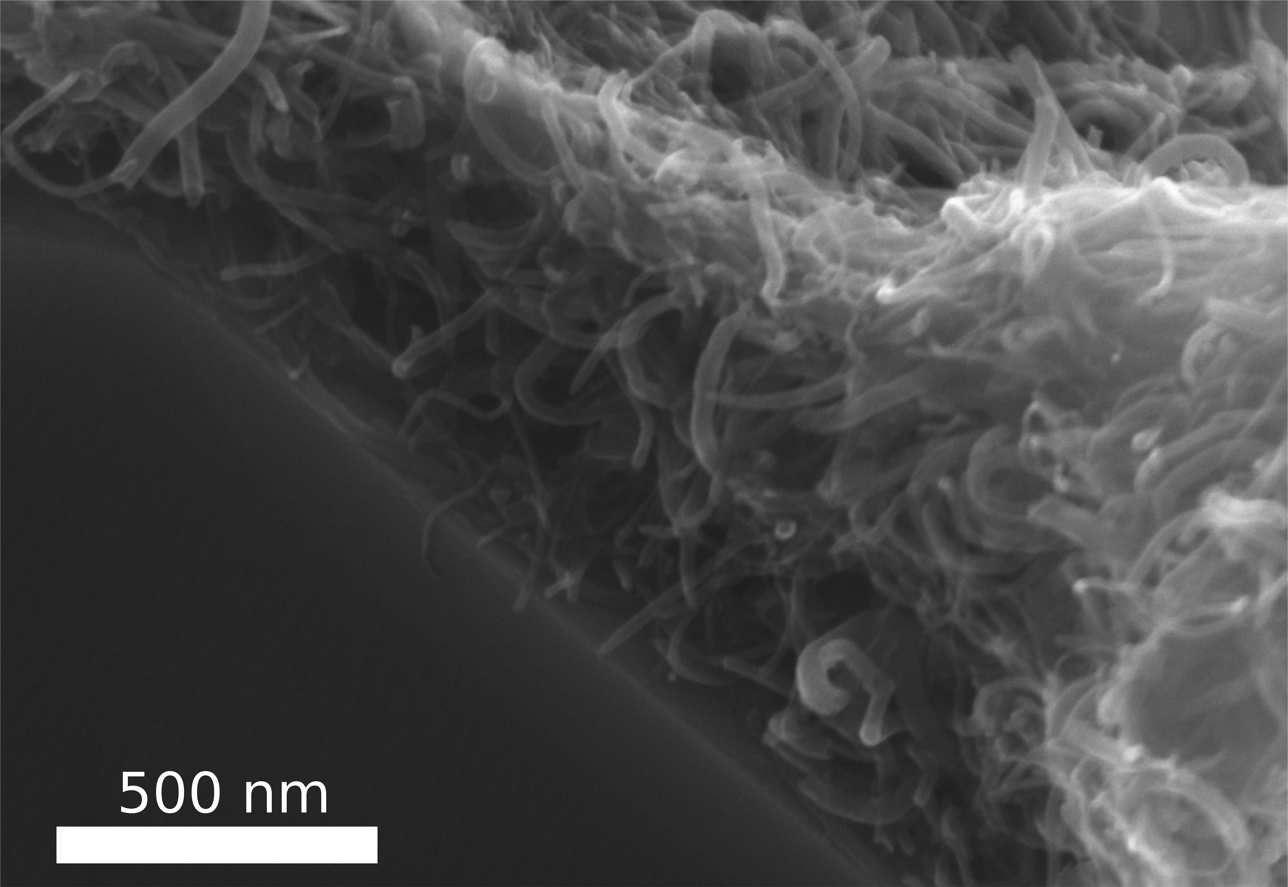
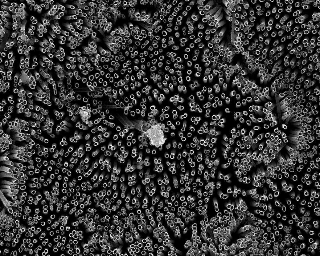
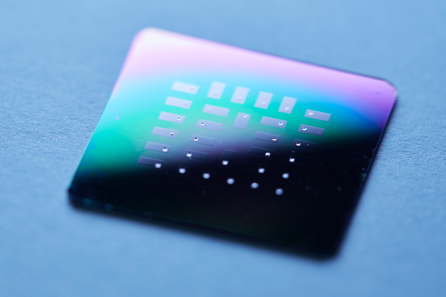
The team sandwiched the electrolyte with an anode and cathode made of platinum, a stable material that readily reacts with glucose. They fabricated 150 individual glucose fuel cells on a chip, each about 400 nanometers thin, and about 300 micrometers wide (about the width of 30 human hairs). They patterned the cells onto silicon wafers, showing that the devices can be paired with a common semiconductor material. They then measured the current produced by each cell as they flowed a solution of glucose over each wafer in a custom-fabricated test station.

They found many cells produced a peak voltage of about 80 millivolts. Given the tiny size of each cell, this output is the highest power density of any existing glucose fuel cell design.

“Excitingly, we are able to draw power and current that’s sufficient to power implantable devices,” Simons says.

“It is the first time that proton conduction in electroceramic materials can be used for glucose-to-power conversion, defining a new type of electrochemstry,” Rupp says. “It extends the material use-cases from hydrogen fuel cells to new, exciting glucose-conversion modes.”

Here’s a link to and a citation for the paper,

A Ceramic-Electrolyte Glucose Fuel Cell for Implantable Electronics by Philipp Simons, Steven A. Schenk, Marco A. Gysel, Lorenz F. Olbrich, Jennifer L. M. Rupp. Advanced Materials https://doi.org/10.1002/adma.202109075 First published: 05 April 2022

This paper is open access.

Electrode-filled elastic fiber for wearable electronics and robots

This work comes out of Switzerland. A May 25, 2018 École Polytechnique Fédérale de Lausanne (EPFL) press release (also on EurekAlert) announces their fibers,

EPFL scientists have found a fast and simple way to make super-elastic, multi-material, high-performance fibers. Their fibers have already been used as sensors on robotic fingers and in clothing. This breakthrough method opens the door to new kinds of smart textiles and medical implants.

It’s a whole new way of thinking about sensors. The tiny fibers developed at EPFL are made of elastomer and can incorporate materials like electrodes and nanocomposite polymers. The fibers can detect even the slightest pressure and strain and can withstand deformation of close to 500% before recovering their initial shape. All that makes them perfect for applications in smart clothing and prostheses, and for creating artificial nerves for robots.

The fibers were developed at EPFL’s Laboratory of Photonic Materials and Fiber Devices (FIMAP), headed by Fabien Sorin at the School of Engineering. The scientists came up with a fast and easy method for embedding different kinds of microstructures in super-elastic fibers. For instance, by adding electrodes at strategic locations, they turned the fibers into ultra-sensitive sensors. What’s more, their method can be used to produce hundreds of meters of fiber in a short amount of time. Their research has just been published in Advanced Materials.

Heat, then stretch
To make their fibers, the scientists used a thermal drawing process, which is the standard process for optical-fiber manufacturing. They started by creating a macroscopic preform with the various fiber components arranged in a carefully designed 3D pattern. They then heated the preform and stretched it out, like melted plastic, to make fibers of a few hundreds microns in diameter. And while this process stretched out the pattern of components lengthwise, it also contracted it crosswise, meaning the components’ relative positions stayed the same. The end result was a set of fibers with an extremely complicated microarchitecture and advanced properties.

Until now, thermal drawing could be used to make only rigid fibers. But Sorin and his team used it to make elastic fibers. With the help of a new criterion for selecting materials, they were able to identify some thermoplastic elastomers that have a high viscosity when heated. After the fibers are drawn, they can be stretched and deformed but they always return to their original shape.

Rigid materials like nanocomposite polymers, metals and thermoplastics can be introduced into the fibers, as well as liquid metals that can be easily deformed. “For instance, we can add three strings of electrodes at the top of the fibers and one at the bottom. Different electrodes will come into contact depending on how the pressure is applied to the fibers. This will cause the electrodes to transmit a signal, which can then be read to determine exactly what type of stress the fiber is exposed to – such as compression or shear stress, for example,” says Sorin.

Artificial nerves for robots

Working in association with Professor Dr. Oliver Brock (Robotics and Biology Laboratory, Technical University of Berlin), the scientists integrated their fibers into robotic fingers as artificial nerves. Whenever the fingers touch something, electrodes in the fibers transmit information about the robot’s tactile interaction with its environment. The research team also tested adding their fibers to large-mesh clothing to detect compression and stretching. “Our technology could be used to develop a touch keyboard that’s integrated directly into clothing, for instance” says Sorin.

The researchers see many other potential applications. Especially since the thermal drawing process can be easily tweaked for large-scale production. This is a real plus for the manufacturing sector. The textile sector has already expressed interest in the new technology, and patents have been filed.

There’s a video of the lead researcher discussing the work as he offers some visual aids,

Here’s a link to and a citation for the paper,

Superelastic Multimaterial Electronic and Photonic Fibers and Devices via Thermal Drawing by Yunpeng Qu, Tung Nguyen‐Dang, Alexis Gérald Page, Wei Yan, Tapajyoti Das Gupta, Gelu Marius Rotaru, René M. Rossi, Valentine Dominique Favrod, Nicola Bartolomei, Fabien Sorin. Advanced Materials First published: 25 May 2018 https://doi.org/10.1002/adma.201707251

This paper is behind a paywall.

Eye implants inspired by glasswing butterflies

Glasswinged butterfly. Greta oto. Credit: David Tiller/CC BY-SA 3.0

My jaw dropped on seeing this image and I still have trouble believing it’s real. (You can find more image of glasswinged butterflies here in an Cot. 25, 2014 posting on thearkinspace. com and there’s a video further down in the post.)

As for the research, an April 30, 2018 news item on phys.org announces work that could improve eye implants,

Inspired by tiny nanostructures on transparent butterfly wings, engineers at Caltech have developed a synthetic analogue for eye implants that makes them more effective and longer-lasting. A paper about the research was published in Nature Nanotechnology.

An April 30, 2018 California Institute of Technology (CalTech) news release (also on EurekAlert) by Robert Perkins, which originated the news item, goes into more detail,

Sections of the wings of a longtail glasswing butterfly are almost perfectly transparent. Three years ago, Caltech postdoctoral researcher Radwanul Hasan Siddique–at the time working on a dissertation involving a glasswing species at Karlsruhe Institute of Technology in Germany–discovered the reason why: the see-through sections of the wings are coated in tiny pillars, each about 100 nanometers in diameter and spaced about 150 nanometers apart. The size of these pillars–50 to 100 times smaller than the width of a human hair–gives them unusual optical properties. The pillars redirect the light that strikes the wings so that the rays pass through regardless of the original angle at which they hit the wings. As a result, there is almost no reflection of the light from the wing’s surface.

In effect, the pillars make the wings clearer than if they were made of just plain glass.

That redirection property, known as angle-independent antireflection, attracted the attention of Caltech’s Hyuck Choo. For the last few years Choo has been developing an eye implant that would improve the monitoring of intra-eye pressure in glaucoma patients. Glaucoma is the second leading cause of blindness worldwide. Though the exact mechanism by which the disease damages eyesight is still under study, the leading theory suggests that sudden spikes in the pressure inside the eye damages the optic nerve. Medication can reduce the increased eye pressure and prevent damage, but ideally it must be taken at the first signs of a spike in eye pressure.

“Right now, eye pressure is typically measured just a couple times a year in a doctor’s office. Glaucoma patients need a way to measure their eye pressure easily and regularly,” says Choo, assistant professor of electrical engineering in the Division of Engineering and Applied Science and a Heritage Medical Research Institute Investigator.

Choo has developed an eye implant shaped like a tiny drum, the width of a few strands of hair. When inserted into an eye, its surface flexes with increasing eye pressure, narrowing the depth of the cavity inside the drum. That depth can be measured by a handheld reader, giving a direct measurement of how much pressure the implant is under.

One weakness of the implant, however, has been that in order to get an accurate measurement, the optical reader has to be held almost perfectly perpendicular–at an angle of 90 degrees (plus or minus 5 degrees)–with respect to the surface of the implant. At other angles, the reader gives an incorrect measurement.

And that’s where glasswing butterflies come into the picture. Choo reasoned that the angle-independent optical property of the butterflies’ nanopillars could be used to ensure that light would always pass perpendicularly through the implant, making the implant angle-insensitive and providing an accurate reading regardless of how the reader is held.

He enlisted Siddique to work in his lab, and the two, working along with Caltech graduate student Vinayak Narasimhan, figured out a way to stud the eye implant with pillars approximately the same size and shape of those on the butterfly’s wings but made from silicon nitride, an inert compound often used in medical implants. Experimenting with various configurations of the size and placement of the pillars, the researchers were ultimately able to reduce the error in the eye implants’ readings threefold.

“The nanostructures unlock the potential of this implant, making it practical for glaucoma patients to test their own eye pressure every day,” Choo says.

The new surface also lends the implants a long-lasting, nontoxic anti-biofouling property.

In the body, cells tend to latch on to the surface of medical implants and, over time, gum them up. One way to avoid this phenomenon, called biofouling, is to coat medical implants with a chemical that discourages the cells from attaching. The problem is that such coatings eventually wear off.

The nanopillars created by Choo’s team, however, work in a different way. Unlike the butterfly’s nanopillars, the lab-made nanopillars are extremely hydrophilic, meaning that they attract water. Because of this, the implant, once in the eye, is soon encased in a coating of water. Cells slide off instead of gaining a foothold.

“Cells attach to an implant by binding with proteins that are adhered to the implant’s surface. The water, however, prevents those proteins from establishing a strong connection on this surface,” says Narasimhan. Early testing suggests that the nanopillar-equipped implant reduces biofouling tenfold compared to previous designs, thanks to this anti-biofouling property.

Being able to avoid biofouling is useful for any implant regardless of its location in the body. The team plans to explore what other medical implants could benefit from their new nanostructures, which can be inexpensively mass produced.

As if the still image wasn’t enough,

Here’s a link to and a citation for the paper,

Multifunctional biophotonic nanostructures inspired by the longtail glasswing butterfly for medical devices by Vinayak Narasimhan, Radwanul Hasan Siddique, Jeong Oen Lee, Shailabh Kumar, Blaise Ndjamen, Juan Du, Natalie Hong, David Sretavan, & Hyuck Choo. Nature Nanotechnology (2018) doi:10.1038/s41565-018-0111-5 Published: 30 April 2018

This paper is behind a paywall.

ETA May 25, 2018:  I’m obsessed. Here’s one more glasswing image,

Caption: The clear wings make this South-American butterfly hard to see in flight, a succesfull defense mechanism. Credit: Eddy Van 3000 from in Flanders fields – Belgiquistan – United Tribes ov Europe Date: 7 October 2007, 14:35 his file is licensed under the Creative Commons Attribution-Share Alike 2.0 Generic license. [downloaded from https://commons.wikimedia.org/wiki/File:E3000_-_the_wings-become-windows_butterfly._(by-sa).jpg]

Felted carbon nanotubes

Parachute (sculpted felt lantern). Artist and artisan felter: Chantal Cardinal. Studio: FELT à la main with LOVE

Scientists from Kiel University (Christian-Albrechts-Universität zu Kiel; Germany) and the University of Trento (Italy) claim to have developed a new method for integrating carbon nanotubes (CNTs) into new materials in a technique they describe as similar to felting according to a November 21, 2017 news item on Nanowerk,

Extremely lightweight, electrically highly conductive, and more stable than steel: due to their unique properties, carbon nanotubes would be ideal for numerous applications, from ultra-lightweight batteries to high-performance plastics, right through to medical implants. However, to date it has been difficult for science and industry to transfer the extraordinary characteristics at the nanoscale into a functional industrial application. The carbon nanotubes either cannot be combined adequately with other materials, or if they can be combined, they then lose their beneficial properties.

Scientists from the Functional Nanomaterials working group at Kiel University (CAU) and the University of Trento have now developed an alternative method, with which the tiny tubes can be combined with other materials, so that they retain their characteristic properties. As such, they “felt” the thread-like tubes into a stable 3D network that is able to withstand extreme forces.

In contrast to the ‘felted’ image which opened this posting, here’s an image of the ‘felted’ carbon nanotubes,

In this new process, the tiny, thread-like carbon nanotubes (CNTs) arrange themselves – almost like felting – to form a stable, tear-resistant layer. Photo/Copyright: Fabian Schütt Courtesy: Kiel University

A November 21, 2017 Kiel University press release (also on EurekAlert), which originated the news item, expands on the theme and adds another analogy,

Industry and science have been intensively researching the significantly less than one hundred nanometre wide carbon tubes (carbon nanotubes, CNTs), in order to make use of the extraordinary properties of rolled graphene. Yet much still remains just theory. “Although carbon nanotubes are flexible like fibre strands, they are also very sensitive to changes,” explained Professor Rainer Adelung, head of the Functional Nanomaterials working group at the CAU. “With previous attempts to chemically connect them with other materials, their molecular structure also changed. This, however, made their properties deteriorate – mostly drastically.”

In contrast, the approach of the research team from Kiel and Trento is based on a simple wet chemical infiltration process. The CNTs are mixed with water and dripped into an extremely porous ceramic material made of zinc oxide, which absorbs the liquid like a sponge. The dripped thread-like CNTs attach themselves to the ceramic scaffolding, and automatically form a stable layer together, similar to a felt. The ceramic scaffolding is coated with nanotubes, so to speak. This has fascinating effects, both for the scaffolding as well as for the coating of nanotubes.

On the one hand, the stability of the ceramic scaffold increases so massively that it can bear 100,000 times its own weight. “With the CNT coating, the ceramic material can hold around 7.5kg, and without it just 50g – as if we had fitted it with a close-fitting pullover made of carbon nanotubes, which provide mechanical support,” summarised first author Fabian Schütt. “The pressure on the material is absorbed by the tensile strength of the CNT felt. Compressive forces are transformed into tensile forces.”

The principle behind this is comparable with bamboo buildings [emphasis mine], such as those widespread in Asia. Here, bamboo stems are bound so tightly with a simple rope that the lightweight material can form extremely stable scaffolding, and even entire buildings. “We do the same at the nano-scale with the CNT threads, which wrap themselves around the ceramic material – only much, much smaller,” said Helge Krüger, co-author of the publication.

The materials scientists were able to demonstrate another major advantage of their process. In a second step, they dissolved the ceramic scaffolding by using a chemical etching process. All that remains is a fine 3D network of tubes, each of which consists of a layer of tiny CNT tubes. In this way, the researchers were able to greatly increase the felt surface, and thus create more opportunities for reactions. “We basically pack the surface of an entire beach volleyball field into a one centimetre cube,” explained Schütt. The huge hollow spaces inside the three-dimensional structure can then be filled with a polymer. As such, CNTs can be connected mechanically with plastics, without their molecular structure – and thus their properties – being modified. “We can specifically arrange the CNTs and manufacture an electrically conductive composite material. To do so only requires a fraction of the usual quantity of CNTs, in order to achieve the same conductivity,” said Schütt.

Applications for use range from battery and filter technology as a filling material for conductive plastics, implants for regenerative medicine, right through to sensors and electronic components at the nano-scale. The good electrical conductivity of the tear-resistant material could in future also be interesting for flexible electronics applications, in functional clothing or in the field of medical technology, for example. “Creating a plastic which, for example, stimulates bone or heart cells to grow is conceivable,” said Adelung. Due to its simplicity, the scientists agree that the process could also be transferred to network structures made of other nanomaterials – which will further expand the range of possible applications.

So, we have ‘felting’ and bamboo buildings. I can appreciate the temptation to use multiple analogies especially since I’ve given into it, on occasion.  But, it’s never considered good style, not even when I do it.

Getting back to the work at hand, here’s a link to and a citation for the paper,

Hierarchical self-entangled carbon nanotube tube networks by Fabian Schütt, Stefano Signetti, Helge Krüger, Sarah Röder, Daria Smazna, Sören Kaps, Stanislav N. Gorb, Yogendra Kumar Mishra, Nicola M. Pugno, & Rainer Adelung. Nature Communications 8, Article number: 1215 (2017) doi:10.1038/s41467-017-01324-7 Published online: 31 October 2017

This is an open access paper.

One final comment, I notice that one of the authors is Nicola Pugno who was last mentioned here in an August 30, 2017 posting titled: Making spider silk stronger by feeding graphene and carbon nanotubes to spiders.

A flexible, organic battery from Northern Ireland

A team from Northern Ireland seems to have made a splash in the race to develop a flexible, environmentally friendly battery. From a Sept. 13, 2017 news item on phys.org,

Experts at Queen’s University Belfast have designed a flexible and organic alternative to the rigid batteries that power up medical implants.

Currently, devices such as pacemakers and defibrillators are fitted with rigid and metal based batteries, which can cause patient discomfort.

Dr Geetha Srinivasan and a team of young researchers from Queen’s University Ionic Liquid Laboratories (QUILL) Research Centre, have now developed a flexible supercapacitor with a longer cycle life, which could power body sensors.

Courtesy: Queen’s University Belfast

A Sept. 13, 2017 Queen’s University Belfast press release (also on EurekAlert), which originated the news item, delves further,

The flexible device is made up of non-flammable electrolytes and organic composites, which are safe to the human body. It can also be easily decomposed without incurring the major costs associated with recycling or disposing off metal based batteries.

The findings, which have been published in Energy Technology and Green Chemistry, show that the device could be manufactured using readily available natural feedstock, rather than sophisticated and expensive metals or semiconductors.

Dr Srinivasan explains: “In modern society, we all increasingly depend on portable electronics such as smartphones and laptops in our everyday lives and this trend has spread to other important areas such as healthcare devices.

“In medical devices such as pacemakers and defibrillators there are two implants, one which is fitted in the heart and another which holds the metal based, rigid batteries – this is implanted under the skin.

“The implant under the skin is wired to the device and can cause patients discomfort as it is rubs against the skin. For this reason batteries need to be compatible to the human body and ideally we would like them to be flexible so that they can adapt to body shapes.”

Dr Srinivasan adds: “At Queen’s University Belfast we have designed a flexible energy storage device, which consists of conducting polymer – biopolymer composites as durable electrodes and ionic liquids as safer electrolytes.

“The device we have created has a longer life-cycle, is non-flammable, has no leakage issues and above all, it is more flexible for placing within the body.”

Environmentally friendly

While the findings show that there are many advantages in the medical world, the organic storage device could also provide solutions in wearable electronics and portable electronic devices, making these more flexible.

Ms Marta Lorenzo, PhD researcher on the project at Queen’s University Belfast, commented: “Although this research could be a potential solution to a global problem, the actual supercapacitor assembly is a straightforward process.”

Dr Srinivasan says: “There is also opportunity to fabricate task-specific supercapacitors. This means that their properties can be tuned and also manufactured using environmentally friendly methods, which is important if they are to be produced on a large scale, for example in powering portable personal electronic devices.”

Here are links and citations to the two papers mentioned in the press release,

Durable Flexible Supercapacitors Utilizing the Multifunctional Role of Ionic Liquids by Marta Lorenzo and Dr Geetha Srinivasan. Energy Technology. DOI: 10.1002/ente.201700407 First published: 23 August 2017

Intrinsically flexible electronic materials for smart device applications by Marta Lorenzo, Biyun Zhu, and Geetha Srinivasan. Green Chem., 2016,18, 3513-3517 DOI: 10.1039/C6GC00826G First published on 20 May 2016

The first paper is open access and the second paper is behind a paywall.

‘Superhemophobic’ medical implants

Counterintuitively, repelling blood is the concept behind a new type of medical implant according to a Jan. 18, 2017 news item on ScienceDaily,

Medical implants like stents, catheters and tubing introduce risk for blood clotting and infection — a perpetual problem for many patients.

Colorado State University engineers offer a potential solution: A specially grown, “superhemophobic” titanium surface that’s extremely repellent to blood. The material could form the basis for surgical implants with lower risk of rejection by the body.

Blood, plasma and water droplets beading on a superomniphobic surface. CSU researchers have created a superhemophobic titanium surface, repellent to blood, that has potential applications for biocompatible medical devices. Courtesy: Colorado State University

A Jan. 18, 2017 Colorado State University news release by Anne Ju Manning, which originated the news item, explains more,

t’s an outside-the-box innovation achieved at the intersection of two disciplines: biomedical engineering and materials science. The work, recently published in Advanced Healthcare Materials, is a collaboration between the labs of Arun Kota, assistant professor of mechanical engineering and biomedical engineering; and Ketul Popat, associate professor in the same departments.

Kota, an expert in novel, “superomniphobic” materials that repel virtually any liquid, joined forces with Popat, an innovator in tissue engineering and bio-compatible materials. Starting with sheets of titanium, commonly used for medical devices, their labs grew chemically altered surfaces that act as perfect barriers between the titanium and blood. Their teams conducted experiments showing very low levels of platelet adhesion, a biological process that leads to blood clotting and eventual rejection of a foreign material.

Chemical compatibility

A material “phobic” (repellent) to blood might seem counterintuitive, the researchers say, as often biomedical scientists use materials “philic” (with affinity) to blood to make them biologically compatible. “What we are doing is the exact opposite,” Kota said. “We are taking a material that blood hates to come in contact with, in order to make it compatible with blood.” The key innovation is that the surface is so repellent, that blood is tricked into believing there’s virtually no foreign material there at all.

The undesirable interaction of blood with foreign materials is an ongoing problem in medical research, Popat said. Over time, stents can form clots, obstructions, and lead to heart attacks or embolisms. Often patients need blood-thinning medications for the rest of their lives – and the drugs aren’t foolproof.

“The reason blood clots is because it finds cells in the blood to go to and attach,” Popat said. “Normally, blood flows in vessels. If we can design materials where blood barely contacts the surface, there is virtually no chance of clotting, which is a coordinated set of events. Here, we’re targeting the prevention of the first set of events.”

nanotubes

Fluorinated nanotubes provided the best superhemophobic surface in the researchers’ experiments.

The researchers analyzed variations of titanium surfaces, including different textures and chemistries, and they compared the extent of platelet adhesion and activation. Fluorinated nanotubes offered the best protection against clotting, and they plan to conduct follow-up experiments.

Growing a surface and testing it in the lab is only the beginning, the researchers say. They want to continue examining other clotting factors, and eventually, to test real medical devices.

Here’s a link to and a citation for the paper,

Hemocompatibility of Superhemophobic Titania Surfaces by Sanli Movafaghi, Victoria Leszczak, Wei Wang, Jonathan A. Sorkin, Lakshmi P. Dasi, Ketul C. Popat, and Arun K. Kota. Advanced Healthcare Materials DOI: 10.1002/adhm.201600717 Version of Record online: 21 DEC 2016

© 2016 WILEY-VCH Verlag GmbH & Co. KGaA, Weinheim

Bacteria and an anti-biofilm coating from Ben Gurion University of the Negev (Israel)

This anti-biofilm acts as an anti-adhesive and is another approach to dealing with unwanted bacteria on medical implants and on marine equipment. From an April 25, 2016 news item about the Israeli research on ScienceDaily,

Researchers at Ben-Gurion University of the Negev (BGU) have developed an innovative anti-biofilm coating, which has significant anti-adhesive potential for a variety of medical and industrial applications.

According to the research published in Advanced Materials Interfaces, anti-adhesive patches that are developed from naturally occurring biomaterials can prevent destructive bacterial biofilm from forming on metal surfaces when they are immersed in water and other damp environments.

An April 25, 2016 American Associates Ben Gurion University of the Negev news release (also on EurekAlert), which originated the news item, describes the research further without adding much detail (Note: A link has been removed),

“Our solution addresses a pervasive need to design environmentally friendly materials to impede dangerous surface bacteria growth,” the BGU researchers from the Avram and Stella Goldstein-Goren Department of Biotechnology Engineering explain. “This holds tremendous potential for averting biofilm formed by surface-anchored bacteria and could have a tremendous impact.”

biofouling

Above: SEM micrographs of A. baumannii, P. aeruginosa (PA14), S. marcescens and P.stuartii biofilm architectures. The untreated control surface shows intricate bacteria densely embedded in the matrix. Biofilms were grown statically on the different surfaces.

The anti-adhesive could be used on medical implants, devices and surgical equipment where bacteria can contribute to chronic diseases, resist antibiotic treatment and thereby compromise the body’s defense system. The prevention of aquatic biofouling on ships and bridges is one of the industrial applications.

Here’s a link to and a citation for the paper,

Novel Anti-Adhesive Biomaterial Patches: Preventing Biofilm with Metal Complex Films (MCF) Derived from a Microalgal Polysaccharide by Karina Golberg, Noa Emuna, T. P. Vinod, Dorit van Moppes, Robert S. Marks, Shoshana Malis Arad, and Ariel Kushmaro. Advanced Materials DOI: 10.1002/admi.201500486 Article first published online: 17 MAR 2016

© 2016 WILEY-VCH Verlag GmbH & Co. KGaA, Weinheim

This article is behind a paywall.

A fatigue-free stretchable conductor for foldable electronics

There’s been a lot of talk about foldable, stretchable, and/or bendable electronics, which is exciting in itself but I find this work on developing a fatigue-free conductor particularly intriguing. After all, who hasn’t purchased something that stretches, folds, etc. only to find that it becomes ‘fatigued’ and is now ‘stretched out’.

A Sept. 23, 2015 news item on Azonano describes the new conductors,

Researchers have discovered a new stretchable, transparent conductor that can be folded or stretched and released, resulting in a large curvature or a significant strain, at least 10,000 times without showing signs of fatigue.

This is a crucial step in creating a new generation of foldable electronics – think a flat-screen television that can be rolled up for easy portability – and implantable medical devices. The work, published Monday [Sept. 21, 2015] in the Proceedings of the National Academy of Sciences, pairs gold nanomesh with a stretchable substrate made with polydimethylsiloxane, or PDMS.

The research is the result of an international collaboration including the University of Houston (US), Harvard University (US), Methodist Research Institute (US), Zhengzhou University (China), Lawrence Berkeley National Laboratory (LBNL; US).

A Sept. 22, 2015 University of Houston news release by Jeannie Kever, which originated the news item, describes this -fatigue-free material in more detail,

The substrate is stretched before the gold nanomesh is placed on it – a process known as “prestretching” – and the material showed no sign of fatigue when cyclically stretched to a strain of more than 50 percent.

The gold nanomesh also proved conducive to cell growth, indicating it is a good material for implantable medical devices.

Fatigue is a common problem for researchers trying to develop a flexible, transparent conductor, making many materials that have good electrical conductivity, flexibility and transparency – all three are needed for foldable electronics – wear out too quickly to be practical, said Zhifeng Ren, a physicist at the University of Houston and principal investigator at the Texas Center for Superconductivity, who was the lead author for the paper.

The new material, produced by grain boundary lithography, solves that problem, he said.

In addition to Ren, other researchers on the project included Chuan Fei Guo and Ching-Wu “Paul” Chu, both from UH; Zhigang Suo, Qihan Liu and Yecheng Wang, all from Harvard University, and Guohui Wang and Zhengzheng Shi, both from the Houston Methodist Research Institute.

In materials science, “fatigue” is used to describe the structural damage to a material caused by repeated movement or pressure, known as “strain cycling.” Bend a material enough times, and it becomes damaged or breaks.    That means the materials aren’t durable enough for consumer electronics or biomedical devices.

“Metallic materials often exhibit high cycle fatigue, and fatigue has been a deadly disease for metals,” the researchers wrote.

“We weaken the constraint of the substrate by making the interface between the Au (gold) nanomesh and PDMS slippery, and expect the Au nanomesh to achieve superstretchability and high fatigue resistance,” they wrote in the paper. “Free of fatigue here means that both the structure and the resistance do not change or have little change after many strain cycles.”

As a result, they reported, “the Au nanomesh does not exhibit strain fatigue when it is stretched to 50 percent for 10,000 cycles.”

Many applications require a less dramatic stretch – and many materials break with far less stretching – so the combination of a sufficiently large range for stretching and the ability to avoid fatigue over thousands of cycles indicates a material that would remain productive over a long period of time, Ren said.

The grain boundary lithography involved a bilayer lift-off metallization process, which included an indium oxide mask layer and a silicon oxide sacrificial layer and offers good control over the dimensions of the mesh structure.

The researchers used mouse embryonic fibroblast cells to determine biocompatibility; that, along with the fact that the stretchability of gold nanomesh on a slippery substrate resembles the bioenvironment of tissue or organ surfaces, suggest the nanomesh “might be implanted in the body as a pacemaker electrode, a connection to nerve endings or the central nervous system, a beating heart, and so on,” they wrote.

Here’s a link to and citation for the paper,

Fatigue-free, superstretchable, transparent, and biocompatible metal electrodes by Chuan Fei Guo, Qihan Liu, Guohui Wang, Yecheng Wang, Zhengzheng Shi, Zhigang Suo, Ching-Wu Chu, and Zhifeng Ren. PNAS (Proceedings of the National Academy of Sciences)  doi: 10.1073/pnas.1516873112 Published online Sept. 21, 2015

This paper appears to be open access.

Preventing deep bone infections with antibiotic-laced polymer layers in implants

I know someone who suffered a deep bone infection after some dental work. Devastatingly, she lost bone material as a consequence and it took years, more than one surgery, and multiple sessions in a hyperbaric chamber to recover, more or less.

While my friend’s infection was due to a dental procedure, the work at the University of Sheffield’s (UK) School of Clinical Dentistry, if successful, will help eliminate incidents of deep bone infection from one potential source, implants. From a May 28, 2015 news item on Azonano,

Leading scientists at the University of Sheffield have discovered nanotechnology could hold the key to preventing deep bone infections, after developing a treatment which prevents bacteria and other harmful microorganisms growing.

The pioneering research, led by the University of Sheffield’s School of Clinical Dentistry, showed applying small quantities of antibiotic to the surface of medical devices, from small dental implants to hip replacements, could protect patients from serious infection.

A May 27, 2015 University of Sheffield press release, which originated the news item, provides more information but few details about how this work is nanotechnology-enabled,

Scientists used revolutionary nanotechnology to work on small polymer layers inside implants which measure between 1 and 100 nanometers (nm) – a human hair is approximately 100,000 nm wide.

Lead researcher Paul Hatton, Professor of Biomaterials Sciences at the University of Sheffield, said: “Microorganisms can attach themselves to implants or replacements during surgery and once they grab onto a non-living surface they are notoriously difficult to treat which causes a lot of problems and discomfort for the patient.

“By making the actual surface of the hip replacement or dental implant inhospitable to these harmful microorganisms, the risk of deep bone infection is substantially reduced.

“Our research shows that applying small quantities of antibiotic to a surface between the polymer layers which make up each device could prevent not only the initial infection but secondary infection – it is like getting between the layers of an onion skin.”

Bone infection affects thousands of patients every year and results in a substantial cost to the NHS.

Treating the surface of medical devices would have a greater impact on patients considered at high risk of infection such as trauma victims from road traffic collisions or combat operations, and those who have had previous bone infections.

Professor Hatton added: “Deep bone infections associated with medical devices are increasing in number, especially among the elderly.

“As well as improving the quality of life, this new application for nanotechnology could save health providers such as the NHS millions of pounds every year.”

Here’s a link to and a citation for the paper,

Functionalised nanoscale coatings using layer-by-layer assembly for imparting antibacterial properties to polylactide-co-glycolide surfaces by Piergiorgio Gentile, ,Maria E. Frongia, Mar Cardellach, Cheryl A. Miller, Graham P. Stafford, Graham J. Leggettc, & Paul V. Hatton. Acta Biomaterialia Volume 21, 15 July 2015, Pages 35–43 doi: 10.1016/j.actbio.2015.04.009

This paper is behind a paywall.

Sweet medical implants courtesy of liquorice

As you have guessed, they are not making implants out of liquorice. Instead, they are using a chemical found in liquorice plants to make nanocoatings that could protect the biological components of medical implants from sterilization procedures. The Oct. 8, 2012 news item on ScienceDaily provides more detail,

Publishing their findings in the latest issue of Materials Today, a team of researchers from Germany and Austria explain how conventional sterilization techniques based on a blast of radiation, or exposure to toxic gas can damage the functional biological components of the device. The coating, containing a component found in liquorice and developed by German biotech company LEUKOCARE AG, protects these sensitive components.

Joachim Koch of the Georg-Speyer Haus, Institute for Biomedical Research in Frankfurt am Main in Germany and colleagues explain how medical devices and implants are increasingly functionalized using pharmacologically active proteins, antibodies and other biomolecules. Harsh sterilization procedures, including beta and gamma irradiation or exposure to toxic ethylene oxide can damage these sensitive molecules and render the device useless. However, without sterilization the patient is at risk of infection when the device is used or implanted.

The team has now successfully evaluated the nano-coating; a technology which employs a composition of stabilizing nano-molecules. One important ingredient is a compound known as glycyrrhizic acid, a natural, sweet-tasting chemical found in liquorice. Unlike other stabilizing approaches used in biopharmaceutical formulations, the nano-coating contains no sugars, sugar-alcohol compounds or proteins that might otherwise interfere with the biological activity of the device.

I found out a little more about the liquorice plant from this essay in Wikipedia (Note: I have removed links and footnotes),

Liquorice or licorice …  is the root of Glycyrrhiza glabra from which a somewhat sweet flavor can be extracted. The liquorice plant is a legume (related to beans and peas) that is native to southern Europe and parts of Asia. It is not botanically related to anise, star anise, or fennel, which are the sources of similar flavouring compounds. The word ‘liquorice’/’licorice’ is derived (via the Old French licoresse), from the Greek γλυκύρριζα (glukurrhiza), meaning “sweet root”,from γλυκύς (glukus), “sweet” + ῥίζα (rhiza), “root”, the name provided by Dioscorides.

Liquorice extract is produced by boiling liquorice root and subsequently evaporating most of the water, and is traded both in solid and syrup form. Its active principle is glycyrrhizin [emphasis mine], a sweetener between 30 to 50 times as sweet as sucrose, and which also has pharmaceutical effects.

Here’s a botanical illustration,

Glycyrrhiza glabra Fabaceae
Original book source: Prof. Dr. Otto Wilhelm Thomé Flora von Deutschland,
Österreich und der Schweiz 1885, Gera, Germany Permission granted to use under GFDL by Kurt Stueber (downloaded from http://en.wikipedia.org/wiki/File:Illustration_Glycyrrhiza_glabra0.jpg)

This undated posting* on the Georg-Speyer-Haus Institute for Biomedical Research website  describes the testing process the team used,

The team has tested the nano-coating by coupling and stabilizing an anti-inflammatory antibody, which may be used in therapy, to a porous polyurethane surface. This carrier acts as a surrogate for a medical device. Such a system might be used as a therapeutic implant to reduce inflammation caused by an overactive immune system in severely ill patients. The researchers found that even if the test device is blasted with radiation to sterilize it entirely, neither the nano-coating nor the proteins are damaged by the radiation and the activity of the device is maintained. “This nano-coating formulation can now be applied for the production of improved biofunctionalized medical devices such as bone implants, vascular stents, and wound dressings and will ease the application of biomedical combination products,” Koch explains.

There’s no indication as to when this nanocoating will appear on the market. For those interested in the technical details, here’s the open access article, Nano-coating protects biofunctional materials by Rupert Tscheliessnig, Martin Zornig, Eva M. Herzig, Katharina Luckerath, Jens Altrichter, Kristina Kemter, Adnana Paunel-Gorgulu, Tim Logters, Joachim Windolf, Silvia Pabisch, Jindrich Cinatl, Holger F. Rabenau, Alois Jungbauer, Peter Muller-Buschbaum, Martin Scholz, and Joachim Koch can be found in Materials Today (2012) 15(9), 394-404.

LEUKOCARE AG, the company the company that developed the liquorice-based coating can be found here.