Tag Archives: molecules

Periodic table of nanomaterials

This charming illustration is the only pictorial representation i’ve seen for Kyoto University’s (Japan) proposed periodic table of nanomaterials, (By the way, 2019 is UNESCO’s [United Nations Educational, Scientific and Cultural Organization] International Year of the Periodic Table of Elements, an event recognizing the table’s 150th anniversary. See my January 8, 2019 posting for information about more events.)

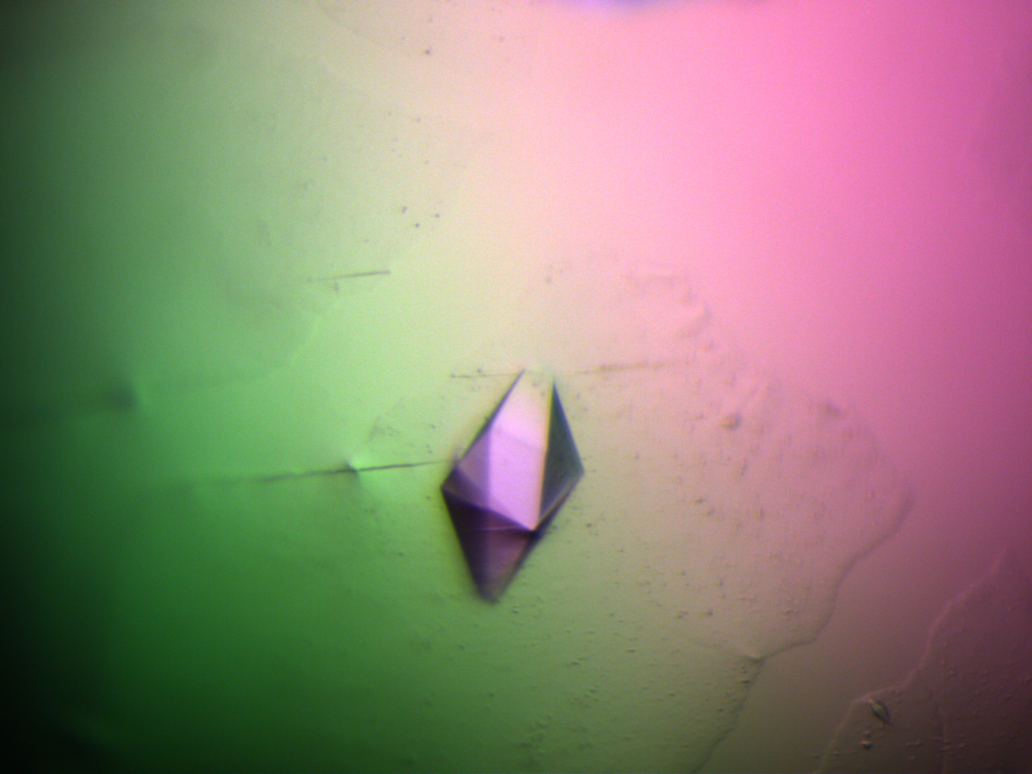
Caption: Molecules interact and align with each other as they self-assemble. This new simulation enables to find what molecules best interact with each other to build nanomaterials, such as materials that work as a nano electrical wire.
Credit Illustration by Izumi Mindy Takamiya

A July 23, 2018 news item on Nanowerk announces the new periodic table (Note: A link has been removed),

The approach was developed by Daniel Packwood of Kyoto University’s Institute for Integrated Cell-Material Sciences (iCeMS) and Taro Hitosugi of the Tokyo Institute of Technology (Nature Communications, “Materials informatics for self-assembly of functionalized organic precursors on metal surfaces”). It involves connecting the chemical properties of molecules with the nanostructures that form as a result of their interaction. A machine learning technique generates data that is then used to develop a diagram that categorizes different molecules according to the nano-sized shapes they form.

This approach could help materials scientists identify the appropriate molecules to use in order to synthesize target nanomaterials.

A July 23, 2018 Kyoto University press release on EurekAlert, which originated the news item, explains further about the computer simulations run by the scientists in pursuit of their specialized periodic table,

Fabricating nanomaterials using a bottom-up approach requires finding ‘precursor molecules’ that interact and align correctly with each other as they self-assemble. But it’s been a major challenge knowing how precursor molecules will interact and what shapes they will form.

Bottom-up fabrication of graphene nanoribbons is receiving much attention due to their potential use in electronics, tissue engineering, construction, and bio-imaging. One way to synthesise them is by using bianthracene precursor molecules that have bromine ‘functional’ groups attached to them. The bromine groups interact with a copper substrate to form nano-sized chains. When these chains are heated, they turn into graphene nanoribbons.

Packwood and Hitosugi tested their simulator using this method for building graphene nanoribbons.

Data was input into the model about the chemical properties of a variety of molecules that can be attached to bianthracene to ‘functionalize’ it and facilitate its interaction with copper. The data went through a series of processes that ultimately led to the formation of a ‘dendrogram’.

This showed that attaching hydrogen molecules to bianthracene led to the development of strong one-dimensional nano-chains. Fluorine, bromine, chlorine, amidogen, and vinyl functional groups led to the formation of moderately strong nano-chains. Trifluoromethyl and methyl functional groups led to the formation of weak one-dimensional islands of molecules, and hydroxide and aldehyde groups led to the formation of strong two-dimensional tile-shaped islands.

The information produced in the dendogram changed based on the temperature data provided. The above categories apply when the interactions are conducted at -73°C. The results changed with warmer temperatures. The researchers recommend applying the data at low temperatures where the effect of the functional groups’ chemical properties on nano-shapes are most clear.

The technique can be applied to other substrates and precursor molecules. The researchers describe their method as analogous to the periodic table of chemical elements, which groups atoms based on how they bond to each other. “However, in order to truly prove that the dendrograms or other informatics-based approaches can be as valuable to materials science as the periodic table, we must incorporate them in a real bottom-up nanomaterial fabrication experiment,” the researchers conclude in their study published in the journal xxx. “We are currently pursuing this direction in our laboratories.”

Here’s a link to and a citation for the paper,

Materials informatics for self-assembly of functionalized organic precursors on metal surfaces by Daniel M. Packwood & Taro Hitosugi. Nature Communicationsvolume 9, Article number: 2469 (2018)DOI: https://doi.org/10.1038/s41467-018-04940-z Published 25 June 2018

This paper is open access.

Humans can distinguish molecular differences by touch

Yesterday, in my December 18, 2017 post about medieval textiles, I posed the question, “How did medieval artisans create nanoscale and microscale gilding when they couldn’t see it?” I realized afterwards that an answer to that question might be in this December 13, 2017 news item on ScienceDaily,

How sensitive is the human sense of touch? Sensitive enough to feel the difference between surfaces that differ by just a single layer of molecules, a team of researchers at the University of California San Diego has shown.

“This is the greatest tactile sensitivity that has ever been shown in humans,” said Darren Lipomi, a professor of nanoengineering and member of the Center for Wearable Sensors at the UC San Diego Jacobs School of Engineering, who led the interdisciplinary project with V. S. Ramachandran, director of the Center for Brain and Cognition and distinguished professor in the Department of Psychology at UC San Diego.

So perhaps those medieval artisans were able to feel the difference before it could be seen in the textiles they were producing?

Getting back to the matter at hand, a December 13, 2017 University of California at San Diego (UCSD) news release (also on EurekAlert) by Liezel Labios offers more detail about the work,

Humans can easily feel the difference between many everyday surfaces such as glass, metal, wood and plastic. That’s because these surfaces have different textures or draw heat away from the finger at different rates. But UC San Diego researchers wondered, if they kept all these large-scale effects equal and changed only the topmost layer of molecules, could humans still detect the difference using their sense of touch? And if so, how?

Researchers say this fundamental knowledge will be useful for developing electronic skin, prosthetics that can feel, advanced haptic technology for virtual and augmented reality and more.

Unsophisticated haptic technologies exist in the form of rumble packs in video game controllers or smartphones that shake, Lipomi added. “But reproducing realistic tactile sensations is difficult because we don’t yet fully understand the basic ways in which materials interact with the sense of touch.”

“Today’s technologies allow us to see and hear what’s happening, but we can’t feel it,” said Cody Carpenter, a nanoengineering Ph.D. student at UC San Diego and co-first author of the study. “We have state-of-the-art speakers, phones and high-resolution screens that are visually and aurally engaging, but what’s missing is the sense of touch. Adding that ingredient is a driving force behind this work.”

This study is the first to combine materials science and psychophysics to understand how humans perceive touch. “Receptors processing sensations from our skin are phylogenetically the most ancient, but far from being primitive they have had time to evolve extraordinarily subtle strategies for discerning surfaces—whether a lover’s caress or a tickle or the raw tactile feel of metal, wood, paper, etc. This study is one of the first to demonstrate the range of sophistication and exquisite sensitivity of tactile sensations. It paves the way, perhaps, for a whole new approach to tactile psychophysics,” Ramachandran said.

Super-Sensitive Touch

In a paper published in Materials Horizons, UC San Diego researchers tested whether human subjects could distinguish—by dragging or tapping a finger across the surface—between smooth silicon wafers that differed only in their single topmost layer of molecules. One surface was a single oxidized layer made mostly of oxygen atoms. The other was a single Teflon-like layer made of fluorine and carbon atoms. Both surfaces looked identical and felt similar enough that some subjects could not differentiate between them at all.

According to the researchers, human subjects can feel these differences because of a phenomenon known as stick-slip friction, which is the jerking motion that occurs when two objects at rest start to slide against each other. This phenomenon is responsible for the musical notes played by running a wet finger along the rim of a wine glass, the sound of a squeaky door hinge or the noise of a stopping train. In this case, each surface has a different stick-slip frequency due to the identity of the molecules in the topmost layer.

In one test, 15 subjects were tasked with feeling three surfaces and identifying the one surface that differed from the other two. Subjects correctly identified the differences 71 percent of the time.

In another test, subjects were given three different strips of silicon wafer, each strip containing a different sequence of 8 patches of oxidized and Teflon-like surfaces. Each sequence represented an 8-digit string of 0s and 1s, which encoded for a particular letter in the ASCII alphabet. Subjects were asked to “read” these sequences by dragging a finger from one end of the strip to the other and noting which patches in the sequence were the oxidized surfaces and which were the Teflon-like surfaces. In this experiment, 10 out of 11 subjects decoded the bits needed to spell the word “Lab” (with the correct upper and lowercase letters) more than 50 percent of the time. Subjects spent an average of 4.5 minutes to decode each letter.

“A human may be slower than a nanobit per second in terms of reading digital information, but this experiment shows a potentially neat way to do chemical communications using our sense of touch instead of sight,” Lipomi said.

Basic Model of Touch

The researchers also found that these surfaces can be differentiated depending on how fast the finger drags and how much force it applies across the surface. The researchers modeled the touch experiments using a “mock finger,” a finger-like device made of an organic polymer that’s connected by a spring to a force sensor. The mock finger was dragged across the different surfaces using multiple combinations of force and swiping velocity. The researchers plotted the data and found that the surfaces could be distinguished given certain combinations of velocity and force. Meanwhile, other combinations made the surfaces indistinguishable from each other.

“Our results reveal a remarkable human ability to quickly home in on the right combinations of forces and swiping velocities required to feel the difference between these surfaces. They don’t need to reconstruct an entire matrix of data points one by one as we did in our experiments,” Lipomi said.

“It’s also interesting that the mock finger device, which doesn’t have anything resembling the hundreds of nerves in our skin, has just one force sensor and is still able to get the information needed to feel the difference in these surfaces. This tells us it’s not just the mechanoreceptors in the skin, but receptors in the ligaments, knuckles, wrist, elbow and shoulder that could be enabling humans to sense minute differences using touch,” he added.

This work was supported by member companies of the Center for Wearable Sensors at UC San Diego: Samsung, Dexcom, Sabic, Cubic, Qualcomm and Honda.

For those who prefer their news by video,

Here’s a link to and a citation for the paper,

Human ability to discriminate surface chemistry by touch by Cody W. Carpenter, Charles Dhong, Nicholas B. Root, Daniel Rodriquez, Emily E. Abdo, Kyle Skelil, Mohammad A. Alkhadra, Julian Ramírez, Vilayanur S. Ramachandran and Darren J. Lipomi. Mater. Horiz., 2018, Advance Article DOI: 10.1039/C7MH00800G

This paper is open access but you do need to have opened a free account on the website.

‘Golden’ protein crystals

Yet another use for gold. From a March 14, 2017 news item on Nanowerk (Note: A link has been removed),

Scientists from the London Centre for Nanotechnology (LCN) have revealed how materials such as gold can help create protein crystals. The team hope their findings, published in the journal Scientific Reports (“Protein crystal nucleation in pores”), could aid the discovery of new medicines and treatments. The Lead author; Professor Naomi Chayen states that “Gold doesn’t react with proteins, due to its inert nature, which makes it an ideal material to create crystals”.

Image: Crystals of an antibody peptide complex related to AIDS research Courtesy: LCN

A March 14, 2017 (?) LCN press release, which originated the news item, expands on the theme,

Proteins are crucial to numerous functions in the body – yet scientists are still in the dark about what most of them look like. This is because the most powerful way of revealing the structure of proteins is to turn them into crystals, and then analyse these with X-rays. However, persuading proteins to turn into useful crystals is notoriously difficult. All crystals start from a conception stage when the first molecules come together; this is called nucleation. But reaching nucleation is often difficult as it requires a lot of energy – and many proteins simply can’t overcome this barrier. Scientists also struggle to create medicines that bind to particular proteins – for instance a protein involved in cancer formation, if they don’t know the protein’s structure.

“How can you target a protein if you have no idea what it looks like? It’s like recognising a face in a crowd – you need a picture,” explained Professor Naomi Chayen, lead author of the research.

Forcing molecules together with gold

One technique for allowing proteins to reach their nucleation point is to trap them in tiny holes. This forces the molecules together, which helps them overcome the energy barrier needed to trigger the first crystal. One material that scientists have found to be effective at growing crystals is gold. Creating many holes in the metal creates a substance called porous gold, which acts as a perfect environment for growing crystals, explained Professor Chayen: “Gold doesn’t react with proteins, due to its inert nature, which makes it an ideal material to create crystals. Creating holes in the metal enable it to act a bit like coral, with each hole providing an ideal environment to harbour crystals.”

Creating crystals

In the latest research, the team investigated the best size hole needed to create crystals. They found that a variety of different sized holes produced the highest quality crystals. Most holes were around 5-10nm, just slightly larger than the width of a human hair. Professor Chayen explained: “Imagine walking down a street with many potholes – some of the holes will be big enough for me to step out of, while some will be too small for my foot to fall into. “However, some will be the exact size of my foot, and will trap me in them. This is the same principle as having different pore sizes – it allows us to trap different size protein molecules, enabling them to form crystals.”

She added that the findings which give a simple explanation of why, and under what conditions porous materials can induce protein crystal nucleation may help scientists design porous materials that would produce the highest quality crystals.

Here’s a link to and a citation for the paper,

Protein crystal nucleation in pores by Christo N. Nanev, Emmanuel Saridakis & Naomi E. Chayen. Scientific Reports 7, Article number: 35821 (2017) doi:10.1038/srep35821 Published online: 16 January 2017

This is an open access article.

Explaining research into matching plasmonic nanoantenna resonances with atoms, molecules, and quantum dots

There’s a very nice explanation of the difficulties associeated with using plasmonic nanoantennas as sensors in a March 21, 2016 news item on phys.org,

Plasmonic nanoantennas are among the hot topics in science at the moment because of their ability to interact strongly with light, which for example makes them useful for different kinds of sensing. But matching their resonances with atoms, molecules or so called quantum dots has been difficult so far because of the very different length scales involved. Thanks to a grant from the Engkvist foundation, Timur Shegai, assistant professor at Chalmers University of Technology, hopes to find a way to do this and by that open doors for applications such as safe long distance communication channels.

A molecule being illuminated by two gold nanoantennas. By: Alexander Ericson Courtesy: Chalmers University of Technology

A molecule being illuminated by two gold nanoantennas. By: Alexander Ericson Courtesy: Chalmers University of Technology

The image, looking like a stylized butterfly or bow tie, above accompanies Karin Weijdegård’s March ??, 2016 Chalmers University of Technology press release, which originated the news item, expands on the research theme,

The diffraction limit makes it very hard for light to interact with the very smallest particles or so called quantum systems such as atoms, molecules or quantum dots. The size of such a particle is simply so much smaller than the wavelength of light that there cannot be a strong interaction between the two. But by using plasmonic nanoantennas, which can be described as metallic nanostructures that are able to focus light very strongly and in wavelengths smaller than those of the visible light, one can build a bridge between the light and the atom, molecule or quantum dot and that is what Timur Shegai is working on.

“Plasmonic nanostructures are themselves smaller than wavelengths of light, but because they have a lot of free electrons they can store the electromagnetic energy in a volume which is actually a lot smaller than the diffraction limit, which helps to bridge the gap between really small objects such as molecules and the larger wavelengths of light,” he says.

Matching the harmonic with the un-harmonic

This might sound easy enough, but the problem with combining the two is that they behave in very different ways. The behaviour of plasmonic nanostructures is very linear, like a harmonic oscillator it will regularly move from side to side no matter how much energy or in other words how many excitations are stored in it. On the other hand, so called quantum systems like atoms, molecules or quantum dots are very much the opposite – their optical properties are highly un-harmonic. Here it makes a big difference if you excite the system with one or two or hundreds of photons.

“Now imagine that you couple together this un-harmonic resonator and a harmonic resonator, and add the possibility to interact with light much stronger than the un-harmonic system alone would have allowed. That opens up very interesting possibilities for quantum technologies and for non-linear optics for example. But as opposed to previous attempts that have been done at very low temperatures and in a vacuum, we will do it at room temperature.”

Communication channels impossible to hack

One possible application where this technology could be useful in the future is to create channels for long distance communications that are impossible to hack. With the current technology this kind of safe communication is only possible if the persons communicating is within a distance of about one hundred kilometres from each other, because that is the maximum distance that an individual photon can run in fibres before it scatters and the signal is lost.

“The kind of ultra small and ultra fast technology we want to develop could be useful in a so called quantum repeater, a device that could be installed across the line from for example New York to London, that would repeat the photon every time it is about to be scattered,” says Timur Shegai.

At the moment though, it is the fundamental aspects of merging plasmons with quantum systems that interest Timur Shegai. To be able to experimentally prove that the there can be interactions between the two systems, he first of all needs to fabricate model systems at the nano level. This is a big challenge, but with the grant of 1,6 million SEK over a period of two years that he just received from the Engkvist foundation, the chances of success have improved.

“Since I am a researcher at the beginning of my career every person is a huge improvement and now I can hire a post doc to work with my group. This means that the project can be divided into sub parts and together we will be able to explore more possibilities about this new technology.”

Thank you Karin Weijdegård for the explanation.

Gold atoms: sometimes they’re a metal and sometimes they’re a molecule

Fascinating work out of Finland shows that a minor change in the number of gold atoms in your gold nanoparticle can mean the difference between a metal and a molecule (coincidentally, this phenomenon is alluded to in my April 14, 2015 post (Nature’s patterns reflected in gold nanoparticles); more about that at the end of this piece. Getting back to Finland and when gold is metal and when it’s a molecule, here’s more from an April 10, 2015 news item on ScienceDaily,

Researchers at the Nanoscience Center at the University of Jyväskylä, Finland, have shown that dramatic changes in the electronic properties of nanometre-sized chunks of gold occur in well-defined size range. Small gold nanoclusters could be used, for instance, in short-term storage of energy or electric charge in the field of molecular electronics. Funded by the Academy of Finland, the researchers have been able to obtain new information which is important, among other things, in developing bioimaging and sensing based on metal-like clusters.

An April 10, 2015 news release (also on EurekAlert) on the Academy of Finland (Suomen Akatemia) website, which originated the news item, describes the work in more detail,

Two recent papers by the researchers at Jyväskylä (1, 2) demonstrate that the electronic properties of two different but still quite similar gold nanoclusters can be drastically different. The clusters were synthesised by chemical methods incorporating a stabilising ligand layer on their surface. The researchers found that the smaller cluster, with up to 102 gold atoms, behaves like a giant molecule while the larger one, with at least 144 gold atoms, already behaves, in principle, like a macroscopic chunk of metal, but in nanosize.

The fundamentally different behaviour of these two differently sized gold nanoclusters was demonstrated by shining a laser light onto solution samples containing the clusters and by monitoring how energy dissipates from the clusters into the surrounding solvent.

“Molecules behave drastically different from metals,” said Professor Mika Pettersson, the principal investigator of the team conducting the experiments. “The additional energy from light, absorbed by the metal-like clusters, transfers to the environment extremely rapidly, in about one hundred billionth of a second, while a molecule-like cluster is excited to a higher energy state and dissipates the energy into the environment with a rate that is at least 100 times slower. This is exactly what we saw: the 102-gold atom cluster is a giant molecule showing even a transient magnetic state while the 144-gold atom cluster is already a metal. We’ve thus managed to bracket an important size region where this fundamentally interesting change in the behaviour takes place.”

“These experimental results go together very well with what our team has seen from computational simulations on these systems,” said Professor Hannu Häkkinen, a co-author of the studies and the scientific director of the nanoscience centre. “My team predicted this kind of behaviour back in 2008-2009 when we saw big differences in the electronic structure of exactly these nanoclusters. It’s wonderful that robust spectroscopic experiments have now proved these phenomena. In fact, the metal-like 144-atom cluster is even more interesting, since we just published a theoretical paper where we saw a big enhancement of the metallic properties of just a few copper atoms mixed with gold.” (3)

Here are links to and citation for the papers,

Ultrafast Electronic Relaxation and Vibrational Cooling Dynamics of Au144(SC2H4Ph)60 Nanocluster Probed by Transient Mid-IR Spectroscopy by Satu Mustalahti, Pasi Myllyperkiö, Tanja Lahtinen, Kirsi Salorinne, Sami Malola, Jaakko Koivisto, Hannu Häkkinen, and Mika Pettersson. J. Phys. Chem. C, 2014, 118 (31), pp 18233–18239 DOI: 10.1021/jp505464z Publication Date (Web): July 3, 2014

Copyright © 2014 American Chemical Society

Copper Induces a Core Plasmon in Intermetallic Au(144,145)–xCux(SR)60 Nanoclusters by Sami Malola, Michael J. Hartmann, and Hannu Häkkinen. J. Phys. Chem. Lett., 2015, 6 (3), pp 515–520 DOI: 10.1021/jz502637b Publication Date (Web): January 22, 2015

Copyright © 2015 American Chemical Society

Molecule-like Photodynamics of Au102(pMBA)44 Nanocluster by Satu Mustalahti, Pasi Myllyperkiö, Sami Malola, Tanja Lahtinen, Kirsi Salorinne, Jaakko Koivisto, Hannu Häkkinen, and Mika Pettersson. ACS Nano, 2015, 9 (3), pp 2328–2335 DOI: 10.1021/nn506711a Publication Date (Web): February 22, 2015

Copyright © 2015 American Chemical Society

These papers are behind paywalls.

As for my April 14, 2015 post (Nature’s patterns reflected in gold nanoparticles), researchers at Carnegie Mellon University were researching patterns in different sized gold nanoparticles when this was noted in passing,

… Normally, gold is one of the best conductors of electrical current, but the size of Au133 is so small that the particle hasn’t yet become metallic. …

The birth of a molecule

This research comes from Korea’s Institute of Basic Science in a Feb. 27, 2015 news item on Azonano,

The research team of the Center for Nanomaterials and Chemical Reactions at the Institute for Basic Science (IBS) has successfully visualized the entire process of bond formation in solution by using femtosecond time-resolved X-ray liquidography (femtosecond TRXL) for the first time in the world.

A Feb. 18, 2015 IBS press release, which originated the news item, provides more details,

Every researcher’s longstanding dream to observe real-time bond formation in chemical reactions has come true. Since this formation takes less than one picosecond, researchers have not been able to visualize the birth of molecules.

The research team has used femtosecond TRXL in order to visualize the formation of a gold trimer complex in real time without being limited by slow diffusion.

They have focused on the process of photoinduced bond formation between gold (Au) atoms dissolved in water. In the ground (S0) state, Au atoms are weakly bound to each other in a bent geometry by van der Waals interactions. On photoexcitation, the S0 state rapidly converts into an excited (S1) state, leading to the formation of covalent Au-Au bonds and bent-to-linear transition. Then, the S1 state changes to a triplet (T1) state with a time constant of 1.6 picosecond, accompanying further bond contraction by 0.1 Å. Later, the T1 state of the trimer transforms to a tetramer on nanosecond time scale, and Au atoms return to their original bent structure.

“By using femtosecond TRXL, we will be able to observe molecular vibration and rotation in the solution phase in real time,” says Hyotcherl Ihee, the group leader of the Center for Nanomaterials at IBS, as well as the professor of the Department of Chemistry at Korea Advanced Institute of Science and Technology.

Here’s a link to and a citation for the paper,

Direct observation of bond formation in solution with femtosecond X-ray scattering by Kyung Hwan Kim, Jong Goo Kim, Shunsuke Nozawa, Tokushi Sato, Key Young Oang, Tae Wu Kim, Hosung Ki, Junbeom Jo, Sungjun Park, Changyong Song, Takahiro Sato, Kanade Ogawa, Tadashi Togashi, Kensuke Tono, Makina Yabashi, Tetsuya Ishikawa, Joonghan Kim, Ryong Ryoo, Jeongho Kim, Hyotcherl Ihee & Shin-ichi Adachi. Nature 518, 385–389 (19 February 2015) doi:10.1038/nature14163 Published online 18 February 2015

This paper is behind a paywall although there is a free preview via ReadCube access.