Tag Archives: nanoscopy

Nanodiamond alternative to organic fluorophores to view inside living human cells

No sooner is a Nobel prize (2014) awarded for nanoscopy which makes use of fluorescence to observe processes in living cells than there is an announcement about a new technique that avoids fluorescence and its attendant shortcomings. From an Oct. 27, 2014 news item on Nanowerk (Note: A link has been removed),

Nanodiamonds are providing scientists with new possibilities for accurate measurements of processes inside living cells with potential to improve drug delivery and cancer therapeutics.

Published in Nature Nanotechnology (“Coherent anti-Stokes Raman scattering microscopy of single nanodiamonds”), researchers from Cardiff University have unveiled a new method for viewing nanodiamonds inside human living cells for purposes of biomedical research.

An Oct. 27, 2014 Cardiff University (Wales) news release, which originated the news item, explains why the use of nanodiamonds is superior to the use of organic flurophores,

Nanodiamonds are very small particles (a thousand times smaller than human hair) and because of their low toxicity they can be used as a carrier to transport drugs inside cells. They also show huge promise as an alternative to the organic fluorophores usually used by scientists to visualise processes inside cells and tissues.

A major limitation of organic fluorophores is that they have the tendency to degrade and bleach over time under light illumination. This makes it difficult to use them for accurate measurements of cellular processes. Moreover, the bleaching and chemical degradation can often be toxic and significantly perturb or even kill cells.

There is a growing consensus among scientists that nanodiamonds are one of the best inorganic material alternatives for use in biomedical research, because of their compatibility with human cells, and due to their stable structural and chemical properties.

Previous attempts by other research teams to visualise nanodiamonds under powerful light microscopes have run into the obstacle that the diamond material per se is transparent to visible light. Locating the nanodiamonds under a microscope had relied on tiny defects in the crystal lattice, which fluoresce under light illumination.

Production of the defects proved both costly and difficult to realise in a controlled way. Furthermore, the fluorescence light emitted by these defects, and in turn the image gleaned from the microscopic exploration of these flawed nanodiamonds, is sometimes also unstable.

In their latest paper, researchers from Cardiff University’s Schools of Biosciences and Physics showed that non-fluorescing nanodiamonds (diamonds without defects) can be imaged optically and far more stably via the interaction between the illuminating light and the vibrating chemical bonds in the diamond lattice structure which results in scattered light at a different colour.

The paper describes how two laser beams beating at a specific frequency are used to drive chemical bonds to vibrate in sync. One of these beams is then used to probe this vibration and generate a light, called coherent anti-Stokes Raman scattering (CARS).

By focusing these laser beams onto the nanodiamond, a high-resolution CARS image is generated. Using an in-house built microscope, the research team was able to measure the intensity of the CARS light on a series of single nanodiamonds of different sizes.

The nanodiamond size was accurately measured by means of electron microscopy and other quantitative optical contrast methods developed within the researcher’s lab. In this way, they were able to quantify the relationship between the CARS light intensity and the nanoparticle size.

Consequently, the calibrated CARS signal enabled the team to analyse the size and number of nanodiamonds that had been delivered into living cells, with a level of accuracy hitherto not achieved by other methods.

Professor Paola Borri from the School of Biosciences, who led the study, said: “This new imaging modality opens the exciting prospect of following complex cellular trafficking pathways quantitatively with important applications in drug delivery. The next step for us will be to push the technique to detect nanodiamonds of even smaller sizes than what we have shown so far and to demonstrate a specific application in drug delivery.”

Here’s a link to and a citation for the paper,

Coherent anti-Stokes Raman scattering microscopy of single nanodiamonds by Iestyn Pope, Lukas Payne, George Zoriniants, Evan Thomas, Oliver Williams, Peter Watson, Wolfgang Langbein, & Paola Borri. Nature Nanotechnology (2014) doi:10.1038/nnano.2014.210 Published online 12 October 2014

The paper is behind a paywall but there is a free preview with ReadCube Access.

For anyone who’d like to read more about fluorescence and its use in nanoscopy there’s my Oct. 8, 2014 posting about the 2014 Nobel Prize in Chemistry and in my Oct. 27, 2014 posting about a specific use for determining how bipolar disorder may affect the brain.

Bipolar disorder at the nanoscale

In all the talk generated by the various brain projects (BRAIN initiative [US], The Human Brain Project [European Union], Brain Canada), there’s remarkably little discussion about mental illness. So, this news is a little unusual.

Using super-high resolution technique scientists at Northwestern University (Chicago, Illinois, US) believe they’ve made a discovery which explains how bipolar disorder affects the brain according to an Oct. 22, 2014 Northwestern University news release (also on EurekAlert and ScienceDaily) by Erin White,

Scientists used a new super-resolution imaging method — the same method recognized with the 2014 Nobel Prize in chemistry — to peer deep into brain tissue from mice with bipolar-like behaviors. In the synapses (where communication between brain cells occurs), they discovered tiny “nanodomain” structures with concentrated levels of ANK3 — the gene most strongly associated with bipolar disorder risk. ANK3 is coding for the protein ankyrin-G.

“We knew that ankyrin-G played an important role in bipolar disease, but we didn’t know how,” said Northwestern Medicine scientist Peter Penzes, corresponding author of the paper. “Through this imaging method we found the gene formed in nanodomain structures in the synapses, and we determined that these structures control or regulate the behavior of synapses.”

Penzes is a professor in physiology and psychiatry and behavioral sciences at Northwestern University Feinberg School of Medicine. The results were published Oct. 22 in the journal Neuron.

High-profile cases, including actress Catherine Zeta-Jones and politician Jesse Jackson, Jr., have brought attention to bipolar disorder. The illness causes unusual shifts in mood, energy, activity levels and the ability to carry out day-to-day tasks. About 3 percent of Americans experience bipolar disorder symptoms, and there is no cure.

Recent large-scale human genetic studies have shown that genes can contribute to disease risk along with stress and other environmental factors. However, how these risk genes affect the brain is not known.

This is the first time any psychiatric risk gene has been analyzed at such a detailed level of resolution. As explained in the paper, Penzes used the Nikon Structured Illumination Super-resolution Microscope to study a mouse model of bipolar disorder. The microscope realizes resolution of up to 115 nanometers. To put that size in perspective, a nanometer is one-tenth of a micron, and there are 25,400 microns in one inch. Very few of these microscopes exist worldwide.

“There is important information about genes and diseases that can only been seen at this level of resolution,” Penzes said. “We provide a neurobiological explanation of the function of the leading risk gene, and this might provide insight into the abnormalities in bipolar disorder.”

The biological framework presented in this paper could be used in human studies of bipolar disorder in the future, with the goal of developing therapeutic approaches to target these genes.

Here’s a link to and a citation for the paper,

Psychiatric Risk Factor ANK3/Ankyrin-G Nanodomains Regulate the Structure and Function of Glutamatergic Synapses by Katharine R. Smith, Katherine J. Kopeikina, Jessica M. Fawcett-Patel, Katherine Leaderbrand, Ruoqi Gao, Britta Schürmann, Kristoffer Myczek, Jelena Radulovic, Geoffrey T. Swanson, and Peter Penzes. Neuron, Volume 84, Issue 2, p399–415, 22 October 2014 DOI: http://dx.doi.org/10.1016/j.neuron.2014.10.010

This paper is behind a paywall.

You can find more about super-high resolution and nanoscopy in my Oct. 8, 2014 post about the 2014 Nobel Chemistry prize winners.

Nanoscopy and a 2014 Nobel Prize for Chemistry

An Oct. 8, 2014 news item on Nanowerk features the 2014 Nobel Prize in Chemistry honourees,

 For a long time optical microscopy was held back by a presumed limitation: that it would never obtain a better resolution than half the wavelength of light. Helped by fluorescent molecules the Nobel Laureates in Chemistry 2014 ingeniously circumvented this limitation.

Their ground-breaking work has brought optical microscopy into the nanodimension.
In what has become known as nanoscopy, scientists visualize the pathways of individual molecules inside living cells. They can see how molecules create synapses between nerve cells in the brain; they can track proteins involved in Parkinson’s, Alzheimer’s and Huntington’s diseases as they aggregate; they follow individual proteins in fertilized eggs as these divide into embryos.

An Oct, 8, 2014 Royal Swedish Academy of Science press release, which originated the news item, expands on the ‘groundbreaking’ theme,

It was all but obvious that scientists should ever be able to study living cells in the tiniest molecular detail. In 1873, the microscopist Ernst Abbe stipulated a physical limit for the maximum resolution of traditional optical microscopy: it could never become better than 0.2 micrometres. Eric Betzig, Stefan W. Hell and William E. Moerner are awarded the Nobel Prize in Chemistry 2014 for having bypassed this limit. Due to their achievements the optical microscope can now peer into the nanoworld.

Two separate principles are rewarded. One enables the method stimulated emission depletion (STED) microscopy, developed by Stefan Hell in 2000. Two laser beams are utilized; one stimulates fluorescent molecules to glow, another cancels out all fluorescence except for that in a nanometre-sized volume. Scanning over the sample, nanometre for nanometre, yields an image with a resolution better than Abbe’s stipulated limit.

Eric Betzig and William Moerner, working separately, laid the foundation for the second method, single-molecule microscopy. The method relies upon the possibility to turn the fluorescence of individual molecules on and off. Scientists image the same area multiple times, letting just a few interspersed molecules glow each time. Superimposing these images yields a dense super-image resolved at the nanolevel. In 2006 Eric Betzig utilized this method for the first time.

Today, nanoscopy is used world-wide and new knowledge of greatest benefit to mankind is produced on a daily basis.

Here’s an image illustrating different microscopy resolutions including one featuring single-molecule microscopy,

The centre image shows lysosome membranes and is one of the first ones taken by Betzig using single-molecule microscopy. To the left, the same image taken using conventional microscopy. To the right, the image of the membranes has been enlarged. Note the scale division of 0.2 micrometres, equivalent to Abbe’s diffraction limit. Image: Science 313:1642–1645. [downloaded from http://www.kva.se/en/pressroom/Press-releases-2014/nobelpriset-i-kemi-2014/]

The centre image shows lysosome membranes and is one of the first ones taken by Betzig using single-molecule microscopy. To the left, the same image taken using conventional microscopy. To the right, the image of the membranes has been enlarged. Note the scale division of 0.2 micrometres, equivalent to Abbe’s diffraction limit. Image: Science 313:1642–1645. [downloaded from http://www.kva.se/en/pressroom/Press-releases-2014/nobelpriset-i-kemi-2014/]

The press release goes on to provide some biographical details about the three honourees and information about the financial size of the award,

Eric Betzig, U.S. citizen. Born 1960 in Ann Arbor, MI, USA. Ph.D. 1988 from Cornell University, Ithaca, NY, USA. Group Leader at Janelia Farm Research Campus, Howard Hughes Medical Institute, Ashburn, VA, USA.

Stefan W. Hell, German citizen. Born 1962 in Arad, Romania. Ph.D. 1990 from the University of Heidelberg, Germany. Director at the Max Planck Institute for Biophysical Chemistry, Göttingen, and Division head at the German Cancer Research Center, Heidelberg, Germany.

William E. Moerner, U.S. citizen. Born 1953 in Pleasanton, CA, USA. Ph.D. 1982 from Cornell University, Ithaca, NY, USA. Harry S. Mosher Professor in Chemistry and Professor, by courtesy, of Applied Physics at Stanford University, Stanford, CA, USA.

Prize amount: SEK 8 million, to be shared equally between the Laureates.

The amount is in Swedish Krona. In USD, it is approximately $1.1M; in CAD, it is approximately $1.2M; and, in pounds sterling (British pounds), it is approximately £689,780.

Congratulations to all three gentlemen!

ETA Oct. 14, 2014: Azonano features an Oct. 14, 2014 news item from the UK’s National Physical Laboratory (NPL)  congratulating the three recipients of the 2014 Nobel Prize for Chemistry. The item also features a description of the recipients’ groundbreaking work along with an update on how this pioneering work has influenced and inspired further research in the field of nanoscopy at the NPL.

French scientists focus optical microscopes down to 30 nm

In fact, the French scientists are using two different imaging techniques to arrive at a resolution of 30 nm for their optical microscopes, according to the May 18, 2012 news item on Nanowerk.

Researchers from the Institut Pasteur and CNRS [Centre national de la recherche scientifique] have set up a new optical microscopy approach that combines two recent imaging techniques in order to visualize molecular assemblies without affecting their biological functions, at a resolution 10 times better than that of traditional microscopes. Using this approach, they were able to observe the AIDS virus and its capsids (containing the HIV genome) within cells at a scale of 30 nanometres, for the first time with light.

More specifically,

A study coordinated by Dr Christophe Zimmer (Institut Pasteur/CNRS), in collaboration with Dr Nathalie Arhel within the lab headed by Pr Pierre Charneau (Institut Pasteur/CNRS), shows that the association of two recent imaging techniques helps obtain unique images of molecular assemblies of HIV-1 capsids, with a resolution around 10 times better than that of traditional microscopes. This new approach, which uses super-resolution imaging and FlAsH labeling, does not affect the virus’ ability to self-replicate. It represents a major step forward in molecular biology studies, enabling the visualisation of microbial complexes at a scale of 30 nm without affecting their function.

The newly developed approach combines super-resolution PALM imaging and fluorescent FlAsH labeling. PALM imaging relies on the acquisition of thousands of low-resolution images, each of which showing only a few fluorescent molecules. The molecular positions are then calculated with high accuracy by computer programs and compiled into a single high-resolution image. FlAsH labeling involves the insertion of a 6-amino-acid peptide into the protein of interest. The binding of the FlAsH fluorophore to the peptide generates a fluorescent signal, thereby enabling the visualization of the protein. For the first time, researchers have combined these two methods in order to obtain high-resolution images of molecular structures in either fixed or living cells.

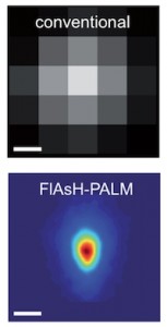
The researchers have supplied an image illustrating the difference between the conventional and new techniques will allow them to view (from the May 16, 2012 press release  [communiqué de presses] on the CNRS website),

© Institut Pasteur Reconstruction optique super-résolutive de la morphologie du VIH. L'image du dessous montre la distribution moyenne de l'enzyme intégrase observée par FlAsH-PALM. La résolution de cette technique (~30 nm) permet de retrouver la taille et la forme conique de la capside. Pour comparaison, la résolution de la microscopie conventionnelle (~200-300 nm), illustrée par l'image du dessus, ne permet pas une description détaillée de cette structure.

The conventional 200 – 300 nm resolution is shown at the top while the new 30 nm resolution achieved by combining the new techniques is shown below. This new technique has already allowed scientists to disprove a popular theory about the AIDS virus, from the May 18, 2012 news item on Nanowerk,

This new method has helped researchers visualise the AIDS Virus and localise its capsids in human cells, at a scale of 30 nm. Capsids are conical structures which contain the HIV genome. These structures must dismantle in order for the viral genome to integrate itself into the host cell’s genome. However, the timing of this disassembly has long been debated. According to a prevailing view, capsids disassemble right after infection of the host cell and, therefore, do not play an important role in the intracellular transport of the virus to the host cell’s nucleus. However, the results obtained by the researchers of the Institut Pasteur and CNRS indicate that numerous capsids remain unaltered until entry of the virus into the nucleus, confirming and strengthening earlier studies based on electron microscopy. Hence, capsids could play a more important role than commonly assumed in the replication cycle of HIV.

I gather excitement about this development is high as the scientists are suggesting that ‘microscopy’ could be known as ‘nanoscopy’ in the future.