Tag Archives: nematodes

Agricultural pest control with nanoparticles derived from plant viruses

As with many of these ‘nanoparticle solutions’ to a problem, it seems the nanoparticles are the delivery system. A September 21, 2023 news item on ScienceDaily announces the research,

A new form of agricultural pest control could one day take root — one that treats crop infestations deep under the ground in a targeted manner with less pesticide.

Engineers at the University of California San Diego have developed nanoparticles, fashioned from plant viruses, that can deliver pesticide molecules to soil depths that were previously unreachable. This advance could potentially help farmers effectively combat parasitic nematodes that plague the root zones of crops, all while minimizing costs, pesticide use and environmental toxicity.

A September 21, 2023 University of California at San Diego news release (also on EurekAlert) by Liezel Labios, which originated the news item, provides more information about the problems along with a nod to nanomedicine as the inspiration for the proposed solution, Note: Links have been removed,

Controlling infestations caused by root-damaging nematodes has long been a challenge in agriculture. One reason is that the types of pesticides used against nematodes tend to cling to the top layers of soil, making it tough to reach the root level where nematodes wreak havoc. As a result, farmers often resort to applying excessive amounts of pesticide, as well as water to wash pesticides down to the root zone. This can lead to contamination of soil and groundwater.

To find a more sustainable and effective solution, a team led by Nicole Steinmetz, a professor of nanoengineering at the UC San Diego Jacobs School of Engineering and founding director of the Center for Nano-ImmunoEngineering, developed plant virus nanoparticles that can transport pesticide molecules deep into the soil, precisely where they are needed. The work is detailed in a paper published in Nano Letters.

Steinmetz’s team drew inspiration from nanomedicine [emphasis mine], where nanoparticles are being created for targeted drug delivery, and adapted this concept to agriculture. This idea of repurposing and redesigning biological materials for different applications is also a focus area of the UC San Diego Materials Research Science and Engineering Center (MRSEC), of which Steinmetz is a co-lead. 

“We’re developing a precision farming approach where we’re creating nanoparticles for targeted pesticide delivery,” said Steinmetz, who is the study’s senior author. “This technology holds the promise of enhancing treatment effectiveness in the field without the need to increase pesticide dosage.”

The star of this approach is the tobacco mild green mosaic virus, a plant virus that has the ability to move through soil with ease. Researchers modified these virus nanoparticles, rendering them noninfectious to crops by removing their RNA. They then mixed these nanoparticles with pesticide solutions in water and heated them, creating spherical virus-like nanoparticles packed with pesticides through a simple one-pot synthesis.

This one-pot synthesis offers several advantages. First, it is cost-effective, with just a few steps and a straightforward purification process. The result is a more scalable method, paving the way toward a more affordable product for farmers, noted Steinmetz. Second, by simply packaging the pesticide inside the nanoparticles, rather than chemically binding it to the surface, this method preserves the original chemical structure of the pesticide.

“If we had used a traditional synthetic method where we link the pesticide molecules to the nanoparticles, we would have essentially created a new compound, which will need to go through a whole new registration and regulatory approval process,” said study first author Adam Caparco, a postdoctoral researcher in Steinmetz’s lab. “But since we’re just encapsulating the pesticide within the nanoparticles, we’re not changing the active ingredient, so we won’t need to get new approval for it. That could help expedite the translation of this technology to the market.”

Moreover, the tobacco mild green mosaic virus is already approved by the Environmental Protection Agency (EPA) for use as an herbicide to control an invasive plant called the tropical soda apple. This existing approval could further streamline the path from lab to market.

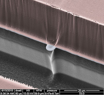
The researchers conducted experiments in the lab to demonstrate the efficacy of their pesticide-packed nanoparticles. The nanoparticles were watered through columns of soil and successfully transported the pesticides to depths of at least 10 centimeters. The solutions were collected from the bottom of the soil columns and were found to contain the pesticide-packed nanoparticles. When the researchers treated nematodes with these solutions, they eliminated at least half of the population in a petri dish.

While the researchers have not yet tested the nanoparticles on nematodes lurking beneath the soil, they note that this study marks a significant step forward.

“Our technology enables pesticides meant to combat nematodes to be used in the soil,” said Caparco. “These pesticides alone cannot penetrate the soil. But with our nanoparticles, they now have soil mobility, can reach the root level, and potentially kill the nematodes.”

Future research will involve testing the nanoparticles on actual infested plants to assess their effectiveness in real-world agricultural scenarios. Steinmetz’s lab will perform these follow-up studies in collaboration with the U.S. Horticultural Research Laboratory. Her team has also established plans for an industry partnership aimed at advancing the nanoparticles into a commercial product.

Here’s a link to and a citation for the paper,

Delivery of Nematicides Using TMGMV-Derived Spherical Nanoparticles by Adam A. Caparco, Ivonne González-Gamboa, Samuel S. Hays, Jonathan K. Pokorski, and Nicole F. Steinmetz. Nano Lett. 2023, 23, 12, 5785–5793 DOI: https://doi.org/10.1021/acs.nanolett.3c01684 Publication Date:June 16, 2023 Copyright © 2023 American Chemical Society

This paper is behind a paywall.

Gently measuring electrical signals in small animals with nano-SPEARs

This work comes from Rice University (Texas, US) according to an April 17, 2017 news item on Nanowerk,

Microscopic probes developed at Rice University have simplified the process of measuring electrical activity in individual cells of small living animals. The technique allows a single animal like a worm to be tested again and again and could revolutionize data-gathering for disease characterization and drug interactions.

The Rice lab of electrical and computer engineer Jacob Robinson has invented “nanoscale suspended electrode arrays” — aka nano-SPEARs — to give researchers access to electrophysiological signals from the cells of small animals without injuring them. Nano-SPEARs replace glass pipette electrodes that must be aligned by hand each time they are used.”

An April 17, 2017 Rice University news release (also on EurekAltert), which originated the news item, details the work,

“One of the experimental bottlenecks in studying synaptic behavior and degenerative diseases that affect the synapse is performing electrical measurements at those synapses,” Robinson said. “We set out to study large groups of animals under lots of different conditions to screen drugs or test different genetic factors that relate to errors in signaling at those synapses.”

Robinson’s early work at Rice focused on high-quality, high-throughput electrical characterization of individual cells. The new platform adapts the concept to probe the surface cells of nematodes, worms that make up 80 percent of all animals on Earth.

Most of what is known about muscle activity and synaptic transmission in the worms comes from the few studies that successfully used manually aligned glass pipettes to measure electrical activity from individual cells, Robinson said. However, this patch clamp technique requires time-consuming and invasive surgery that could negatively affect the data that is gathered from small research animals.

The platform developed by Robinson’s team works something like a toll booth for traveling worms. As each animal passes through a narrow channel, it is temporarily immobilized and pressed against one or several nano-SPEARS that penetrate its body-wall muscle and record electrical activity from nearby cells. That animal is then released, the next is captured and measured, and so on. Robinson said the device proved much faster to use than traditional electrophysiological cell measurement techniques.

The nano-SPEARs are created using standard thin-film deposition procedures and electron-beam or photolithography and can be made from less than 200 nanometers to more than 5 microns thick, depending on the size of animal to be tested. Because the nano-SPEARs can be fabricated on either silicon or glass, the technique easily combines with fluorescence microscopy, Robinson said.

The animals suitable for probing with a nano-SPEAR can be as large as several millimeters, like hydra, cousins of the jellyfish and the subject of an upcoming study. But nematodes known as Caenorhabditis elegans were practical for several reasons: First, Robinson said, they’re small enough to be compatible with microfluidic devices and nanowire electrodes. Second, there were a lot of them down the hall at the lab of Rice colleague Weiwei Zhong, who studies nematodes as transparent, easily manipulated models for signaling pathways that are common to all animals.

“I used to shy away from measuring electrophysiology because the conventional method of patch clamping is so technically challenging,” said Zhong, an assistant professor of biochemistry and cell biology and co-author of the paper. “Only a few graduate students or postdocs can do it. With Jacob’s device, even an undergraduate student can measure electrophysiology.”

“This meshes nicely with the high-throughput phenotyping she does,” Robinson said. “She can now correlate locomotive phenotypes with activity at the muscle cells. We believe that will be useful to study degenerative diseases centered around neuromuscular junctions.”

In fact, the labs have begun doing so. “We are now using this setup to profile worms with neurodegenerative disease models such as Parkinson’s and screen for drugs that reduce the symptoms,” Zhong said. “This would not be possible using the conventional method.”

Initial tests on C. elegans models for amyotrophic lateral sclerosis and Parkinson’s disease revealed for the first time clear differences in electrophysiological responses between the two, the researchers reported.

Testing the efficacy of drugs will be helped by the new ability to study small animals for long periods. “What we can do, for the first time, is look at electrical activity over a long period of time and discover interesting patterns of behavior,” Robinson said.

Some worms were studied for up to an hour, and others were tested on multiple days, said lead author Daniel Gonzales, a Rice graduate student in Robinson’s lab who took charge of herding nematodes through the microfluidic devices.

“It was in some way easier than working with isolated cells because the worms are larger and fairly sturdy,” Gonzales said. “With cells, if there’s too much pressure, they die. If they hit a wall, they die. But worms are really sturdy, so it was just a matter of getting them up against the electrodes and keeping them there.”

The team constructed microfluidic arrays with multiple channels that allowed testing of many nematodes at once. In comparison with patch-clamping techniques that limit labs to studying about one animal per hour, Robinson said his team measured as many as 16 nematodes per hour.

“Because this is a silicon-based technology, making arrays and producing recording chambers in high numbers becomes a real possibility,” he said.

A scanning electron micrograph shows a nano-SPEAR suspended midway between layers of silicon (grey) and photoresist material (pink) that form a recording chamber for immobilized nematodes. The high-throughput technology developed at Rice University can be adapted for other small animals and could enhance data-gathering for disease characterization and drug interactions. Courtesy of the Robinson Lab

Here’s a link to and a citation for the paper,

Scalable electrophysiology in intact small animals with nanoscale suspended electrode arrays by Daniel L. Gonzales, Krishna N. Badhiwala, Daniel G. Vercosa, Benjamin W. Avants, Zheng Liu, Weiwei Zhong, & Jacob T. Robinson. Nature Nanotechnology (2017) doi:10.1038/nnano.2017.55 Published online 17 April 2017

This paper is behind a paywall.