Tag Archives: nerve cells

Artificial organic neuron mimics characteristics of biological nerve cells

There’s a possibility that in the future, artificial neurons could be used for medical treatment according to a January 12, 2023 news item on phys.org,

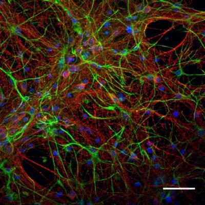
Researchers at Linköping University (LiU), Sweden, have created an artificial organic neuron that closely mimics the characteristics of biological nerve cells. This artificial neuron can stimulate natural nerves, making it a promising technology for various medical treatments in the future.

Work to develop increasingly functional artificial nerve cells continues at the Laboratory for Organic Electronics, LOE. In 2022, a team of scientists led by associate professor Simone Fabiano demonstrated how an artificial organic neuron could be integrated into a living carnivorous plant [emphasis mine] to control the opening and closing of its maw. This synthetic nerve cell met two of the 20 characteristics that differentiate it from a biological nerve cell.

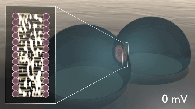
I wasn’t expecting a carnivorous plant, living or otherwise. Sadly, they don’t seem to have been able to include it in this image although the ‘green mitts’ are evocative,

Caption: Artificial neurons created by the researchers at Linköping University. Credit: Thor Balkhed

A January 13, 2023 Linköping University (LiU) press release by Mikael Sönne (also on EurkeAlert but published January 12, 2023), which originated the news item, delves further into the work,

In their latest study, published in the journal Nature Materials, the same researchers at LiU have developed a new artificial nerve cell called “conductance-based organic electrochemical neuron” or c-OECN, which closely mimics 15 out of the 20 neural features that characterise biological nerve cells, making its functioning much more similar to natural nerve cells.

“One of the key challenges in creating artificial neurons that effectively mimic real biological neurons is the ability to incorporate ion modulation. Traditional artificial neurons made of silicon can emulate many neural features but cannot communicate through ions. In contrast, c-OECNs use ions to demonstrate several key features of real biological neurons”, says Simone Fabiano, principal investigator of the Organic Nanoelectronics group at LOE.

In 2018, this research group at Linköping University was one of the first to develop organic electrochemical transistors based on n-type conducting polymers, which are materials that can conduct negative charges. This made it possible to build printable complementary organic electrochemical circuits. Since then, the group has been working to optimise these transistors so that they can be printed in a printing press on a thin plastic foil. As a result, it is now possible to print thousands of transistors on a flexible substrate and use them to develop artificial nerve cells.

In the newly developed artificial neuron, ions are used to control the flow of electronic current through an n-type conducting polymer, leading to spikes in the device’s voltage. This process is similar to that which occurs in biological nerve cells. The unique material in the artificial nerve cell also allows the current to be increased and decreased in an almost perfect bell-shaped curve that resembles the activation and inactivation of sodium ion channels found in biology.

“Several other polymers show this behaviour, but only rigid polymers are resilient to disorder, enabling stable device operation”, says Simone Fabiano

In experiments carried out in collaboration with Karolinska Institute (KI), the new c-OECN neurons were connected to the vagus nerve of mice. The results show that the artificial neuron could stimulate the mice’s nerves, causing a 4.5% change in their heart rate.

The fact that the artificial neuron can stimulate the vagus nerve itself could, in the long run, pave the way for essential applications in various forms of medical treatment. In general, organic semiconductors have the advantage of being biocompatible, soft, and malleable, while the vagus nerve plays a key role, for example, in the body’s immune system and metabolism.

The next step for the researchers will be to reduce the energy consumption of the artificial neurons, which is still much higher than that of human nerve cells. Much work remains to be done to replicate nature artificially.

“There is much we still don’t fully understand about the human brain and nerve cells. In fact, we don’t know how the nerve cell makes use of many of these 15 demonstrated features. Mimicking the nerve cells can enable us to understand the brain better and build circuits capable of performing intelligent tasks. We’ve got a long road ahead, but this study is a good start,” says Padinhare Cholakkal Harikesh, postdoc and main author of the scientific paper.

Here’s a link to and a citation for the paper,

Ion-tunable antiambipolarity in mixed ion–electron conducting polymers enables biorealistic organic electrochemical neurons by Padinhare Cholakkal Harikesh, Chi-Yuan Yang, Han-Yan Wu, Silan Zhang, Mary J. Donahue, April S. Caravaca, Jun-Da Huang, Peder S. Olofsson, Magnus Berggren, Deyu Tu & Simone Fabiano. Nature Materials volume 22, pages 242–248 (2023) DOI: https://doi.org/10.1038/s41563-022-01450-8 Published online: 12 January 2023 Issue Date: February 2023

This paper is open access.

A lipid-based memcapacitor,for neuromorphic computing

Caption: Researchers at ORNL’s Center for Nanophase Materials Sciences demonstrated the first example of capacitance in a lipid-based biomimetic membrane, opening nondigital routes to advanced, brain-like computation. Credit: Michelle Lehman/Oak Ridge National Laboratory, U.S. Dept. of Energy

The last time I wrote about memcapacitors (June 30, 2014 posting: Memristors, memcapacitors, and meminductors for faster computers), the ideas were largely theoretical; I believe this work is the first research I’ve seen on the topic. From an October 17, 2019 news item on ScienceDaily,

Researchers at the Department of Energy’s Oak Ridge National Laboratory ]ORNL], the University of Tennessee and Texas A&M University demonstrated bio-inspired devices that accelerate routes to neuromorphic, or brain-like, computing.

Results published in Nature Communications report the first example of a lipid-based “memcapacitor,” a charge storage component with memory that processes information much like synapses do in the brain. Their discovery could support the emergence of computing networks modeled on biology for a sensory approach to machine learning.

An October 16, 2019 ORNL news release (also on EurekAlert but published Oct. 17, 2019), which originated the news item, provides more detail about the work,

“Our goal is to develop materials and computing elements that work like biological synapses and neurons—with vast interconnectivity and flexibility—to enable autonomous systems that operate differently than current computing devices and offer new functionality and learning capabilities,” said Joseph Najem, a recent postdoctoral researcher at ORNL’s Center for Nanophase Materials Sciences, a DOE Office of Science User Facility, and current assistant professor of mechanical engineering at Penn State.

The novel approach uses soft materials to mimic biomembranes and simulate the way nerve cells communicate with one another.

The team designed an artificial cell membrane, formed at the interface of two lipid-coated water droplets in oil, to explore the material’s dynamic, electrophysiological properties. At applied voltages, charges build up on both sides of the membrane as stored energy, analogous to the way capacitors work in traditional electric circuits.

But unlike regular capacitors, the memcapacitor can “remember” a previously applied voltage and—literally—shape how information is processed. The synthetic membranes change surface area and thickness depending on electrical activity. These shapeshifting membranes could be tuned as adaptive filters for specific biophysical and biochemical signals.

“The novel functionality opens avenues for nondigital signal processing and machine learning modeled on nature,” said ORNL’s Pat Collier, a CNMS staff research scientist.

A distinct feature of all digital computers is the separation of processing and memory. Information is transferred back and forth from the hard drive and the central processor, creating an inherent bottleneck in the architecture no matter how small or fast the hardware can be.

Neuromorphic computing, modeled on the nervous system, employs architectures that are fundamentally different in that memory and signal processing are co-located in memory elements—memristors, memcapacitors and meminductors.

These “memelements” make up the synaptic hardware of systems that mimic natural information processing, learning and memory.

Systems designed with memelements offer advantages in scalability and low power consumption, but the real goal is to carve out an alternative path to artificial intelligence, said Collier.

Tapping into biology could enable new computing possibilities, especially in the area of “edge computing,” such as wearable and embedded technologies that are not connected to a cloud but instead make on-the-fly decisions based on sensory input and past experience.

Biological sensing has evolved over billions of years into a highly sensitive system with receptors in cell membranes that are able to pick out a single molecule of a specific odor or taste. “This is not something we can match digitally,” Collier said.

Digital computation is built around digital information, the binary language of ones and zeros coursing through electronic circuits. It can emulate the human brain, but its solid-state components do not compute sensory data the way a brain does.

“The brain computes sensory information pushed through synapses in a neural network that is reconfigurable and shaped by learning,” said Collier. “Incorporating biology—using biomembranes that sense bioelectrochemical information—is key to developing the functionality of neuromorphic computing.”

While numerous solid-state versions of memelements have been demonstrated, the team’s biomimetic elements represent new opportunities for potential “spiking” neural networks that can compute natural data in natural ways.

Spiking neural networks are intended to simulate the way neurons spike with electrical potential and, if the signal is strong enough, pass it on to their neighbors through synapses, carving out learning pathways that are pruned over time for efficiency.

A bio-inspired version with analog data processing is a distant aim. Current early-stage research focuses on developing the components of bio-circuitry.

“We started with the basics, a memristor that can weigh information via conductance to determine if a spike is strong enough to be broadcast through a network of synapses connecting neurons,” said Collier. “Our memcapacitor goes further in that it can actually store energy as an electric charge in the membrane, enabling the complex ‘integrate and fire’ activity of neurons needed to achieve dense networks capable of brain-like computation.”

The team’s next steps are to explore new biomaterials and study simple networks to achieve more complex brain-like functionalities with memelements.

Here’s a link to and a citation for the paper,

Dynamical nonlinear memory capacitance in biomimetic membranes by Joseph S. Najem, Md Sakib Hasan, R. Stanley Williams, Ryan J. Weiss, Garrett S. Rose, Graham J. Taylor, Stephen A. Sarles & C. Patrick Collier. Nature Communications volume 10, Article number: 3239 (2019) DOI: DOIhttps://doi.org/10.1038/s41467-019-11223-8 Published July 19, 2019

This paper is open access.

One final comment, you might recognize one of the authors (R. Stanley Williams) who in 2008 helped launch ‘memristor’ research.

Neuronal regenerative-interfaces made of cross-linked carbon nanotube films

If I understand this research rightly, they are creating a film made of carbon nanotubes that can stimulate the growth of nerve cells (neurons) thus creating a ‘living/nonliving’ hybrid or as they call it in the press release a ‘biosynthetic hybrid’.

An August 2, 2019 news item on Nanowerk introduces the research (Note 1: There seem to be some translation issues; Note 2: Links have been removed),

Carbon nanotubes able to take on the desired shapes thanks to a special chemical treatment, called crosslinking and, at the same time, able to function as substrata for the growth of nerve cells, finely tuning their growth and activity.

The research published in ACS Nano (“Chemically Cross-Linked Carbon Nanotube Films Engineered to Control Neuronal Signaling”), is a new and important step towards the construction of neuronal regenerative-interfaces to repair spinal injuries.

The study is the new achievement of a long-term and, in terms of results, successful collaboration between the scientists Laura Ballerini of SISSA (Scuola Internazionale Superiore di Studi Avanzati), Trieste, and Maurizio Prato of the University of Trieste. The work team has also been assisted by CIC biomaGUNE of San Sebastián, Spain.

Caption: Carbon nanotubes able to take on the desired shapes thanks to a special chemical treatment, called crosslinking and, at the same time, able to function as substrata for the growth of nerve cells, finely tuning their growth and activity. Credit: Rossana Rauti

An August 2, 2019 SISSA press release (also on EurekAlert), which originated the news item, adds detail,

The carbon nanotubes used in the research have been modified by appropriate chemical treatments: “For many years, in our laboratories we have been working on the chemical reactivity of carbon nanotubes, a fascinating but very difficult material to work. Thanks to our experience, we have crosslinked them or, to say it more clearly, we have treated the nanotubes so they could link themselves to one another thanks to specific chemical reactions. We have discovered that this procedure gives the material very interesting characteristics. For example, the material organises itself in a stable manner according to a precise shape, we choose: a tissue where nerve cells need to be planted, for example. Or around some electrodes” explains Professor Prato. “We know from previous research that nerve cells grow well on carbon nanotubes so they could be used as a surface to build hybrid devices to regenerate nerve tissues. It was necessary to ensure that this chemical modification did not compromise this process and study whether the interaction with neurons was altered”.

Towards biosynthetic hybrids

Professor Ballerini continues: “We have discovered that the chemical process has important effects because through this treatment we can modulate the activity of neurons, in terms of growth, adhesion and survival. These materials can also regulate the communication between neurons. We can say that the carpet of crosslinked carbon nanotubes interacts intensely and constructively with the nerve cells”. This interaction depends on how much the different carbon nanotubes are linked to each other, or rather crosslinked. The lower the link number among the nanotubes the higher the activity of neurons that grow on their surface. Through the chemical control of their properties, and of the links between them, it is possible to regulate the response of the neurons. Ballerini and Prato explain: “This is an intriguing result that emerges from the important and fruitful collaboration between our research groups involving advanced research in chemistry, nanoscience and neurobiology . This study provides a further step in the design of future biosynthetic hybrids to recover injured nerve tissues functions”.

Here’s a link to and a citation for the paper,

Chemically Cross-Linked Carbon Nanotube Films Engineered to Control Neuronal Signaling by Myriam Barrejón, Rossana Rauti, Laura Ballerini, Maurizio Prato. ACS Nano2019 XXXXXXXXXX-XXX Publication Date:July 22, 2019 DOI: https://doi.org/10.1021/acsnano.9b02429 Copyright © 2019 American Chemical Society

This paper is behind a paywall.

Carbon nanotubes to repair nerve fibres (cyborg brains?)

Can cyborg brains be far behind now that researchers are looking at ways to repair nerve fibers with carbon nanotubes (CNTs)? A June 26, 2017 news item on ScienceDaily describes the scheme using carbon nanotubes as a material for repairing nerve fibers,

Carbon nanotubes exhibit interesting characteristics rendering them particularly suited to the construction of special hybrid devices — consisting of biological issue and synthetic material — planned to re-establish connections between nerve cells, for instance at spinal level, lost on account of lesions or trauma. This is the result of a piece of research published on the scientific journal Nanomedicine: Nanotechnology, Biology, and Medicine conducted by a multi-disciplinary team comprising SISSA (International School for Advanced Studies), the University of Trieste, ELETTRA Sincrotrone and two Spanish institutions, Basque Foundation for Science and CIC BiomaGUNE. More specifically, researchers have investigated the possible effects on neurons of the interaction with carbon nanotubes. Scientists have proven that these nanomaterials may regulate the formation of synapses, specialized structures through which the nerve cells communicate, and modulate biological mechanisms, such as the growth of neurons, as part of a self-regulating process. This result, which shows the extent to which the integration between nerve cells and these synthetic structures is stable and efficient, highlights the great potentialities of carbon nanotubes as innovative materials capable of facilitating neuronal regeneration or in order to create a kind of artificial bridge between groups of neurons whose connection has been interrupted. In vivo testing has actually already begun.

The researchers have included a gorgeous image to illustrate their work,

Caption: Scientists have proven that these nanomaterials may regulate the formation of synapses, specialized structures through which the nerve cells communicate, and modulate biological mechanisms, such as the growth of neurons, as part of a self-regulating process. Credit: Pixabay

A June 26, 2017 SISSA press release (also on EurekAlert), which originated the news item, describes the work in more detail while explaining future research needs,

“Interface systems, or, more in general, neuronal prostheses, that enable an effective re-establishment of these connections are under active investigation” explain Laura Ballerini (SISSA) and Maurizio Prato (UniTS-CIC BiomaGUNE), coordinating the research project. “The perfect material to build these neural interfaces does not exist, yet the carbon nanotubes we are working on have already proved to have great potentialities. After all, nanomaterials currently represent our best hope for developing innovative strategies in the treatment of spinal cord injuries”. These nanomaterials are used both as scaffolds, a supportive framework for nerve cells, and as means of interfaces releasing those signals that empower nerve cells to communicate with each other.

Many aspects, however, still need to be addressed. Among them, the impact on neuronal physiology of the integration of these nanometric structures with the cell membrane. “Studying the interaction between these two elements is crucial, as it might also lead to some undesired effects, which we ought to exclude”. Laura Ballerini explains: “If, for example, the mere contact provoked a vertiginous rise in the number of synapses, these materials would be essentially unusable”. “This”, Maurizio Prato adds, “is precisely what we have investigated in this study where we used pure carbon nanotubes”.

The results of the research are extremely encouraging: “First of all we have proved that nanotubes do not interfere with the composition of lipids, of cholesterol in particular, which make up the cellular membrane in neurons. Membrane lipids play a very important role in the transmission of signals through the synapses. Nanotubes do not seem to influence this process, which is very important”.

There is more, however. The research has also highlighted the fact that the nerve cells growing on the substratum of nanotubes, thanks to this interaction, develop and reach maturity very quickly, eventually reaching a condition of biological homeostasis. “Nanotubes facilitate the full growth of neurons and the formation of new synapses. This growth, however, is not indiscriminate and unlimited since, as we proved, after a few weeks a physiological balance is attained. Having established the fact that this interaction is stable and efficient is an aspect of fundamental importance”. Maurizio Prato and Laura Ballerini conclude as follows: “We are proving that carbon nanotubes perform excellently in terms of duration, adaptability and mechanical compatibility with the tissue. Now we know that their interaction with the biological material, too, is efficient. Based on this evidence, we are already studying the in vivo application, and preliminary results appear to be quite promising also in terms of recovery of the lost neurological functions”.

Here’s a link to and a citation for the paper,

Sculpting neurotransmission during synaptic development by 2D nanostructured interfaces by Niccolò Paolo Pampaloni, Denis Scaini, Fabio Perissinotto, Susanna Bosi, Maurizio Prato, Laura Ballerini. Nanomedicine: Nanotechnology, Biology and Medicine, DOI: http://dx.doi.org/10.1016/j.nano.2017.01.020 Published online: May 25, 2017

This paper is open access.

Bioelectronics: creating components that speak the body’s own language

This is work is still in its early stages but the idea that the body could be stimulated to release more of its own pain relievers is exciting. From a Nov. 2, 2016 news item on ScienceDaily,

With a microfabricated ion pump built from organic electronic components, ions can be sent to nerve or muscle cells at the speed of the nervous system and with a precision of a single cell. “Now we can start to develop components that speak the body’s own language,” says Daniel Simon, head of bioelectronics research at the Laboratory of Organic Electronics, Linköping University, Campus Norrköping.

A Nov. 2, 2016 Linköping University press release (also on EurekAlert), which originated the news item, discusses the research in more detail,

Our nerve and muscle cells send signals to each other using ions and molecules. Certain substances, such as the neurotransmitter GABA (gamma aminobutyric acid), are important signal substances throughout the central nervous system. Eighteen months ago, researchers at the Laboratory of Organic Electronics demonstrated an ion pump which researchers at the Karolinska Institutet could use to reduce the sensation of pain in awake, freely-moving rats. The ion pump delivered GABA directly to the rat´s spinal cord. The news that researchers could deliver the body’s own neurotransmitters was published in Science Advances and garnered intense interest all over the world.

The research group at the Laboratory of Organic Electronics has now achieved another major advance and developed a significantly smaller and more rapid ion pump that transmits signals nearly as rapidly as the cells themselves, and with a precision on the scale of an individual cell. …

“Our skilled doctoral students, Amanda Jonsson and Theresia Arbring Sjöström, have succeeded with the last important part of the puzzle in the development of the ion pump. When a signal passes between two synapses it takes 1-10 milliseconds, and we are now very close to the nervous system’s own speed,” says Magnus Berggren, professor of organic electronics and director of the Laboratory of Organic Electronics.

“We conclude that we have produced artificial nerves that can communicate seamlessly with the nervous system. After more than 10 years’ research we have finally got all the parts of the puzzle in place,” he says.

Amanda Jonsson, who together with Theresia Arbring Sjöström is principal author of the article in Science Advances, has developed the pain-alleviating ion pump as part of her doctoral studies. She proudly presents a glass disk with many of the new miniaturized ion pumps. Some pumps have only a single outlet, but others have six tiny point outlets.

“We can make them with several outlets, it’s just as easy as making one. And all of the outlets can be individually controlled. Previously we could only transport ions horizontally and from all outputs at the same time. Now, however, we can deliver the ions vertically, which makes the distance they have to be transported as short as a micrometre,” she explains.

All of the outputs of the ion pump can also be rapidly switched on or off with the aid of micrometre-sized ion diodes.

“The ions are released rapidly by an electrical signal, in the same way that the neurotransmitter is released in a synapse,” says Theresia Arbring Sjöström.

Organic electronic components have a major advantage here: they can conduct both ions and electricity. In this case, the material PEDOT:PSS enables the electrical signals to be converted to chemical signals that the body understands.

The ion diode has recently been developed, as has the material that forms the basis of the new rapid ion pump.

“The new material makes it possible to build with a precision and reliability not possible in previous versions of the ion pump,” says Daniel Simon.

The new ion pump has so far only been tested in the laboratory. The next step will be to test it with live cells and the researchers hope eventually to, for example alleviate pain, stop epileptic seizures, and reduce the symptoms of Parkinsons disease, using exactly the required dose at exactly the affected cells. Communication using the cell´s own language, and the cell´s own speed.

Here’s a link to and a citation for the paper,

Chemical delivery array with millisecond neurotransmitter release by Amanda Jonsson, Theresia Arbring Sjöström, Klas Tybrandt, Magnus Berggren, and Daniel T. Simon. Science Advances  02 Nov 2016: Vol. 2, no. 11, e1601340 DOI: 10.1126/sciadv.1601340

This paper is open access.

Graphene and neurons in a UK-Italy-Spain collaboration

There’s been a lot of talk about using graphene-based implants in the brain due to the material’s flexibility along with its other properties. A step forward has been taking according to a Jan. 29, 2016 news item on phys.org,

Researchers have successfully demonstrated how it is possible to interface graphene – a two-dimensional form of carbon – with neurons, or nerve cells, while maintaining the integrity of these vital cells. The work may be used to build graphene-based electrodes that can safely be implanted in the brain, offering promise for the restoration of sensory functions for amputee or paralysed patients, or for individuals with motor disorders such as epilepsy or Parkinson’s disease.

A Jan. 29, 2016 Cambridge University press release (also on EurekAlert), which originated the news item, provides more detail,

Previously, other groups had shown that it is possible to use treated graphene to interact with neurons. However the signal to noise ratio from this interface was very low. By developing methods of working with untreated graphene, the researchers retained the material’s electrical conductivity, making it a significantly better electrode.

“For the first time we interfaced graphene to neurons directly,” said Professor Laura Ballerini of the University of Trieste in Italy. “We then tested the ability of neurons to generate electrical signals known to represent brain activities, and found that the neurons retained their neuronal signalling properties unaltered. This is the first functional study of neuronal synaptic activity using uncoated graphene based materials.”

Our understanding of the brain has increased to such a degree that by interfacing directly between the brain and the outside world we can now harness and control some of its functions. For instance, by measuring the brain’s electrical impulses, sensory functions can be recovered. This can be used to control robotic arms for amputee patients or any number of basic processes for paralysed patients – from speech to movement of objects in the world around them. Alternatively, by interfering with these electrical impulses, motor disorders (such as epilepsy or Parkinson’s) can start to be controlled.

Scientists have made this possible by developing electrodes that can be placed deep within the brain. These electrodes connect directly to neurons and transmit their electrical signals away from the body, allowing their meaning to be decoded.

However, the interface between neurons and electrodes has often been problematic: not only do the electrodes need to be highly sensitive to electrical impulses, but they need to be stable in the body without altering the tissue they measure.

Too often the modern electrodes used for this interface (based on tungsten or silicon) suffer from partial or complete loss of signal over time. This is often caused by the formation of scar tissue from the electrode insertion, which prevents the electrode from moving with the natural movements of the brain due to its rigid nature.

Graphene has been shown to be a promising material to solve these problems, because of its excellent conductivity, flexibility, biocompatibility and stability within the body.

Based on experiments conducted in rat brain cell cultures, the researchers found that untreated graphene electrodes interfaced well with neurons. By studying the neurons with electron microscopy and immunofluorescence the researchers found that they remained healthy, transmitting normal electric impulses and, importantly, none of the adverse reactions which lead to the damaging scar tissue were seen.

According to the researchers, this is the first step towards using pristine graphene-based materials as an electrode for a neuro-interface. In future, the researchers will investigate how different forms of graphene, from multiple layers to monolayers, are able to affect neurons, and whether tuning the material properties of graphene might alter the synapses and neuronal excitability in new and unique ways. “Hopefully this will pave the way for better deep brain implants to both harness and control the brain, with higher sensitivity and fewer unwanted side effects,” said Ballerini.

“We are currently involved in frontline research in graphene technology towards biomedical applications,” said Professor Maurizio Prato from the University of Trieste. “In this scenario, the development and translation in neurology of graphene-based high-performance biodevices requires the exploration of the interactions between graphene nano- and micro-sheets with the sophisticated signalling machinery of nerve cells. Our work is only a first step in that direction.”

“These initial results show how we are just at the tip of the iceberg when it comes to the potential of graphene and related materials in bio-applications and medicine,” said Professor Andrea Ferrari, Director of the Cambridge Graphene Centre. “The expertise developed at the Cambridge Graphene Centre allows us to produce large quantities of pristine material in solution, and this study proves the compatibility of our process with neuro-interfaces.”

The research was funded by the Graphene Flagship [emphasis mine],  a European initiative which promotes a collaborative approach to research with an aim of helping to translate graphene out of the academic laboratory, through local industry and into society.

Here’s a link to and a citation for the paper,

Graphene-Based Interfaces Do Not Alter Target Nerve Cells by Alessandra Fabbro, Denis Scaini, Verónica León, Ester Vázquez, Giada Cellot, Giulia Privitera, Lucia Lombardi, Felice Torrisi, Flavia Tomarchio, Francesco Bonaccorso, Susanna Bosi, Andrea C. Ferrari, Laura Ballerini, and Maurizio Prato. ACS Nano, 2016, 10 (1), pp 615–623 DOI: 10.1021/acsnano.5b05647 Publication Date (Web): December 23, 2015

Copyright © 2015 American Chemical Society

This paper is behind a paywall.

There are a couple things I found a bit odd about this project. First, all of the funding is from the Graphene Flagship initiative. I was expecting to see at least some funding from the European Union’s other mega-sized science initiative, the Human Brain Project. Second, there was no mention of Spain nor were there any quotes from the Spanish researchers. For the record, the Spanish institutions represented were: University of Castilla-La Mancha, Carbon Nanobiotechnology Laboratory, and the Basque Foundation for Science.

Synthesizing nerve tissues with 3D printers and cellulose nanocrystals (CNC)

There are lots of stories about bioprinting and tissue engineering here and I think it’s time (again) for one which one has some good, detailed descriptions and, bonus, it features cellulose nanocrystals (CNC) and graphene. From a May 13, 2015 news item on Azonano,

The printer looks like a toaster oven with the front and sides removed. Its metal frame is built up around a stainless steel circle lit by an ultraviolet light. Stainless steel hydraulics and thin black tubes line the back edge, which lead to an inner, topside box made of red plastic.

In front, the metal is etched with the red Bio Bot logo. All together, the gray metal frame is small enough to fit on top of an old-fashioned school desk, but nothing about this 3D printer is old school. In fact, the tissue-printing machine is more like a sci-fi future in the flesh—and it has very real medical applications.

Researchers at Michigan Technological University hope to use this newly acquired 3D bioprinter to make synthesized nerve tissue. The key is developing the right “bioink” or printable tissue. The nanotechnology-inspired material could help regenerate damaged nerves for patients with spinal cord injuries, says Tolou Shokuhfar, an assistant professor of mechanical engineering and biomedical engineering at Michigan Tech.

Shokuhfar directs the In-Situ Nanomedicine and Nanoelectronics Laboratory at Michigan Tech, and she is an adjunct assistant professor in the Bioengineering Department and the College of Dentistry at the University of Illinois at Chicago.

In the bioprinting research, Shokuhfar collaborates with Reza Shahbazian-Yassar, the Richard and Elizabeth Henes Associate Professor in the Department of Mechanical Engineering-Engineering Mechanics at Michigan Tech. Shahbazian-Yassar’s highly interdisciplinary background on cellulose nanocrystals as biomaterials, funded by the National Science Foundation’s (NSF) Biomaterials Program, helped inspire the lab’s new 3D printing research. “Cellulose nanocrystals with extremely good mechanical properties are highly desirable for bioprinting of scaffolds that can be used for live tissues,” says Shahbazian-Yassar. [emphases mine]

A May 11, 2015 Michigan Technological University (MTU) news release by Allison Mills, which originated the news item, explains the ‘why’ of the research,

“We wanted to target a big issue,” Shokuhfar says, explaining that nerve regeneration is a particularly difficult biomedical engineering conundrum. “We are born with all the nerve cells we’ll ever have, and damaged nerves don’t heal very well.”

Other facilities are trying to address this issue as well. Many feature large, room-sized machines that have built-in cell culture hoods, incubators and refrigeration. The precision of this equipment allows them to print full organs. But innovation is more nimble at smaller scales.

“We can pursue nerve regeneration research with a simpler printer set-up,” says Shayan Shafiee, a PhD student working with Shokuhfar. He gestures to the small gray box across the lab bench.

He opens the red box under the top side of the printer’s box. Inside the plastic casing, a large syringe holds a red jelly-like fluid. Shafiee replenishes the needle-tipped printer, pulls up his laptop and, with a hydraulic whoosh, he starts to print a tissue scaffold.

The news release expands on the theme,

At his lab bench in the nanotechnology lab at Michigan Tech, Shafiee holds up a petri dish. Inside is what looks like a red gummy candy, about the size of a half-dollar.

Here’s a video from MTU illustrating the printing process,

Back to the news release, which notes graphene could be instrumental in this research,

“This is based on fractal geometry,” Shafiee explains, pointing out the small crenulations and holes pockmarking the jelly. “These are similar to our vertebrae—the idea is to let a nerve pass through the holes.”

Making the tissue compatible with nerve cells begins long before the printer starts up. Shafiee says the first step is to synthesize a biocompatible polymer that is syrupy—but not too thick—that can be printed. That means Shafiee and Shokuhfar have to create their own materials to print with; there is no Amazon.com or even a specialty shop for bioprinting nerves.

Nerves don’t just need a biocompatible tissue to act as a carrier for the cells. Nerve function is all about electric pulses. This is where Shokuhfar’s nanotechnology research comes in: Last year, she was awarded a CAREER grant from NSF for her work using graphene in biomaterials research. [emphasis mine] “Graphene is a wonder material,” she says. “And it has very good electrical conductivity properties.”

The team is extending the application of this material for nerve cell printing. “Our work always comes back to the question, is it printable or not?” Shafiee says, adding that a successful material—a biocompatible, graphene-bound polymer—may just melt, mush or flat out fail under the pressure of printing. After all, imagine building up a substance more delicate than a soufflé using only the point of a needle. And in the nanotechnology world, a needlepoint is big, even clumsy.

Shafiee and Shokuhfar see these issues as mechanical obstacles that can be overcome.

“It’s like other 3D printers, you need a design to work from,” Shafiee says, adding that he will tweak and hone the methodology for printing nerve cells throughout his dissertation work. He is also hopeful that the material will have use beyond nerve regeneration.

This looks like a news release designed to publicize work funded at MTU by the US National Science Foundation (NSF) which is why there is no mention of published work.

One final comment regarding cellulose nanocrystals (CNC). They have also been called nanocrystalline cellulose (NCC), which you will still see but it seems CNC is emerging as the generic term. NCC has been trademarked by CelluForce, a Canadian company researching and producing CNC (or if you prefer, NCC) from forest products.

Nanocellulose as scaffolding for nerve cells

Swedish scientists have announced success with growing nerve cells using nanocellulose as the scaffolding. From the March 19, 2012 news item on Naowerk,

Researchers from Chalmers and the University of Gothenburg have shown that nanocellulose stimulates the formation of neural networks. This is the first step toward creating a three-dimensional model of the brain. Such a model could elevate brain research to totally new levels, with regard to Alzheimer’s disease and Parkinson’s disease, for example.

“This has been a great challenge,” says Paul Gatenholm, Professor of Biopolymer Technology at Chalmers.?Until recently the cells were dying after a while, since we weren’t able to get them to adhere to the scaffold. But after many experiments we discovered a method to get them to attach to the scaffold by making it more positively charged. Now we have a stable method for cultivating nerve cells on nanocellulose.”

When the nerve cells finally attached to the scaffold they began to develop and generate contacts with one another, so-called synapses. A neural network of hundreds of cells was produced. The researchers can now use electrical impulses and chemical signal substances to generate nerve impulses, that spread through the network in much the same way as they do in the brain. They can also study how nerve cells react with other molecules, such as pharmaceuticals.

I found the original March 19, 2012 press release  and an image on the University of Chalmers website,

Nerve cells growing on a three-dimensional nanocellulose scaffold. One of the applications the research group would like to study is destruction of synapses between nerve cells, which is one of the earliest signs of Alzheimer’s disease. Synapses are the connections between nerve cells. In the image, the functioning synapses are yellow and the red spots show where synapses have been destroyed. Illustration: Philip Krantz, Chalmers

This latest research from Gatenholm and his team will be presented at the American Chemical Society annual meeting in San Diego, March 25, 2012.

The research team from Chalmers University and its partners are working on other applications for nanocellulose including one for artificial ears. From the Chalmers University Jan. 22, 2012 press release,

As the first group in the world, researchers from Chalmers will build up body parts using nanocellulose and the body’s own cells. Funding will be from the European network for nanomedicine, EuroNanoMed.

Professor Paul Gatenholm at Chalmers is leading and co-ordinating this European research programme, which will construct an outer ear using nanocellulose and a mixture of the patient’s own cartilage cells and stem cells.

Previously, Paul Gatenholm and his colleagues succeeded, in close co-operation with Sahlgrenska University Hospital, in developing artificial blood vessels using nanocellulose, where small bacteria “spin” the cellulose.

In the new programme , the researchers will build up a three-dimensional nanocellulose network that is an exact copy of the patient’s healthy outer ear and construct an exact mirror image of the ear. It will have sufficient mechanical stability for it to be used as a bioreactor, which means that the patient’s own cartilage and stem cells can be cultivated directly inside the body or on the patient, in this case on the head. [Presumably the patient has one ear that is healthy and the researchers are attempting to repair or replace an unhealthy ear on the other side of the head.]

As for the Swedish perspective on nanocellulose (from the 2010 press release),

Cellulose-based material is of strategic significance to Sweden and materials science is one of Chalmers eight areas of advance. Biopolymers are highly interesting as they are renewable and could be of major significance in the development of future materials.

Further research into using the forest as a resource for new materials is continuing at Chalmers within the new research programme that is being built up with different research groups at Chalmers and Swerea – IVF. The programme is part of the Wallenberg Wood Science Center, which is being run jointly by the Royal Institute of Technology in Stockholm and Chalmers under the leadership of Professor Lars Berglund at the Royal Institute of Technology.

The 2012 press release announcing the work on nerve cells had this about nanocellulose,

Nanocellulose is a material that consists of nanosized cellulose fibers. Typical dimensions are widths of 5 to 20 nanometers and lengths of up to 2,000 nanometers. Nanocellulose can be produced by bacteria that spin a close-meshed structure of cellulose fibers. It can also be isolated from wood pulp through processing in a high-pressure homogenizer.

I last wrote about the Swedes and nanocellulose in a Feb. 15, 2012 posting about recovering it (nanocellulose) from wood-based sludge.

As for anyone interested in the Canadian scene, there is an article by David Manly in the Jan.-Feb. 2012 issue of Canadian Biomass Magazine that focuses largely on economic impacts and value-added products as they pertain to nanocellulose manufacturing production in Canada. You can also search this blog as I have covered the nanocellulose story in Canada and elsewhere as extensively as I can.