Tag Archives: neurodegenerative diseases

Cortical spheroids (like mini-brains) could unlock (larger) brain’s mysteries

A March 19, 2021 Northwestern University news release on EurekAlert announces the creation of a device designed to monitor brain organoids (for anyone unfamiliar with brain organoids there’s more information after the news),

A team of scientists, led by researchers at Northwestern University, Shirley Ryan AbilityLab and the University of Illinois at Chicago (UIC), has developed novel technology promising to increase understanding of how brains develop, and offer answers on repairing brains in the wake of neurotrauma and neurodegenerative diseases.

Their research is the first to combine the most sophisticated 3-D bioelectronic systems with highly advanced 3-D human neural cultures. The goal is to enable precise studies of how human brain circuits develop and repair themselves in vitro. The study is the cover story for the March 19 [March 17, 2021 according to the citation] issue of Science Advances.

The cortical spheroids used in the study, akin to “mini-brains,” were derived from human-induced pluripotent stem cells. Leveraging a 3-D neural interface system that the team developed, scientists were able to create a “mini laboratory in a dish” specifically tailored to study the mini-brains and collect different types of data simultaneously. Scientists incorporated electrodes to record electrical activity. They added tiny heating elements to either keep the brain cultures warm or, in some cases, intentionally overheated the cultures to stress them. They also incorporated tiny probes — such as oxygen sensors and small LED lights — to perform optogenetic experiments. For instance, they introduced genes into the cells that allowed them to control the neural activity using different-colored light pulses.

This platform then enabled scientists to perform complex studies of human tissue without directly involving humans or performing invasive testing. In theory, any person could donate a limited number of their cells (e.g., blood sample, skin biopsy). Scientists can then reprogram these cells to produce a tiny brain spheroid that shares the person’s genetic identity. The authors believe that, by combining this technology with a personalized medicine approach using human stem cell-derived brain cultures, they will be able to glean insights faster and generate better, novel interventions.

“The advances spurred by this research will offer a new frontier in the way we study and understand the brain,” said Shirley Ryan AbilityLab’s Dr. Colin Franz, co-lead author on the paper who led the testing of the cortical spheroids. “Now that the 3-D platform has been developed and validated, we will be able to perform more targeted studies on our patients recovering from neurological injury or battling a neurodegenerative disease.”

Yoonseok Park, postdoctoral fellow at Northwestern University and co-lead author, added, “This is just the beginning of an entirely new class of miniaturized, 3-D bioelectronic systems that we can construct to expand the capacity of the regenerative medicine field. For example, our next generation of device will support the formation of even more complex neural circuits from brain to muscle, and increasingly dynamic tissues like a beating heart.”

Current electrode arrays for tissue cultures are 2-D, flat and unable to match the complex structural designs found throughout nature, such as those found in the human brain. Moreover, even when a system is 3-D, it is extremely challenging to incorporate more than one type of material into a small 3-D structure. With this advance, however, an entire class of 3-D bioelectronics devices has been tailored for the field of regenerative medicine.

“Now, with our small, soft 3-D electronics, the capacity to build devices that mimic the complex biological shapes found in the human body is finally possible, providing a much more holistic understanding of a culture,” said Northwestern’s John Rogers, who led the technology development using technology similar to that found in phones and computers. “We no longer have to compromise function to achieve the optimal form for interfacing with our biology.”

As a next step, scientists will use the devices to better understand neurological disease, test drugs and therapies that have clinical potential, and compare different patient-derived cell models. This understanding will then enable a better grasp of individual differences that may account for the wide variation of outcomes seen in neurological rehabilitation.

“As scientists, our goal is to make laboratory research as clinically relevant as possible,” said Kristen Cotton, research assistant in Dr. Franz’s lab. “This 3-D platform opens the door to new experiments, discovery and scientific advances in regenerative neurorehabilitation medicine that have never been possible.”
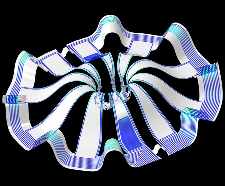
Caption: Three dimensional multifunctional neural interfaces for cortical spheroids and engineered assembloids Credit: Northwestern University

As for what brain ogranoids might be, Carl Zimmer in an Aug. 29, 2019 article for the New York Times provides an explanation,

Organoids Are Not Brains. How Are They Making Brain Waves?

Two hundred and fifty miles over Alysson Muotri’s head, a thousand tiny spheres of brain cells were sailing through space.

The clusters, called brain organoids, had been grown a few weeks earlier in the biologist’s lab here at the University of California, San Diego. He and his colleagues altered human skin cells into stem cells, then coaxed them to develop as brain cells do in an embryo.

The organoids grew into balls about the size of a pinhead, each containing hundreds of thousands of cells in a variety of types, each type producing the same chemicals and electrical signals as those cells do in our own brains.

In July, NASA packed the organoids aboard a rocket and sent them to the International Space Station to see how they develop in zero gravity.

Now the organoids were stowed inside a metal box, fed by bags of nutritious broth. “I think they are replicating like crazy at this stage, and so we’re going to have bigger organoids,” Dr. Muotri said in a recent interview in his office overlooking the Pacific.

What, exactly, are they growing into? That’s a question that has scientists and philosophers alike scratching their heads.

On Thursday, Dr. Muotri and his colleagues reported that they  have recorded simple brain waves in these organoids. In mature human brains, such waves are produced by widespread networks of neurons firing in synchrony. Particular wave patterns are linked to particular forms of brain activity, like retrieving memories and dreaming.

As the organoids mature, the researchers also found, the waves change in ways that resemble the changes in the developing brains of premature babies.

“It’s pretty amazing,” said Giorgia Quadrato, a neurobiologist at the University of Southern California who was not involved in the new study. “No one really knew if that was possible.”

But Dr. Quadrato stressed it was important not to read too much into the parallels. What she, Dr. Muotri and other brain organoid experts build are clusters of replicating brain cells, not actual brains.

If you have the time, I recommend reading Zimmer’s article in its entirety. Perhaps not coincidentally, Zimmer has an excerpt titled “Lab-Grown Brain Organoids Aren’t Alive. But They’re Not Not Alive, Either.” published in Slate.com,

From Life’s Edge: The Search For What It Means To Be Alive by Carl Zimmer, published by Dutton, an imprint of Penguin Publishing Group, a division of Penguin Random House, LLC. Copyright © 2021 by Carl Zimmer.

Cleber Trujillo led me to a windowless room banked with refrigerators, incubators, and microscopes. He extended his blue-gloved hands to either side and nearly touched the walls. “This is where we spend half our day,” he said.

In that room Trujillo and a team of graduate students raised a special kind of life. He opened an incubator and picked out a clear plastic box. Raising it above his head, he had me look up at it through its base. Inside the box were six circular wells, each the width of a cookie and filled with what looked like watered-down grape juice. In each well 100 pale globes floated, each the size of a housefly head.

Getting back to the research about monitoring brain organoids, here’s a link to and a citation for the paper about cortical spheroids,

Three-dimensional, multifunctional neural interfaces for cortical spheroids and engineered assembloids by Yoonseok Park, Colin K. Franz, Hanjun Ryu, Haiwen Luan, Kristen Y. Cotton, Jong Uk Kim, Ted S. Chung, Shiwei Zhao, Abraham Vazquez-Guardado, Da Som Yang, Kan Li, Raudel Avila, Jack K. Phillips, Maria J. Quezada, Hokyung Jang, Sung Soo Kwak, Sang Min Won, Kyeongha Kwon, Hyoyoung Jeong, Amay J. Bandodkar, Mengdi Han, Hangbo Zhao, Gabrielle R. Osher, Heling Wang, KunHyuck Lee, Yihui Zhang, Yonggang Huang, John D. Finan and John A. Rogers. Science Advances 17 Mar 2021: Vol. 7, no. 12, eabf9153 DOI: 10.1126/sciadv.abf9153

This paper appears to be open access.

According to a March 22, 2021 posting on the Shirley Riley AbilityLab website, the paper is featured on the front cover of Science Advances (vol. 7 no. 12).

Transplanting healthy neurons could be possible with walking molecules and 3D printing

A February 23, 2021 news item on ScienceDaily announces work which may lead to healing brain injuries and diseases,

Imagine if surgeons could transplant healthy neurons into patients living with neurodegenerative diseases or brain and spinal cord injuries. And imagine if they could “grow” these neurons in the laboratory from a patient’s own cells using a synthetic, highly bioactive material that is suitable for 3D printing.

By discovering a new printable biomaterial that can mimic properties of brain tissue, Northwestern University researchers are now closer to developing a platform capable of treating these conditions using regenerative medicine.

A February 22, 2021 Northwestern University news release (also received by email and available on EurekAlert) by Lila Reynolds, which originated the news item, delves further into self-assembling ‘walking’ molecules and the nanofibers resulting in a new material designed to promote the growth of healthy neurons,

A key ingredient to the discovery is the ability to control the self-assembly processes of molecules within the material, enabling the researchers to modify the structure and functions of the systems from the nanoscale to the scale of visible features. The laboratory of Samuel I. Stupp published a 2018 paper in the journal Science which showed that materials can be designed with highly dynamic molecules programmed to migrate over long distances and self-organize to form larger, “superstructured” bundles of nanofibers.

Now, a research group led by Stupp has demonstrated that these superstructures can enhance neuron growth, an important finding that could have implications for cell transplantation strategies for neurodegenerative diseases such as Parkinson’s and Alzheimer’s disease, as well as spinal cord injury.

“This is the first example where we’ve been able to take the phenomenon of molecular reshuffling we reported in 2018 and harness it for an application in regenerative medicine,” said Stupp, the lead author on the study and the director of Northwestern’s Simpson Querrey Institute. “We can also use constructs of the new biomaterial to help discover therapies and understand pathologies.

Walking molecules and 3D printing

The new material is created by mixing two liquids that quickly become rigid as a result of interactions known in chemistry as host-guest complexes that mimic key-lock interactions among proteins, and also as the result of the concentration of these interactions in micron-scale regions through a long scale migration of “walking molecules.”

The agile molecules cover a distance thousands of times larger than themselves in order to band together into large superstructures. At the microscopic scale, this migration causes a transformation in structure from what looks like an uncooked chunk of ramen noodles into ropelike bundles.

“Typical biomaterials used in medicine like polymer hydrogels don’t have the capabilities to allow molecules to self-assemble and move around within these assemblies,” said Tristan Clemons, a research associate in the Stupp lab and co-first author of the paper with Alexandra Edelbrock, a former graduate student in the group. “This phenomenon is unique to the systems we have developed here.”

Furthermore, as the dynamic molecules move to form superstructures, large pores open that allow cells to penetrate and interact with bioactive signals that can be integrated into the biomaterials.

Interestingly, the mechanical forces of 3D printing disrupt the host-guest interactions in the superstructures and cause the material to flow, but it can rapidly solidify into any macroscopic shape because the interactions are restored spontaneously by self-assembly. This also enables the 3D printing of structures with distinct layers that harbor different types of neural cells in order to study their interactions.

Signaling neuronal growth

The superstructure and bioactive properties of the material could have vast implications for tissue regeneration. Neurons are stimulated by a protein in the central nervous system known as brain-derived neurotrophic factor (BDNF), which helps neurons survive by promoting synaptic connections and allowing neurons to be more plastic. BDNF could be a valuable therapy for patients with neurodegenerative diseases and injuries in the spinal cord but these proteins degrade quickly in the body and are expensive to produce.

One of the molecules in the new material integrates a mimic of this protein that activates its receptor known as Trkb, and the team found that neurons actively penetrate the large pores and populate the new biomaterial when the mimetic signal is present. This could also create an environment in which neurons differentiated from patient-derived stem cells mature before transplantation.

Now that the team has applied a proof of concept to neurons, Stupp believes he could now break into other areas of regenerative medicine by applying different chemical sequences to the material. Simple chemical changes in the biomaterials would allow them to provide signals for a wide range of tissues.

“Cartilage and heart tissue are very difficult to regenerate after injury or heart attacks, and the platform could be used to prepare these tissues in vitro from patient-derived cells,” Stupp said. “These tissues could then be transplanted to help restore lost functions. Beyond these interventions, the materials could be used to build organoids to discover therapies or even directly implanted into tissues for regeneration since they are biodegradable.”

Here’s a link to and a citation for the paper,

Superstructured Biomaterials Formed by Exchange Dynamics and Host–Guest Interactions in Supramolecular Polymers by Alexandra N. Edelbrock, Tristan D. Clemons, Stacey M. Chin, Joshua J. W. Roan, Eric P. Bruckner, Zaida Álvarez, Jack F. Edelbrock, Kristen S. Wek, Samuel I. Stupp. Advanced Science DOI: https://doi.org/10.1002/advs.202004042 First published: 22 February 2021

This paper is open access.

Repairing brain circuits using nanotechnology

A July 30, 2019 news item on Nanowerk announces some neuroscience research (they used animal models) that could prove helpful with neurodegenerative diseases,

Working with mouse and human tissue, Johns Hopkins Medicine researchers report new evidence that a protein pumped out of some — but not all — populations of “helper” cells in the brain, called astrocytes, plays a specific role in directing the formation of connections among neurons needed for learning and forming new memories.

Using mice genetically engineered and bred with fewer such connections, the researchers conducted proof-of-concept experiments that show they could deliver corrective proteins via nanoparticles to replace the missing protein needed for “road repairs” on the defective neural highway.

Since such connective networks are lost or damaged by neurodegenerative diseases such as Alzheimer’s or certain types of intellectual disability, such as Norrie disease, the researchers say their findings advance efforts to regrow and repair the networks and potentially restore normal brain function.

A July 30, 2019 Johns Hopkins University School of Medicine news release (also on EurekAlert) provides more detail about the work (Note: A link has been removed),

“We are looking at the fundamental biology of how astrocytes function, but perhaps have discovered a new target for someday intervening in neurodegenerative diseases with novel therapeutics,” says Jeffrey Rothstein, M.D., Ph.D., the John W. Griffin Director of the Brain Science Institute and professor of neurology at the Johns Hopkins University School of Medicine.

“Although astrocytes appear to all look alike in the brain, we had an inkling that they might have specialized roles in the brain due to regional differences in the brain’s function and because of observed changes in certain diseases,” says Rothstein. “The hope is that learning to harness the individual differences in these distinct populations of astrocytes may allow us to direct brain development or even reverse the effects of certain brain conditions, and our current studies have advanced that hope.”

In the brain, astrocytes are the support cells that act as guides to direct new cells, promote chemical signaling, and clean up byproducts of brain cell metabolism.

Rothstein’s team focused on a particular astrocyte protein, glutamate transporter-1, which previous studies suggested was lost from astrocytes in certain parts of brains with neurodegenerative diseases. Like a biological vacuum cleaner, the protein normally sucks up the chemical “messenger” glutamate from the spaces between neurons after a message is sent to another cell, a step required to end the transmission and prevent toxic levels of glutamate from building up.

When these glutamate transporters disappear from certain parts of the brain — such as the motor cortex and spinal cord in people with amyotrophic lateral sclerosis (ALS) — glutamate hangs around much too long, sending messages that overexcite and kill the cells.

To figure out how the brain decides which cells need the glutamate transporters, Rothstein and colleagues focused on the region of DNA in front of the gene that typically controls the on-off switch needed to manufacture the protein. They genetically engineered mice to glow red in every cell where the gene is activated.

Normally, the glutamate transporter is turned on in all astrocytes. But, by using between 1,000- and 7,000-bit segments of DNA code from the on-off switch for glutamate, all the cells in the brain glowed red, including the neurons. It wasn’t until the researchers tried the largest sequence of an 8,300-bit DNA code from this location that the researchers began to see some selection in red cells. These red cells were all astrocytes but only in certain layers of the brain’s cortex in mice.

Because they could identify these “8.3 red astrocytes,” the researchers thought they might have a specific function different than other astrocytes in the brain. To find out more precisely what these 8.3 red astrocytes do in the brain, the researchers used a cell-sorting machine to separate the red astrocytes from the uncolored ones in mouse brain cortical tissue, and then identified which genes were turned on to much higher than usual levels in the red compared to the uncolored cell populations. The researchers found that the 8.3 red astrocytes turn on high levels of a gene that codes for a different protein known as Norrin.

Rothstein’s team took neurons from normal mouse brains, treated them with Norrin, and found that those neurons grew more of the “branches” — or extensions — used to transmit chemical messages among brain cells. Then, Rothstein says, the researchers looked at the brains of mice engineered to lack Norrin, and saw that these neurons had fewer branches than in healthy mice that made Norrin.

In another set of experiments, the research team took the DNA code for Norrin plus the 8,300 “location” DNA and assembled them into deliverable nanoparticles. When they injected the Norrin nanoparticles into the brains of mice engineered without Norrin, the neurons in these mice began to quickly grow many more branches, a process suggesting repair to neural networks. They repeated these experiments with human neurons too.

Rothstein notes that mutations in the Norrin protein that reduce levels of the protein in people cause Norrie disease — a rare, genetic disorder that can lead to blindness in infancy and intellectual disability. Because the researchers were able to grow new branches for communication, they believe it may one day be possible to use Norrin to treat some types of intellectual disabilities such as Norrie disease.

For their next steps, the researchers are investigating if Norrin can repair connections in the brains of animal models with neurodegenerative diseases, and in preparation for potential success, Miller [sic] and Rothstein have submitted a patent for Norrin.

Here’s a link to and a citation for the paper,

Molecularly defined cortical astroglia subpopulation modulates neurons via secretion of Norrin by Sean J. Miller, Thomas Philips, Namho Kim, Raha Dastgheyb, Zhuoxun Chen, Yi-Chun Hsieh, J. Gavin Daigle, Malika Datta, Jeannie Chew, Svetlana Vidensky, Jacqueline T. Pham, Ethan G. Hughes, Michael B. Robinson, Rita Sattler, Raju Tomer, Jung Soo Suk, Dwight E. Bergles, Norman Haughey, Mikhail Pletnikov, Justin Hanes & Jeffrey D. Rothstein. Nature Neuroscience volume 22, pages741–752 (2019) DOI: https://doi.org/10.1038/s41593-019-0366-7 Published: 01 April 2019 Issue Date: May 2019

This paper is behind a paywall.

Structure of tunneling nanotubes (TNTs) challenges the dogma of the cell

There is a video that accompanies the news but I strongly advise reading the press release first, unless you already know a lot about cells and tunneling nanotubes.

A January 30, 2019 Institut Pasteur press release (also on EurekAlert but published Jan.31, 2019) announces the work,

Cells in our bodies have the ability to speak with one another much like humans do. This communication allows organs in our bodies to work synchronously, which in turn, enables us to perform the remarkable range of tasks we meet on a daily basis. One of this mean of communication is ‘tunneling nanotubes’ or TNTs. In an article published in Nature Communications, researchers from the Institut Pasteur leaded by Chiara Zurzolo discovered, thanks to advanced imaging techniques, that the structure of these nanotubes challenged the very concept of cell.

As their name implies, TNTs are tiny tunnels that link two (or more cells) and allow the transport of a wide variety of cargoes between them, including ions, viruses, and entire organelles. Previous research by the same team (Membrane Traf?c and Pathogenesis Unit) at the Institut Pasteur have shown that TNTs are involved in the intercellular spreading of pathogenic amyloid proteins involved in Alzheimer and Parkinson’s disease. This led researchers to propose that they serve as a major avenue for the spreading of neurodegenerative diseases in the brain and therefore represent a novel therapeutic target to stop the progression of these incurable diseases. TNTs also appear to play a major role in cancer resistance to therapy. But as scientists still know very little about TNTs and how they relate or differ from other cellular protrusions such as filopodia, they decided to pursue their research to deal with these tiny tubular connections in depth.

The dogma of cell unit questioned

A better understanding of these tiny tubular connections is therefore required as TNTs might have tremendous implications in human health and disease. Addressing this issue has been very difficult due to the fragile and transitory nature of these structures, which do not survive classical microscopic techniques. In order to overcome these obstacles, researchers combined various state-of-the-art electron microscopy approaches, and imaged TNTs at below-freezing temperatures.

Using this imaging strategy, researchers were able to decipher the structure of TNTs in high detail. Specifically, they show that most TNTs – previously shown to be single connections – are in fact made up of multiple, smaller, individual tunneling nanotubes (iTNTs). Their images also show the existence of thin wires that connect iTNTs, which could serve to increase their mechanical stability. They demonstrate the functionality of iTNTs by showing the transport of organelles using time-lapse imaging. Finally, researchers employed a type of microscopy known as ‘FIB-SEM’ to produce 3D images with sufficient resolution to clearly identify that TNTs are ‘open’ at both ends, and thus create continuity between two cells. “This discovery challenges the dogma of cells as individual units, showing that cells can open up to neighbors and exchange materials without a membrane barrier” explains Chiara Zurzolo, head of the Membrane Traf?c and Pathogenesis Unit at the Institut Pasteur.

A news step in cell-to-cell communication decoding

By applying an imaging work-flow that improves upon, and avoids, previous limitations of tools used to study the anatomy of TNTs, researchers provide the first structural description of TNTs. Importantly, they provide the absolute demonstration that these are novel cellular organelles with a defined structure, very different from known cell protrusions. “The description of the structure allows the understanding of the mechanisms involved in their formation and provides a better comprehension of their function in transferring material directly between (the cytosol of) two connected cells” says Chiara Zurzolo. Furthermore, their strategy, which preserves these delicate structures, will be useful for studying the role TNTs play in other physiological and pathological conditions

This work is an essential step toward understanding cell-to-cell communication via TNTs and lays the groundwork for investigations into their physiological functions and their role in spreading of particles linked to diseases such as viruses, bacteria, and misfolded proteins.

The researchers have kindly produced a version of the video in English,

Here’s a link to and a citation for the paper,

Correlative cryo-electron microscopy reveals the structure of TNTs in neuronal cells by Anna Sartori-Rupp, Diégo Cordero Cervantes, Anna Pepe, Karine Gousset, Elise Delage, Simon Corroyer-Dulmont, Christine Schmitt, Jacomina Krijnse-Locker & Chiara Zurzolo. Nature Communications volume 10, Article number: 342 (2019) DOI https://doi.org/10.1038/s41467-018-08178-7 Published 21 January 2019

This paper is open access.

Café Scientifique Vancouver talk on January 30, 2018 and a couple of February 2018 art/sci events in Toronto

Vancouver

This could be a first for Café Scientifique Vancouver. From a January 28, 2018 Café Scientifique Vancouver announcement (received via email)

This is a reminder that our next café with biotech entrepreneur Dr.Andrew Tait (TUESDAY, JANUARY 30TH [2018] at 7:30PM) in the back room of YAGGER'S DOWNTOWN (433 W Pender).

COMBINING TRADITIONAL NATURAL MEDICINES WITH SCIENTIFIC RESEARCH: UNVEILING THE POTENTIAL OF THE MANDARIN ORANGE PEEL

The orange peel is something most of us may think of as a throw-away compost item, but it is so much more. Travel back in time 9,000 years to China, where orange peel was found in the first fermented alcoholic beverage, and return to today, where mandarin orange peel remains one of China’s top selling herbs that promotes digestion. Now meet Tait Laboratories Inc., a company that was founded based on one chemistry Ph.D. student’s idea, that mandarin orange peel has the potential to reverse incurable neurodegenerative diseases like multiple sclerosis. You will learn about the company’s journey through a scientific lens, from its early days to the present, having developed a mandarin orange peel product sold across Canada in over 1,000 stores including 400 Rexall pharmacies. You will leave with a basic understanding of how herbal products like the company’s mandarin orange peel-based product are developed and brought to market in Canada, and about the science that is required to substantiate health claims on this and other exciting new botanical products.

Bio:

Dr. Andrew Tait is the founder of Tait Laboratories Inc., a company devoted to developing natural medicines from agricultural bi-products. After a B.Sc. in Biochemistry and M.Sc. in Chemistry from Concordia University (Montreal), he completed a Ph.D. in Chemistry at the
University of British Columbia [UBC].

Inspired by his thesis work on multiple sclerosis, he subsequently identified Traditional Chinese Medicines as having potential to treat a wide range of chronic diseases; he founded the company while finishing his graduate studies.

In 2012, he was invited to Ottawa to be awarded the NSERC [{Canada} Natural Sciences and Engineering Research Council] Innovation Challenge Award, for successfully translating his Ph.D. research to an entrepreneurial venture. In 2014, he was awarded the BC Food Processors Association “Rising Star” award.

Dr. Tait is a regularly invited speaker on the topics of entrepreneurship and the science supporting natural health products; he was keynote speaker in 2012 at the Annual Symposium of the Boucher Institute of Naturopathic Medicine (Vancouver) and in 2016 at the
Functional Foods and Natural Health Products Graduate Research Symposium (Winnipeg).

Supported by the Futurpreneur Canada, the Bank of Development of Canada, the UBC’s Entrepreneurship@UBC program, and the NSERC  and NRC  [{Canada} National Research Council] Industry Research Assistance Program (IRAP), he works with industrial and academic researchers developing safe, affordable, and clinically proven medicines. He successfully launched MS+ Mandarin Skin PlusÒ, a patent-pending digestive product now on shelf in over 1000 pharmacies and health food stores across Canada, including 400 Rexall pharmacies.

Dr. Tait mentors young companies as an Entrepreneur in Residence at both SFU [Simon Fraser University] Coast Capital Savings Venture Connection and also the Health Tech Innovation Hub and he also volunteers his time to mentor students of the Student Biotechnology Network.

Lest it be forgotten, many drugs and therapeutic agents are based on natural remedies; a fact often ignored in the discussion about drugs and natural remedies. In any event, I am surprised this talk is being hosted by Café Scientifique Vancouver which has tended to more ‘traditional’ (i.e., university academic) presentations without any hint of ‘alternative’ or ‘entrepreneurial’ aspects. I wonder if this is the harbinger of new things to come from the Café Scientifique Vancouver community.

Meanwhile, interested parties can find out more about Tait Laboratories on their company website. They are selling one product at this time (from the MS+ [Mandarin Skin Plus] product webpage,

MS+™ (Mandarin Skin Plus) is a revolutionary natural health product that aids with digestion and promotes gastrointestinal health. It is a patent-pending proprietary extract based on dry-aged mandarin orange peel, an ancient Traditional Chinese Medicine. This remedy has been safely used for centuries to relieve bloating, indigestion, diarrhea, nausea, upset stomach, cough with phlegm. Experience ULTIMATE DIGESTIVE RELIEF and top gastrointestinal health for only about a dollar a day!

Directions: take one capsule twice a day, up to six capsules per day. Swallow capsule directly OR dissolve powder in water.
60 vegan capsules for ~ 1 month supply

I would have liked to have seen a list of research papers and discussion of human clinical trials regarding their ‘digestive’ product. Will Tait be discussing his research and results into what seems to be a new direction (i.e., the use of mandarin skin peel-derived therapeutics for neurodegenerative diseases)?

I don’t think I’m going to make it to the talk but should anyone who attends care to answer the question, please feel free to add a comment.

ArtSci Salon in Toronto

2018 is proving to be an active year for the ArtSci Salon folks in Toronto. They’ve just finished hosting a January 24-25, 2018 workshop and January 26, 2018 panel discussion on the gene-editing tool CRISPR/CAS9 (see my January 10, 2018 posting for a description).

Now they’ve announced another workshop and panel discussion on successive nights in February, the topic being: cells. From a January 29, 2018 ArtSci Salon announcement (received via email), Note: The panel discussion is listed first, then the workshop, then the artists’ biographies,

FROM CELL TO CANVAS: CREATIVE EXPLORATIONS OF THE MICROSCOPIC [panel discussion]

From the complex forms of the cell to the colonies created by the microbiota; from the undetectable chemical reactions activated by enzymes and natural processes to the environmental information captured through data visualization, the five local and international artists presenting tonight have developed a range of very diverse practices all inspired by the invisible, the undetectable and the microscopic.

We invite you to an evening of artist talks and discussion on the creative process of exploring the microscopic and using living organisms in art, on its potentials and implication for science and its popular dissemination, as well as on its ethics.

WITH:
Robyn Crouch
Mellissa Fisher
JULIA KROLIK
SHAVON MADDEN
TOSCA TERAN

FRIDAY, FEB 9, 2018
6:00-8:00 PM
THE FIELDS INSTITUTE
222 COLLEGE STREET,
RM 230

[Go to this page for access to registration]

FROM CELL TO CANVAS: CREATIVE EXPLORATIONS OF THE MICROSCOPIC [workshop]

THE EVENT WILL BE FOLLOWED BY A WORKSHOP BY: MELLISSA FISHER, SHAVON MADDEN AND JULIA KROLIK
FEB. 10, 2018
11:00AM-5:00PM
AT HACKLAB,
1266 Queen St West

[Go to this page for access to registration]

Workshop:

Design My Microbiome

Artist Mellissa Fisher invites participants to mould parts of her body in agar to create their own microbial version of her, alongside producing their own microbial portrait with painting techniques.

Cooking with the Invasive

Artist Shavon Madden invites participants to discuss invasive species like garlic mustard and cook invasive species whilst exploring, do species which we define and brand as invasive simply have no benefits?

Intoduction to Biological Staining

Artist & Scientist Julia Krolik invites participants to learn about 3 different types of biological staining and have a chance to try staining procedures.

BIOS:

ROBYN CROUCH
The symbolic imagery that comes through Robyn’s work invites one’s gaze inward to the cellular realms. There, one discovers playful depictions of chemical processes; the unseen lattice upon which our macro­cosmic world is constructed. Technological advancements create windows into this molecular realm, and human consciousness acts as the interface between the seen and the unseen worlds. In her functional ceramic work, the influence of Chinese and Japanese tea ceremony encourages contem­plation and appreciation of a quiet
moment. The viewer-participant can lose their train of thought while meandering through geometry and biota, con­nected by strands of double-helical DNA. A flash of recognition, a momentary mirror.

MELLISSA FISHER
Mellissa Fisher is a British Bio Artist based in Kent. Her practice explores the invisible world on our skin by using living organisms and by creating sculptures made with agar to show the public what the surface of our skin really looks like. She is best known for her work with bacteria and works extensively with collaborators in microbiology and immunology. She has exhibited an installation _ “Microbial Me”_with Professor Mark Clements and Dr Richard Harvey at The Eden Project for their permanent exhibition _“The Invisible You: The Human
Microbiome”._The installation included a living portrait in bacteria of the artists face as well as a time-lapse film of the sculpture growing.

JULIA KROLIK
Julia Krolik is a creative director, entrepreneur, scientist and award-winning artist. Her diverse background enables a rare cross-disciplinary empathy, and she continuously advocates for both art and science through several initiatives. Julia is the founder of Art the Science, a non-profit organization dedicated to facilitating artist residencies in scientific research laboratories to foster Canadian science-art culture and expand scientific knowledge communication to benefit the public. Through her consulting agency Pixels and Plans, Julia works with private and public organizations, helping them with strategy, data visualization and knowledge mobilization, often utilizing creative technology and skills-transfer workshops.

SHAVON MADDEN
Shavon Madden is a Brampton based artist, specializing in sculptural, performance and instillation based work exploring the social injustices inflicted on the environment and its creatures. Her work focuses on challenging social-environmental and political ethics, through the embodied experience and feelings of self. She graduated from the University of Toronto Specializing in Art and Art History, along with studies in Environmental Science and will be on her way to Edinburgh for her MFA. Shavon has had works shown at Shelly Peterson, the Burlington Art Gallery and the Art Gallery of Mississauga, among many others. Website: www.greenheartartistry.com [4]

TOSCA TERAN
Working with metal for over 30+ years, Tosca was introduced to glass as an artistic medium in 2004. Through developing bodies of work incorporating metal + glass Tosca has been awarded scholarships at The Corning Museum of Glass, Pilchuck Glass School and The Penland school of Crafts. Her work has been featured at SOFA New York, Culture Canada,
Metalsmith Magazine, The Toronto Design Exchange, and the Memphis Metal Museum. She has been awarded residencies at Gullkistan, Nes, and the Ayatana Research Program. A long-term guest artist instructor at the Ontario Science Centre, Tosca continues to explore materials, code, BioArt, SciArt and teach Metal + Glass courses out of her studio in Toronto.

It seems that these February events and the two events with Marta de Menezes are part of the FACTT (transdisciplinary and transnational festival of art and science) Toronto, from the FACTT Toronto webpage,

FACTT Toronto – Festival of Art & Science posted in: blog, events

The Arte Institute, in partnership with Cultivamos Cultura and ArtSi Salon, has the pleasure to announce FACTT – Festival of Art & Science in Toronto.

The Festival took place in Lisbon, New York, Mexico, Berlin and will continue in Toronto.
Exhibition: The Cabinet Project/ Art Sci Salon / FACTT

Artists:

Andrew Carnie
Elaine Whittaker
Erich Berger
Joana Ricou
Ken Rinaldo
Laura Beloff and Maria Antonia Gonzalez Valerio
Marta de Menezes and Luís Graça
Pedro Cruz

Dates: Jan 26- feb 15 [2018 {sic}]

Where: Meet us on Jan 26 [2018] in the Lobby of the Physics Department, 255 Huron Street
University of Toronto
When: 4:45 PM

You may want to keep an eye on the ArtSci Salon website although I find their posting schedule a bit erratic. Sometimes, I get email notices for events that aren’t yet listed on their website.

Breathing nanoparticles into your brain

Thanks to Dexter Johnson and his Sept. 8, 2016 posting (on the Nanoclast blog on the IEEE [Institute for Electrical and Electronics Engineers]) for bringing this news about nanoparticles in the brain to my attention (Note: Links have been removed),

An international team of researchers, led by Barbara Maher, a professor at Lancaster University, in England, has found evidence that suggests that the nanoparticles that were first detected in the human brain over 20 years ago may have an external rather an internal source.

These magnetite nanoparticles are an airborne particulate that are abundant in urban environments and formed by combustion or friction-derived heating. In other words, they have been part of the pollution in the air of our cities since the dawn of the Industrial Revolution.

However, according to Andrew Maynard, a professor at Arizona State University, and a noted expert on the risks associated with nanomaterials,  the research indicates that this finding extends beyond magnetite to any airborne nanoscale particles—including those deliberately manufactured.

“The findings further support the possibility of these particles entering the brain via the olfactory nerve if inhaled.  In this respect, they are certainly relevant to our understanding of the possible risks presented by engineered nanomaterials—especially those that are iron-based and have magnetic properties,” said Maynard in an e-mail interview with IEEE Spectrum. “However, ambient exposures to airborne nanoparticles will typically be much higher than those associated with engineered nanoparticles, simply because engineered nanoparticles will usually be manufactured and handled under conditions designed to avoid release and exposure.”

A Sept. 5, 2016 University of Lancaster press release made the research announcement,

Researchers at Lancaster University found abundant magnetite nanoparticles in the brain tissue from 37 individuals aged three to 92-years-old who lived in Mexico City and Manchester. This strongly magnetic mineral is toxic and has been implicated in the production of reactive oxygen species (free radicals) in the human brain, which are associated with neurodegenerative diseases including Alzheimer’s disease.

Professor Barbara Maher, from Lancaster Environment Centre, and colleagues (from Oxford, Glasgow, Manchester and Mexico City) used spectroscopic analysis to identify the particles as magnetite. Unlike angular magnetite particles that are believed to form naturally within the brain, most of the observed particles were spherical, with diameters up to 150 nm, some with fused surfaces, all characteristic of high-temperature formation – such as from vehicle (particularly diesel) engines or open fires.

The spherical particles are often accompanied by nanoparticles containing other metals, such as platinum, nickel, and cobalt.

Professor Maher said: “The particles we found are strikingly similar to the magnetite nanospheres that are abundant in the airborne pollution found in urban settings, especially next to busy roads, and which are formed by combustion or frictional heating from vehicle engines or brakes.”

Other sources of magnetite nanoparticles include open fires and poorly sealed stoves within homes. Particles smaller than 200 nm are small enough to enter the brain directly through the olfactory nerve after breathing air pollution through the nose.

“Our results indicate that magnetite nanoparticles in the atmosphere can enter the human brain, where they might pose a risk to human health, including conditions such as Alzheimer’s disease,” added Professor Maher.

Leading Alzheimer’s researcher Professor David Allsop, of Lancaster University’s Faculty of Health and Medicine, said: “This finding opens up a whole new avenue for research into a possible environmental risk factor for a range of different brain diseases.”

Damian Carrington’s Sept. 5, 2016 article for the Guardian provides a few more details,

“They [the troubling magnetite particles] are abundant,” she [Maher] said. “For every one of [the crystal shaped particles] we saw about 100 of the pollution particles. The thing about magnetite is it is everywhere.” An analysis of roadside air in Lancaster found 200m magnetite particles per cubic metre.

Other scientists told the Guardian the new work provided strong evidence that most of the magnetite in the brain samples come from air pollution but that the link to Alzheimer’s disease remained speculative.

For anyone who might be concerned about health risks, there’s this from Andrew Maynard’s comments in Dexter Johnson’s Sept. 8, 2016 posting,

“In most workplaces, exposure to intentionally made nanoparticles is likely be small compared to ambient nanoparticles, and so it’s reasonable to assume—at least without further data—that this isn’t a priority concern for engineered nanomaterial production,” said Maynard.

While deliberate nanoscale manufacturing may not carry much risk, Maynard does believe that the research raises serious questions about other manufacturing processes where exposure to high concentrations of airborne nanoscale iron particles is common—such as welding, gouging, or working with molten ore and steel.

It seems everyone is agreed that the findings are concerning but I think it might be good to remember that the percentage of people who develop Alzheimer’s Disease is much smaller than the population of people who have crystals in their brains. In other words, these crystals might (they don’t know) be a factor and likely there would have to be one or more factors to create the condition for developing Alzheimer’s.

Here’s a link to and a citation for the paper,

Magnetite pollution nanoparticles in the human brain by Barbara A. Maher, Imad A. M. Ahmed, Vassil Karloukovski, Donald A. MacLaren, Penelope G. Fouldsd, David Allsop, David M. A. Mann, Ricardo Torres-Jardón, and Lilian Calderon-Garciduenas. PNAS [Proceedings of the National Academy of Sciences] doi: 10.1073/pnas.1605941113

This paper is behind a paywall but Dexter’s posting offers more detail for those who are still curious.

Nanotubes tunnel between neurons in Parkinson’s disease

An Aug. 22, 2016 news item on ScienceDaily describes how scientists from the Institut Pasteur (France) have developed insight into one of the processes in Parkinson’s disease,

Scientists have demonstrated the role of lysosomal vesicles in transporting alpha-synuclein aggregates, responsible for Parkinson’s and other neurodegenerative diseases, between neurons. These proteins move from one neuron to the next in lysosomal vesicles which travel along the ‘tunneling nanotubes’ between cells.

An Aug. 22, 2016 Institut Pasteur press release (also on EurekAlert), expands on the theme,

Synucleinopathies, a group of neurodegenerative diseases including Parkinson’s disease, are characterized by the pathological deposition of aggregates of the misfolded α-synuclein protein into inclusions throughout the central and peripheral nervous system. Intercellular propagation (from one neuron to the next) of α-synuclein aggregates contributes to the progression of the neuropathology, but little was known about the mechanism by which spread occurs.

In this study, scientists from the Membrane Traffic and Pathogenesis Unit, directed by Chiara Zurzolo at the Institut Pasteur, used fluorescence microscopy to demonstrate that pathogenic α-synuclein fibrils travel between neurons in culture, inside lysosomal vesicles through tunneling nanotubes (TNTs), a new mechanism of intercellular communication.

After being transferred via TNTs, α-synuclein fibrils are able to recruit and induce aggregation of the soluble α-synuclein protein in the cytosol of cells receiving the fibrils, thus explaining the propagation of the disease. The scientists propose that cells overloaded with α-synuclein aggregates in lysosomes dispose of this material by hijacking TNT-mediated intercellular trafficking. However, this results in the disease being spread to naive neurons.

This study demonstrates that TNTs play a significant part in the intercellular transfer of α-synuclein fibrils and reveals the specific role of lysosomes in this process. This represents a major breakthrough in understanding the mechanisms underlying the progression of synucleinopathies.

These compelling findings, together with previous reports from the same team, point to the general role of TNTs in the propagation of prion-like proteins in neurodegenerative diseases and identify TNTs as a new therapeutic target to combat the progression of these incurable diseases.

Here’s a link to and a citation for the paper,

Tunneling nanotubes spread fibrillar α‐synuclein by intercellular trafficking of lysosomes by Saïda Abounit, Luc Bousset, Frida Loria, Seng Zhu, Fabrice de Chaumont, Laura Pieri, Jean-Christophe Olivo-Marin, Ronald Melki, Chiara Zurzolo. The EMBO Journal (2016) e201593411 DOI 10.15252/embj.201593411 Published online 22.08.2016

This paper is behind a paywall.

Device detects molecules associated with neurodegenerative diseases

It’s nice to get notice of research in South America, an area for which I rarely stumble across any news releases. Brazilian researchers have developed a device that could help diagnose neurodegenerative diseases such as Alzheimer’s and and Parkinson’s as well as some cancers according to a May 20, 2016 news item on Nanotechnology Now,

A biosensor developed by researchers at the National Nanotechnology Laboratory (LNNano) in Campinas, São Paulo State, Brazil, has been proven capable of detecting molecules associated with neurodegenerative diseases and some types of cancer.

The device is basically a single-layer organic nanometer-scale transistor on a glass slide. It contains the reduced form of the peptide glutathione (GSH), which reacts in a specific way when it comes into contact with the enzyme glutathione S-transferase (GST), linked to Parkinson’s, Alzheimer’s and breast cancer, among other diseases. The GSH-GST reaction is detected by the transistor, which can be used for diagnostic purposes.

The project focuses on the development of point-of-care devices by researchers in a range of knowledge areas, using functional materials to produce simple sensors and microfluidic systems for rapid diagnosis.

A May 19, 2016 Fundação de Amparo à Pesquisa do Estado de São Paulo (FAPESP) press release, which originated the news item, provides more detail,

“Platforms like this one can be deployed to diagnose complex diseases quickly, safely and relatively cheaply, using nanometer-scale systems to identify molecules of interest in the material analyzed,” explained Carlos Cesar Bof Bufon, Head of LNNano’s Functional Devices & Systems Lab (DSF) and a member of the research team for the project, whose principal investigator is Lauro Kubota, a professor at the University of Campinas’s Chemistry Institute (IQ-UNICAMP).

In addition to portability and low cost, the advantages of the nanometric biosensor include its sensitivity in detecting molecules, according to Bufon.

“This is the first time organic transistor technology has been used in detecting the pair GSH-GST, which is important in diagnosing degenerative diseases, for example,” he explained. “The device can detect such molecules even when they’re present at very low levels in the examined material, thanks to its nanometric sensitivity.” A nanometer (nm) is one billionth of a meter (10-9 meter), or one millionth of a millimeter.

The system can be adapted to detect other substances, such as molecules linked to different diseases and elements present in contaminated material, among other applications. This requires replacing the molecules in the sensor with others that react with the chemicals targeted by the test, which are known as analytes.

The team is working on paper-based biosensors to lower the cost even further and to improve portability and facilitate fabrication as well as disposal.

The challenge is that paper is an insulator in its usual form. Bufon has developed a technique to make paper conductive and capable of transporting sensing data by impregnating cellulose fibers with polymers that have conductive properties.

The technique is based on in situ synthesis of conductive polymers. For the polymers not to remain trapped on the surface of the paper, they have to be synthesized inside and between the pores of the cellulose fibers. This is done by gas-phase chemical polymerization: a liquid oxidant is infiltrated into the paper, which is then exposed to monomers in the gas phase. A monomer is a molecule of low molecular weight capable of reacting with identical or different molecules of low molecular weight to form a polymer.

The monomers evaporate under the paper and penetrate the pores of the fibers at the submicrometer scale. Inside the pores, they blend with the oxidant and begin the polymerization process right there, impregnating the entire material.

The polymerized paper acquires the conductive properties of the polymers. This conductivity can be adjusted by manipulating the element embedded in the cellulose fibers, depending on the application for which the paper is designed. Thus, the device can be electrically conductive, allowing current to flow without significant losses, or semiconductive, interacting with specific molecules and functioning as a physical, chemical or electrochemical sensor.

There’s no mention of a published paper.