Tag Archives: neurological diseases

Gently measuring electrical signals in small animals with nano-SPEARs

This work comes from Rice University (Texas, US) according to an April 17, 2017 news item on Nanowerk,

Microscopic probes developed at Rice University have simplified the process of measuring electrical activity in individual cells of small living animals. The technique allows a single animal like a worm to be tested again and again and could revolutionize data-gathering for disease characterization and drug interactions.

The Rice lab of electrical and computer engineer Jacob Robinson has invented “nanoscale suspended electrode arrays” — aka nano-SPEARs — to give researchers access to electrophysiological signals from the cells of small animals without injuring them. Nano-SPEARs replace glass pipette electrodes that must be aligned by hand each time they are used.”

An April 17, 2017 Rice University news release (also on EurekAltert), which originated the news item, details the work,

“One of the experimental bottlenecks in studying synaptic behavior and degenerative diseases that affect the synapse is performing electrical measurements at those synapses,” Robinson said. “We set out to study large groups of animals under lots of different conditions to screen drugs or test different genetic factors that relate to errors in signaling at those synapses.”

Robinson’s early work at Rice focused on high-quality, high-throughput electrical characterization of individual cells. The new platform adapts the concept to probe the surface cells of nematodes, worms that make up 80 percent of all animals on Earth.

Most of what is known about muscle activity and synaptic transmission in the worms comes from the few studies that successfully used manually aligned glass pipettes to measure electrical activity from individual cells, Robinson said. However, this patch clamp technique requires time-consuming and invasive surgery that could negatively affect the data that is gathered from small research animals.

The platform developed by Robinson’s team works something like a toll booth for traveling worms. As each animal passes through a narrow channel, it is temporarily immobilized and pressed against one or several nano-SPEARS that penetrate its body-wall muscle and record electrical activity from nearby cells. That animal is then released, the next is captured and measured, and so on. Robinson said the device proved much faster to use than traditional electrophysiological cell measurement techniques.

The nano-SPEARs are created using standard thin-film deposition procedures and electron-beam or photolithography and can be made from less than 200 nanometers to more than 5 microns thick, depending on the size of animal to be tested. Because the nano-SPEARs can be fabricated on either silicon or glass, the technique easily combines with fluorescence microscopy, Robinson said.

The animals suitable for probing with a nano-SPEAR can be as large as several millimeters, like hydra, cousins of the jellyfish and the subject of an upcoming study. But nematodes known as Caenorhabditis elegans were practical for several reasons: First, Robinson said, they’re small enough to be compatible with microfluidic devices and nanowire electrodes. Second, there were a lot of them down the hall at the lab of Rice colleague Weiwei Zhong, who studies nematodes as transparent, easily manipulated models for signaling pathways that are common to all animals.

“I used to shy away from measuring electrophysiology because the conventional method of patch clamping is so technically challenging,” said Zhong, an assistant professor of biochemistry and cell biology and co-author of the paper. “Only a few graduate students or postdocs can do it. With Jacob’s device, even an undergraduate student can measure electrophysiology.”

“This meshes nicely with the high-throughput phenotyping she does,” Robinson said. “She can now correlate locomotive phenotypes with activity at the muscle cells. We believe that will be useful to study degenerative diseases centered around neuromuscular junctions.”

In fact, the labs have begun doing so. “We are now using this setup to profile worms with neurodegenerative disease models such as Parkinson’s and screen for drugs that reduce the symptoms,” Zhong said. “This would not be possible using the conventional method.”

Initial tests on C. elegans models for amyotrophic lateral sclerosis and Parkinson’s disease revealed for the first time clear differences in electrophysiological responses between the two, the researchers reported.

Testing the efficacy of drugs will be helped by the new ability to study small animals for long periods. “What we can do, for the first time, is look at electrical activity over a long period of time and discover interesting patterns of behavior,” Robinson said.

Some worms were studied for up to an hour, and others were tested on multiple days, said lead author Daniel Gonzales, a Rice graduate student in Robinson’s lab who took charge of herding nematodes through the microfluidic devices.

“It was in some way easier than working with isolated cells because the worms are larger and fairly sturdy,” Gonzales said. “With cells, if there’s too much pressure, they die. If they hit a wall, they die. But worms are really sturdy, so it was just a matter of getting them up against the electrodes and keeping them there.”

The team constructed microfluidic arrays with multiple channels that allowed testing of many nematodes at once. In comparison with patch-clamping techniques that limit labs to studying about one animal per hour, Robinson said his team measured as many as 16 nematodes per hour.

“Because this is a silicon-based technology, making arrays and producing recording chambers in high numbers becomes a real possibility,” he said.
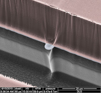
A scanning electron micrograph shows a nano-SPEAR suspended midway between layers of silicon (grey) and photoresist material (pink) that form a recording chamber for immobilized nematodes. The high-throughput technology developed at Rice University can be adapted for other small animals and could enhance data-gathering for disease characterization and drug interactions. Courtesy of the Robinson Lab

Here’s a link to and a citation for the paper,

Scalable electrophysiology in intact small animals with nanoscale suspended electrode arrays by Daniel L. Gonzales, Krishna N. Badhiwala, Daniel G. Vercosa, Benjamin W. Avants, Zheng Liu, Weiwei Zhong, & Jacob T. Robinson. Nature Nanotechnology (2017) doi:10.1038/nnano.2017.55 Published online 17 April 2017

This paper is behind a paywall.

Montreal Neuro creates a new paradigm for technology transfer?

It’s one heck of a Christmas present. Canadian businessmen Larry Tannenbaum and his wife Judy have given the Montreal Neurological Institute (Montreal Neuro), which is affiliated with McGill University, a $20M donation. From a Dec. 16, 2016 McGill University news release,

The Prime Minister of Canada, Justin Trudeau, was present today at the Montreal Neurological Institute and Hospital (MNI) for the announcement of an important donation of $20 million by the Larry and Judy Tanenbaum family. This transformative gift will help to establish the Tanenbaum Open Science Institute, a bold initiative that will facilitate the sharing of neuroscience findings worldwide to accelerate the discovery of leading edge therapeutics to treat patients suffering from neurological diseases.

‟Today, we take an important step forward in opening up new horizons in neuroscience research and discovery,” said Mr. Larry Tanenbaum. ‟Our digital world provides for unprecedented opportunities to leverage advances in technology to the benefit of science.  That is what we are celebrating here today: the transformation of research, the removal of barriers, the breaking of silos and, most of all, the courage of researchers to put patients and progress ahead of all other considerations.”

Neuroscience has reached a new frontier, and advances in technology now allow scientists to better understand the brain and all its complexities in ways that were previously deemed impossible. The sharing of research findings amongst scientists is critical, not only due to the sheer scale of data involved, but also because diseases of the brain and the nervous system are amongst the most compelling unmet medical needs of our time.

Neurological diseases, mental illnesses, addictions, and brain and spinal cord injuries directly impact 1 in 3 Canadians, representing approximately 11 million people across the country.

“As internationally-recognized leaders in the field of brain research, we are uniquely placed to deliver on this ambitious initiative and reinforce our reputation as an institution that drives innovation, discovery and advanced patient care,” said Dr. Guy Rouleau, Director of the Montreal Neurological Institute and Hospital and Chair of McGill University’s Department of Neurology and Neurosurgery. “Part of the Tanenbaum family’s donation will be used to incentivize other Canadian researchers and institutions to adopt an Open Science model, thus strengthening the network of like-minded institutes working in this field.”

What they don’t mention in the news release is that they will not be pursuing any patents (for five years according to one of the people in the video but I can’t find text to substantiate that time limit*; there are no time limits noted elsewhere) on their work. For this detail and others, you have to listen to the video they’ve created,

The CBC (Canadian Broadcasting Corporation) news online Dec. 16, 2016 posting (with files from Sarah Leavitt and Justin Hayward) adds a few personal details about Tannenbaum,

“Our goal is simple: to accelerate brain research and discovery to relieve suffering,” said Tanenbaum.

Tanenbaum, a Canadian businessman and chairman of Maple Leaf Sports and Entertainment, said many of his loved ones suffered from neurological disorders.

“I lost my mother to Alzheimer’s, my father to a stroke, three dear friends to brain cancer, and a brilliant friend and scientist to clinical depression,” said Tanenbaum.

He hopes the institute will serve as the template for science research across the world, a thought that Trudeau echoed.

“This vision around open science, recognizing the role that Canada can and should play, the leadership that Canadians can have in this initiative is truly, truly exciting,” said Trudeau.

The Neurological Institute says the pharmaceutical industry is supportive of the open science concept because it will provide crucial base research that can later be used to develop drugs to fight an array of neurological conditions.

Jack Stilgoe in a Dec. 16, 2016 posting on the Guardian blogs explains what this donation could mean (Note: Links have been removed),

With the help of Tanenbaum’s gift of 20 million Canadian dollars (£12million) the ‘Neuro’, the Montreal Neurological Institute and Hospital, is setting up an experiment in experimentation, an Open Science Initiative with the express purpose of finding out the best way to realise the potential of scientific research.

Governments in science-rich countries are increasingly concerned that they do not appear to reaping the economic returns they feel they deserve from investments in scientific research. Their favoured response has been to try to bridge what they see as a ‘valley of death’ between basic scientific research and industrial applications. This has meant more funding for ‘translational research’ and the flowering of technology transfer offices within universities.

… There are some success stories, particularly in the life sciences. Patents from the work of Richard Axel at Columbia University at one point brought the university almost $100 million per year. The University of Florida received more than $150 million for inventing Gatorade in the 1960s. The stakes are high in the current battle between Berkely and MIT/Harvard over who owns the rights to the CRISPR/Cas9 system that has revolutionised genetic engineering and could be worth billions.

Policymakers imagine a world in which universities pay for themselves just as a pharmaceutical research lab does. However, for critics of technology transfer, such stories blind us to the reality of university’s entrepreneurial abilities.

For most universities, evidence of their money-making prowess is, to put it charitably, mixed. A recent Bloomberg report shows how quickly university patent incomes plunge once we look beyond the megastars. In 2014, just 15 US universities earned 70% of all patent royalties. British science policy researchers Paul Nightingale and Alex Coad conclude that ‘Roughly 9/10 US universities lose money on their technology transfer offices… MIT makes more money from selling T-shirts than it does from licensing’. A report from the Brookings institute concluded that the model of technology transfer ‘is unprofitable for most universities and sometimes even risks alienating the private sector’. In the UK, the situation is even worse. Businesses who have dealings with universities report that their technology transfer offices are often unrealistic in negotiations. In many cases, academics are, like a small child who refuses to let others play with a brand new football, unable to make the most of their gifts. And areas of science outside the life sciences are harder to patent than medicines, sports drinks and genetic engineering techniques. Trying too hard to force science towards the market may be, to use the phrase of science policy professor Keith Pavitt, like pushing a piece of string.

Science policy is slowly waking up to the realisation that the value of science may lie in people and places rather than papers and patents. It’s an idea that the Neuro, with the help of Tanenbaum’s gift, is going to test. By sharing data and giving away intellectual property, the initiative aims to attract new private partners to the institute and build Montreal as a hub for knowledge and innovation. The hypothesis is that this will be more lucrative than hoarding patents.

This experiment is not wishful thinking. It will be scientifically measured. It is the job of Richard Gold, a McGill University law professor, to see whether it works. He told me that his first task is ‘to figure out what to counts… There’s going to be a gap between what we would like to measure and what we can measure’. However, he sees an open-mindedness among his colleagues that is unusual. Some are evangelists for open science; some are sceptics. But they share a curiosity about new approaches and a recognition of a problem in neuroscience: ‘We haven’t come up with a new drug for Parkinson’s in 30 years. We don’t even understand the biological basis for many of these diseases. So whatever we’re doing at the moment doesn’t work’. …

Montreal Neuro made news on the ‘open science’ front in January 2016 when it formally announced its research would be freely available and that researchers would not be pursuing patents (see my January 22, 2016 posting).

I recommend reading Stilgoe’s posting in its entirety and for those who don’t know or have forgotten, Prime Minister’s Trudeau’s family has some experience with mental illness. His mother has been very open about her travails. This makes his presence at the announcement perhaps a bit more meaningful than the usual political presence at a major funding announcement.

*The five-year time limit is confirmed in a Feb. 17, 2017 McGill University news release about their presentations at the AAAS (American Association for the Advancement of Science) 2017 annual meeting) on EurekAlert,

umpstarting Neurological Research through Open Science – MNI & McGill University

Friday, February 17, 2017, 1:30-2:30 PM/ Room 208

Neurological research is advancing too slowly according to Dr. Guy Rouleau, director of the Montreal Neurological Institute (MNI) of McGill University. To speed up discovery, MNI has become the first ever Open Science academic institution in the world. In a five-year experiment, MNI is opening its books and making itself transparent to an international group of social scientists, policymakers, industrial partners, and members of civil society. They hope, by doing so, to accelerate research and the discovery of new treatments for patients with neurological diseases, and to encourage other leading institutions around the world to consider a similar model. A team led by McGill Faculty of Law’s Professor Richard Gold will monitor and evaluate how well the MNI Open Science experiment works and provide the scientific and policy worlds with insight into 21st century university-industry partnerships. At this workshop, Rouleau and Gold will discuss the benefits and challenges of this open-science initiative.

Electrochemical measurements of biomolecules

This work comes from Finland and features some new nano shapes. From a Nov. 10, 2016 news item on phys.org,

Tomi Laurila’s research topic has many quirky names.

“Nanodiamond, nanohorn, nano-onion…,” lists off the Aalto University Professor, recounting the many nano-shapes of carbon. Laurila is using these shapes to build new materials: tiny sensors, only a few hundred nanometres across, that can achieve great things due to their special characteristics.

For one, the sensors can be used to enhance the treatment of neurological conditions. That is why Laurila, University of Helsinki Professor Tomi Taira and experts from HUS (the Hospital District of Helsinki and Uusimaa) are looking for ways to use the sensors for taking electrochemical measurements of biomolecules. Biomolecules are e.g. neurotransmitters such as glutamate, dopamine and opioids, which are used by nerve cells to communicate with each other.

A Nov. 10, 2016 Aalto University press release, which originated the news item, expands on the theme,

Most of the drugs meant for treating neurological diseases change the communication between nerve cells that is based on neurotransmitters. If we had real time and individual information on the operation of the neurotransmitter system, it would make it much easier to for example plan precise treatments’, explains Taira.

Due to their small size, carbon sensors can be taken directly next to a nerve cell, where the sensors will report what kind of neurotransmitter the cell is emitting and what kind of reaction it is inducing in other cells.

‘In practice, we are measuring the electrons that are moving in oxidation and reduction reactions’, Laurila explains the operating principle of the sensors.

‘The advantage of the sensors developed by Tomi and the others is their speed and small size. The probes used in current measurement methods can be compared to logs on a cellular scale – it’s impossible to use them and get an idea of the brain’s dynamic’, summarizes Taira.

Feedback system and memory traces

For the sensors, the journey from in vitro tests conducted in glass dishes and test tubes to in vivo tests and clinical use is long. However, the researchers are highly motivated.

‘About 165 million people are suffering from various neurological diseases in Europe alone. And because they are so expensive to treat, neurological diseases make up as much as 80 per cent of health care costs’, tells Taira.

Tomi Laurila believes that carbon sensors will have applications in fields such as optogenetics. Optogenetics is a recently developed method where a light-sensitive molecule is brought into a nerve cell so that the cell’s electric operation can then be turned on or off by stimulating it with light. A few years ago, a group of scientists proved in the scientific journal Nature that they had managed to use optogenetics to activate a memory trace that had been created previously due to learning. Using the same technique, researchers were able to demonstrate that with a certain type of Alzheimer’s, the problem is not that there are no memory traces being created, but that the brain cannot read the traces.

‘So the traces exist, and they can be activated by boosting them with light stimuli’, explains Taira but stresses that a clinical application is not yet a reality. However, clinical applications for other conditions may be closer by. One example is Parkinson’s disease. In Parkinson’s disease, the amount of dopamine starts to decrease in the cells of a particular brain section, which causes the typical symptoms such as tremors, rigidity and slowness of movement. With the sensors, the level of dopamine could be monitored in real time.

‘A sort of feedback system could be connected to it, so that it would react by giving an electric or optical stimulus to the cells, which would in turn release more dopamine’, envisions Taira.

‘Another application that would have an immediate clinical use is monitoring unconscious and comatose patients. With these patients, the level of glutamate fluctuates very much, and too much glutamate damages the nerve cell – online monitoring would therefore improve their treatment significantly.

Atom by atom

Manufacturing carbon sensors is definitely not a mass production process; it is slow and meticulous handiwork.

‘At this stage, the sensors are practically being built atom by atom’, summarises Tomi Laurila.

‘Luckily, we have many experts on carbon materials of our own. For example, the nanobuds of Professor Esko Kauppinen and the carbon films of Professor Jari Koskinen help with the manufacturing of the sensors. Carbon-based materials are mainly very compatible with the human body, but there is still little information about them. That’s why a big part of the work is to go through the electrochemical characterisation that has been done on different forms of carbon.’

The sensors are being developed and tested by experts from various fields, such as chemistry, materials science, modelling, medicine and imaging. Twenty or so articles have been published on the basic properties of the materials. Now, the challenge is to build them into geometries that are functional in a physiological environment. And taking measurements is not simple, either.

‘Brain tissue is delicate and doesn’t appreciate having objects being inserted in it. But if this were easy, someone would’ve already done it’, conclude the two.

I wish the researchers good luck.

Calming a synapse (part of a neuron) with graphene flakes

As we continue to colonize our own brains, there’s more news of graphene and neurons (see my Feb. 1, 2016 post featuring research from the same team in Italy featured in this post). A May 10, 2016 news item on ScienceDaily highlights work that could be used for epilepsy,

Innovative graphene technology to buffer the activity of synapses– this is the idea behind a recently-published study in the journal ACS Nano coordinated by the International School for Advanced Studies in Trieste (SISSA) and the University of Trieste. In particular, the study showed how effective graphene oxide flakes are at interfering with excitatory synapses, an effect that could prove useful in new treatments for diseases like epilepsy.

I guess the press release took a while to make its way through translation, here’s more from the April 10, 2016 SISSA (International School for Advanced Studies) press release (also on EurekAlert),

The laboratory of SISSA’s Laura Ballerini in collaboration with the University of Trieste, the University of Manchester and the University of Castilla -la Mancha, has discovered a new approach to modulating synapses. This methodology could be useful for treating diseases in which electrical nerve activity is altered. Ballerini and Maurizio Prato (University of Trieste) are the principal investigators of the project within the European flagship on graphene, a far-reaching 10-year international collaboration (one billion euros in funding) that studies innovative uses of the material.

Traditional treatments for neurological diseases generally include drugs that act on the brain or neurosurgery. Today however, graphene technology is showing promise for these types of applications, and is receiving increased attention from the scientific community. The method studied by Ballerini and colleagues uses “graphene nano-ribbons” (flakes) which buffer activity of synapses simply by being present.

“We administered aqueous solutions of graphene flakes to cultured neurons in ‘chronic’ exposure conditions, repeating the operation every day for a week. Analyzing functional neuronal electrical activity, we then traced the effect on synapses” says Rossana Rauti, SISSA researcher and first author of the study.

In the experiments, size of the flakes varied (10 microns or 80 nanometers) as well as the type of graphene: in one condition graphene was used, in another, graphene oxide. “The ‘buffering’ effect on synaptic activity happens only with smaller flakes of graphene oxide and not in other conditions,” says Ballerini. “The effect, in the system we tested, is selective for the excitatory synapses, while it is absent in inhibitory ones”

A Matter of Size

What is the origin of this selectivity? “We know that in principle graphene does not interact chemically with synapses in a significant way- its effect is likely due to the mere presence of synapses,” explains SISSA researcher and one of the study’s authors, Denis Scaini. “We do not yet have direct evidence, but our hypothesis is that there is a link with the sub-cellular organization of the synaptic space.”

A synapse is a contact point between one neuron and another where the nervous electrical signal “jumps” between a pre and post-synaptic unit. [emphasis mine] There is a small gap or discontinuity where the electrical signal is “translated” by a neurotransmitter and released by pre-synaptic termination into the extracellular space and reabsorbed by the postsynaptic space, to be translated again into an electrical signal. The access to this space varies depending on the type of synapses: “For the excitatory synapses, the structure’s organization allows higher exposure for the graphene flakes interaction, unlike inhibitory synapses, which are less physically accessible in this experimental model,” says Scaini.

Another clue that distance and size could be crucial in the process is found in the observation that graphene performs its function only in the oxidized form. “Normal graphene looks like a stretched and stiff sheet while graphene oxide appears crumpled, and thus possibly favoring interface with the synaptic space, ” adds Rauti.

Administering graphene flake solutions leaves the neurons alive and intact. For this reason the team thinks they could be used in biomedical applications for treating certain diseases. “We may imagine to target a drug by exploiting the apparent flakes’ selectivity for synapses, thus targeting directly the basic functional unit of neurons”concludes Ballerini.

That’s a nice description of neurons, synapses, and neurotransmitters.

Here’s a link to and a citation for the paper,

Graphene Oxide Nanosheets Reshape Synaptic Function in Cultured Brain Networks by Rossana Rauti, Neus Lozano, Veronica León, Denis Scaini†, Mattia Musto, Ilaria Rago, Francesco P. Ulloa Severino, Alessandra Fabbro, Loredana Casalis, Ester Vázquez, Kostas Kostarelos, Maurizio Prato, and Laura Ballerini. ACS Nano, 2016, 10 (4), pp 4459–4471
DOI: 10.1021/acsnano.6b00130 Publication Date (Web): March 31, 2016

Copyright © 2016 American Chemical Society

This paper is behind a paywall.

Better neuroprostheses for brain diseases and mental illneses

I don’t often get news releases from Sweden but I do on occasion and, sometimes, they even come in their original Swedish versions. In this case, Lund University sent me an English language version about their latest work making brain implants (neural prostheses) safer and effective. From a Sept. 29, 2015 Lund University news release (also on EurekAlert),

Neurons thrive and grow in a new type of nanowire material developed by researchers in Nanophysics and Ophthalmology at Lund University in Sweden. In time, the results might improve both neural and retinal implants, and reduce the risk of them losing their effectiveness over time, which is currently a problem

By implanting electrodes in the brain tissue one can stimulate or capture signals from different areas of the brain. These types of brain implants, or neuro-prostheses as they are sometimes called, are used to treat Parkinson’s disease and other neurological diseases.

They are currently being tested in other areas, such as depression, severe cases of autism, obsessive-compulsive disorders and paralysis. Another research track is to determine whether retinal implants are able to replace light-sensitive cells that die in cases of Retinitis Pigmentosa and other eye diseases.

However, there are severe drawbacks associated with today’s implants. One problem is that the body interprets the implants as foreign objects, resulting in an encapsulation of the electrode, which in turn leads to loss of signal.

One of the researchers explains the approach adopted by the research team (from the news release),

“Our nanowire structure prevents the cells that usually encapsulate the electrodes – glial cells – from doing so”, says Christelle Prinz, researcher in Nanophysics at Lund University in Sweden, who developed this technique together with Maria Thereza Perez, a researcher in Ophthalmology.

“I was very pleasantly surprised by these results. In previous in-vitro experiments, the glial cells usually attach strongly to the electrodes”, she says.

To avoid this, the researchers have developed a small substrate where regions of super thin nanowires are combined with flat regions. While neurons grow and extend processes on the nanowires, the glial cells primarily occupy the flat regions in between.

“The different types of cells continue to interact. This is necessary for the neurons to survive because the glial cells provide them with important molecules.”

So far, tests have only been done with cultured cells (in vitro) but hopefully they will soon be able to continue with experiments in vivo.

The substrate is made from the semiconductor material gallium phosphide where each outgrowing nanowire has a diameter of only 80 nanometres (billionths of a metre).

Here’s a link to and a citation for the paper,

Support of Neuronal Growth Over Glial Growth and Guidance of Optic Nerve Axons by Vertical Nanowire Arrays by Gaëlle Piret, Maria-Thereza Perez, and Christelle N. Prinz. ACS Appl. Mater. Interfaces, 2015, 7 (34), pp 18944–18948 DOI: 10.1021/acsami.5b03798 Publication Date (Web): August 11, 2015

Copyright © 2015 American Chemical Society

This paper appears to be open access as I was able to link to the PDF version.