Tag Archives: Northeastern University

Therapeutic nanoparticles for COVID-19 (disease caused by severe acute respiratory syndrome coronavirus 2 [SARS-CoV-2])—don’t hold your breath!

Last week (specifically, Tuesday, March 3, 2020), someone moved away from me during a class. I’d sneezed.

The irony of the situation is that of the two of us, with my lung issues I’d be the one most at risk of getting very ill and/or dying from COVID-19. ([1] Yes, I confirmed that was the reason she’d moved. [2] The therapeutic nanoparticles news item is coming later) Here are the risk factors to take into account (from the US Centers for Disease Control’s People at Risk for Serious Illness from COVID-19 webpage,

  • Older adults [Note: In one report the age range was stated as ‘people over 70’]
  • People who have serious chronic medical conditions like:
    • Heart disease
    • Diabetes
    • Lung disease

I’m not suggesting that all precautions be abandoned but it would seem that panic might not be called for. Jeremy Samuel Faust, an emergency medicine physician at Brigham and Women’s Hospital in Boston, faculty in its division of health policy and public health, and an instructor at Harvard Medical School, has written a calming March 4, 2020 article (COVID-19 Isn’t As Deadly As We Think; Don’t hoard masks and food. Figure out how to help seniors and the immunosuppressed stay healthy.) for Slate.com (Note: Links have been removed],

There are many compelling reasons to conclude that SARS-CoV-2, the virus that causes COVID-19, is not nearly as deadly as is currently feared. But COVID-19 panic has set in nonetheless. You can’t find hand sanitizer in stores, and N95 face masks are being sold online for exorbitant prices, never mind that neither is the best way to protect against the virus (yes, just wash your hands). The public is behaving as if this epidemic is the next Spanish flu, which is frankly understandable given that initial reports have staked COVID-19 mortality at about 2–3 percent, quite similar to the 1918 pandemic that killed tens of millions of people.

Allow me to be the bearer of good news. These frightening numbers are unlikely to hold. The true case fatality rate, known as CFR, of this virus is likely to be far lower than current reports suggest. Even some lower estimates, such as the 1 percent death rate recently mentioned by the directors of the National Institutes of Health and the Centers for Disease Control and Prevention, likely substantially overstate the case. [emphases mine]

But the most straightforward and compelling evidence that the true case fatality rate of SARS-CoV-2 is well under 1 percent comes not from statistical trends and methodological massage, but from data from the Diamond Princess cruise outbreak and subsequent quarantine off the coast of Japan.

A quarantined boat is an ideal—if unfortunate—natural laboratory to study a virus. Many variables normally impossible to control are controlled. We know that all but one patient boarded the boat without the virus. We know that the other passengers were healthy enough to travel. We know their whereabouts and exposures. While the numbers coming out of China are scary, we don’t know how many of those patients were already ill for other reasons. How many were already hospitalized for another life-threatening illness and then caught the virus? How many were completely healthy, caught the virus, and developed a critical illness? In the real world, we just don’t know.

Here’s the problem with looking at mortality numbers in a general setting: In China, 9 million people die per year, which comes out to 25,000 people every single day, or around 1.5 million people over the past two months alone. A significant fraction of these deaths results from diseases like emphysema/COPD, lower respiratory infections, and cancers of the lung and airway whose symptoms are clinically indistinguishable from the nonspecific symptoms seen in severe COVID-19 cases. And, perhaps unsurprisingly, the death rate from COVID-19 in China spiked precisely among the same age groups in which these chronic diseases first become common. During the peak of the outbreak in China in January and early February, around 25 patients per day were dying with SARS-CoV-2. Most were older patients in whom the chronic diseases listed above are prevalent. Most deaths occurred in Hubei province, an area in which lung cancer and emphysema/COPD are significantly higher than national averages in China, a country where half of all men smoke. How were doctors supposed to sort out which of those 25 out of 25,000 daily deaths were solely due to coronavirus, and which were more complicated? What we need to know is how many excess deaths this virus causes.

This all suggests that COVID-19 is a relatively benign disease for most young people, and a potentially devastating one for the old and chronically ill, albeit not nearly as risky as reported. Given the low mortality rate among younger patients with coronavirus—zero in children 10 or younger among hundreds of cases in China, and 0.2-0.4 percent in most healthy nongeriatric adults (and this is still before accounting for what is likely to be a high number of undetected asymptomatic cases)—we need to divert our focus away from worrying about preventing systemic spread among healthy people—which is likely either inevitable, or out of our control—and commit most if not all of our resources toward protecting those truly at risk of developing critical illness and even death: everyone over 70, and people who are already at higher risk from this kind of virus.

This still largely comes down to hygiene and isolation. But in particular, we need to focus on the right people and the right places. Nursing homes, not schools. Hospitals, not planes. We need to up the hygienic and isolation ante primarily around the subset of people who can’t simply contract SARS-CoV-2 and ride it out the way healthy people should be able to.

Curtis Kim of Vancouver, Canada, has created a website dedicated to tracking the statistics and information about COVID-19 in Canada and around the world. Here’s more about Kim and the website from a March 8, 2020 article by Megan Devlin for the Daily Hive,

Curtis Kim, who studied Computer Systems Technology at the British Columbia Institute of Technology [BCIT], launched the site this week after getting frustrated he was spending so much time on various websites looking for daily coronavirus updates.

The site breaks down the number of cases in Canada, the number of deaths (zero in Canada so far), and the number of people who have recovered. Further down, it provides the same stats for global COVID-19 cases.

There’s also a colour-coded map showing where cases are distributed, and a feed of latest news articles about the virus. Kim also included information about symptoms and how to contact Canadian public health services.

Kim is looking for work and given what I’ve seen of his COVID-19 website, he should have no difficulty. Although I think it might be an idea for him to explain how the ‘lethality’ rate on his website has been obtained since Faust who seems to have more directly relevant experience suggests in his article that the numbers are highly problematic,

My name is Curtis, recently graduated from BCIT. I thought it would be a serious worldwide issue considering the speed of the spread of this virus ever since this COVID-19 occurred. I frequently googled to check up the current status by going through many websites and felt I was wasting time repeatedly searching with same keywords and for sure I wasn’t the only one feeling this way. That’s why I started creating this application. It provides up-to-date information on the COVID-19 broken by province and country around the world, key contact information, and latest news. I like to help people, and want them to understand this situation easily using this application. Hopefully this situation improves soon.

If you have any further inquries about the information on this web application, Please reach me at curtisk808@gmail.com

At about 11:45 am (PT) on March 9, 2020, Kim’s COVID-19 website was updated to include one death in Canada. As you might expect, ti was a resident in a long term care home. Wanyee Li’s March 9, 2020 article for The Star presents the news,

A resident at a long-term care home experiencing a COVID-19 outbreak in North Vancouver has died after contracting the virus, B.C. health officials confirmed Monday [March 9, 2020].

It is the first reported death in Canada linked to the virus.

The outbreak at the Lynn Valley Care Centre has so far been linked to three community transmission cases of the virus.

Provincial Health Officer Dr. Bonnie Henry confirmed five new cases of COVID-19 in B.C. on Monday [March 9, 2020], putting the total in the province at 32.

The five new cases include one health-care worker, two people who are close contacts of an existing case, one person who recently returned from travel to Iran and another who was in Italy recently.

Officials are conducting an investigation into the three community transmission cases at the long-term care home to determine how a health care worker contracted the virus.

I looked up the population figures for the province of British Columbia (BC; Wikipedia entry for Demographics of British Columbia). As of the 2016 census, there were 4,648,055 people in the province. Assuming that population number holds, 67 cases in all of Canada (with 27 cases in BC) of COVID-19 don’t seem like big numbers.

We should definitely take precautions and be careful but there’s no need to panic.

Nanoparticles and a COVID-19 treatment?

Don’t hold you breath. This March 5, 2020 news item on Nanowerk is speculative,

There is no vaccine or specific treatment for COVID-19, the disease caused by the severe acute respiratory syndrome coronavirus 2, or SARS-CoV-2.

Since the outbreak began in late 2019, researchers have been racing to learn more about SARS-CoV-2, which is a strain from a family of viruses known as coronavirus for their crown-like shape.

Northeastern Ûniversity] chemical engineer Thomas Webster, who specializes in developing nano-scale medicine and technology to treat diseases, is part of a contingency of scientists that are contributing ideas and technology to the Centers for Disease Control and Prevention to fight the COVID-19 outbreak.

The idea of using nanoparticles, Webster says, is that the virus behind COVID-19 consists of a structure of a similar scale as his nanoparticles. At that scale, matter is ultra-small, about ten thousand times smaller than the width of a single strand of hair.

..
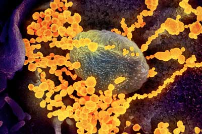
This scanning electron microscope image shows SARS-CoV-2 (round gold objects) emerging from the surface of cells cultured in the lab. SARS-CoV-2, also known as 2019-nCoV, is the virus that causes COVID-19. The virus shown was isolated from a patient in the U.S. (Image: NIAID-RML)

A March 4, 2020 Northeastern University news release by Roberto Molar Candanosa, which originated the news item, delves further into Webster’s thinking process,

Webster is proposing particles of similar sizes that could attach to SARS-CoV-2 viruses, disrupting their structure with a combination of infrared light treatment. That structural change would then halt the ability of the virus to survive and reproduce in the body.

“You have to think in this size range,” says Webster, Art Zafiropoulo Chair of chemical engineering at Northeastern. “In the nanoscale size range, if you want to detect viruses, if you want to deactivate them.”

Finding and neutralizing viruses with nanomedicine is at the core of what Webster and other researchers call theranostics, which focuses on combining therapy and diagnosis. Using that approach, his lab has specialized in nanoparticles to fight the microbes that cause influenza and tuberculosis. 

“It’s not just having one approach to detect whether you have a virus and another approach to use it as a therapy,” he says, “but having the same particle, the same approach, for both your detection and therapy.”

I wish Webster good luck. As for the rest us, let’s wash our hands and keep calm.
Improving batteries with cellulosic nanomaterials

This is a cellulose nanocrystal (CNC) story and in this story it’s derived from trees as opposed to banana skins or carrots or … A February 19, 2020 news item on Nanowerk announces CNC research from Northeastern University (Massachusetts, US),

Nature isn’t always generous with its secrets. That’s why some researchers look into unusual places for solutions to our toughest challenges, from powerful antibiotics hiding in the guts of tiny worms, to swift robots inspired by bats.

Now, Northeastern researchers have taken to the trees to look for ways to make new sustainable materials from abundant natural resources—specifically, within the chemical structure of microfibers that make up wood.

A team led by Hongli (Julie) Zhu, an assistant professor of mechanical and industrial engineering at Northeastern, is using unique nanomaterials derived from cellulose to improve the large and expensive kind of batteries needed to store renewable energy harnessed from sources such as sunlight and the wind.

A February 18, 2020 Northeastern University news release by Roberto Molar Candanosa, which originated the news item, provides more detail (Note: Links have been removed),

Cellulose, the most abundant natural polymer on Earth, is also the most important structural component of plants. It contains important molecular structures to improve batteries, reduce plastic pollution, and power the sort of electrical grids that could support entire communities with renewable energy, Zhu says.  

“We try to use polymers from wood, from bark, from seeds, from flowers, bacteria, green tea—from these kinds of plants to replace plastic,”  Zhu says.

One of the main challenges in storing energy from the sun, wind, and other types of renewables is that variation in factors such as the weather lead to inconsistent sources of power. 

That’s where batteries with large capacity come in. But storing the large amounts of energy that sunlight and the wind are able to provide requires a special kind of device.

The most advanced batteries to do that are called flow batteries, and are made with vanadium ions dissolved in acid in two separate tanks—one with a substance of negatively charged ions, and one with positive ones. The two solutions are continuously pumped from the tank into a cell, which functions like an engine for the battery. 

These substances are always separated by a special membrane that ensures that they exchange positive hydrogen ions without flowing into each other. That selective exchange of ions is the basis for the ability of the battery to charge and discharge energy. 

Flow batteries are ideal devices in which to store solar and wind energy because they can be tweaked to increase the amount of energy stored without compromising the amount of energy that can be generated. The bigger the tanks, the more energy the battery can store from non-polluting and practically inexhaustible resources.

But manufacturing them requires several moving pieces of hardware. As the membrane separating the two flowing substances decays, it can cause the vanadium ions from the solution to mix. That crossover reduces the stability of a battery, along with its capacity to store energy.

Zhu says the limited efficiency of that membrane, combined with  its high cost, are the main factors keeping flow batteries from being widely used in large-scale grids.

In a recent paper, Zhu reported that a new membrane made with cellulose nanocrystals demonstrates superior efficiency compared to other membranes used commonly in the market. The team tested different membranes made from cellulose nanocrystals to make flow batteries cheaper.

“The cost of our membrane per square meter is 147.68 US dollars, ” Zhu says, adding that her calculations do not include costs associated with marketing. “The price quote for the commercialized Nafion membrane is $1,321 per square meter.”

Their tests also showed that the membranes, made with support from the Rogers Corporation and its Innovation Center at Northeastern’s Kostas Research Institute, can offer substantially longer battery lifetimes than other membranes. 

Zhu’s naturally derived membrane is especially efficient because its cellular structure contains thousands of hydroxyl groups, which involve bonds of hydrogen and oxygen that make it easy for water to be transported in plants and trees. 

In flow batteries, that molecular makeup speeds the transport of protons as they flow through the membrane.

The membrane also consists of another polymer known as poly(vinylidene fluoride-hexafluoropropylene), which prevents the negatively and positively charged acids from mixing with each other. 

“For these materials, one of the challenges is that it is difficult to find a polymer that is proton conductive and that is also a material that is very stable in the flowing acid,” Zhu says. 

Because these materials are practically everywhere, membranes made with it can be easily put together at large scales needed for complex power grids. 

Unlike other expensive artificial materials that need to be concocted in a lab, cellulose can be extracted from natural sources including algae, solid waste, and bacteria. 

“A lot of material in nature is a composite, and if we disintegrate its components, we can use it to extract cellulose,” Zhu says. “Like waste from our yard, and a lot of solid waste that we don’t always know what to do with.” 

Here’s a link to and a citation for the paper mentioned in the news release,

Stable and Highly Ion-Selective Membrane Made from Cellulose Nanocrystals for Aqueous Redox Flow Batteries by Alolika Mukhopadhyay, Zheng Cheng, Avi Natan, Yi Ma, Yang Yang, Daxian Cao, Wei Wan, Hongli Zhu. Nano Lett. 2019, 19, 12, 8979-8989 DOI: https://doi.org/10.1021/acs.nanolett.9b03964Publication Date:November 8, 2019 Copyright © 2019 American Chemical Society

This paper is behind a paywall.

Audio map of 24 emotions

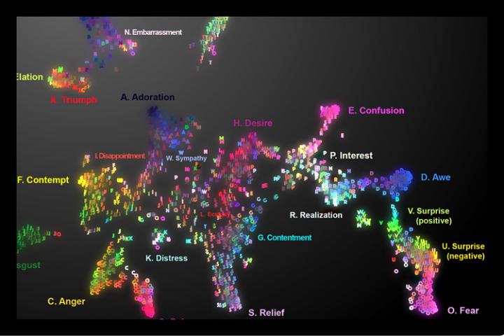
Caption: Audio map of vocal bursts across 24 emotions. To visit the online map and hear the sounds, go to https://s3-us-west-1.amazonaws.com/vocs/map.html# and move the cursor across the map. Credit: Courtesy of Alan Cowen

The real map, not the the image of the map you see above, offers a disconcerting (for me, anyway) experience. Especially since I’ve just finished reading Lisa Feldman Barrett’s 2017 book, How Emotions are Made, where she presents her theory of ‘constructed emotion. (There’s more about ‘constructed emotion’ later in this post.)

Moving on to the story about the ‘auditory emotion map’ in the headline, a February 4, 2019 University of California at Berkeley news release by Yasmin Anwar (also on EurekAlert but published on Feb. 5, 2019) describes the work,

Ooh, surprise! Those spontaneous sounds we make to express everything from elation (woohoo) to embarrassment (oops) say a lot more about what we’re feeling than previously understood, according to new research from the University of California, Berkeley.

Proving that a sigh is not just a sigh [a reference to the song, As Time Goes By? The lyric is “a kiss is still a kiss, a sigh is just a sigh …”], UC Berkeley scientists conducted a statistical analysis of listener responses to more than 2,000 nonverbal exclamations known as “vocal bursts” and found they convey at least 24 kinds of emotion. Previous studies of vocal bursts set the number of recognizable emotions closer to 13.

The results, recently published online in the American Psychologist journal, are demonstrated in vivid sound and color on the first-ever interactive audio map of nonverbal vocal communication.

“This study is the most extensive demonstration of our rich emotional vocal repertoire, involving brief signals of upwards of two dozen emotions as intriguing as awe, adoration, interest, sympathy and embarrassment,” said study senior author Dacher Keltner, a psychology professor at UC Berkeley and faculty director of the Greater Good Science Center, which helped support the research.

For millions of years, humans have used wordless vocalizations to communicate feelings that can be decoded in a matter of seconds, as this latest study demonstrates.

“Our findings show that the voice is a much more powerful tool for expressing emotion than previously assumed,” said study lead author Alan Cowen, a Ph.D. student in psychology at UC Berkeley.

On Cowen’s audio map, one can slide one’s cursor across the emotional topography and hover over fear (scream), then surprise (gasp), then awe (woah), realization (ohhh), interest (ah?) and finally confusion (huh?).

Among other applications, the map can be used to help teach voice-controlled digital assistants and other robotic devices to better recognize human emotions based on the sounds we make, he said.

As for clinical uses, the map could theoretically guide medical professionals and researchers working with people with dementia, autism and other emotional processing disorders to zero in on specific emotion-related deficits.

“It lays out the different vocal emotions that someone with a disorder might have difficulty understanding,” Cowen said. “For example, you might want to sample the sounds to see if the patient is recognizing nuanced differences between, say, awe and confusion.”

Though limited to U.S. responses, the study suggests humans are so keenly attuned to nonverbal signals – such as the bonding “coos” between parents and infants – that we can pick up on the subtle differences between surprise and alarm, or an amused laugh versus an embarrassed laugh.

For example, by placing the cursor in the embarrassment region of the map, you might find a vocalization that is recognized as a mix of amusement, embarrassment and positive surprise.

A tour through amusement reveals the rich vocabulary of laughter and a spin through the sounds of adoration, sympathy, ecstasy and desire may tell you more about romantic life than you might expect,” said Keltner.

Researchers recorded more than 2,000 vocal bursts from 56 male and female professional actors and non-actors from the United States, India, Kenya and Singapore by asking them to respond to emotionally evocative scenarios.

Next, more than 1,000 adults recruited via Amazon’s Mechanical Turk online marketplace listened to the vocal bursts and evaluated them based on the emotions and meaning they conveyed and whether the tone was positive or negative, among several other characteristics.

A statistical analysis of their responses found that the vocal bursts fit into at least two dozen distinct categories including amusement, anger, awe, confusion, contempt, contentment, desire, disappointment, disgust, distress, ecstasy, elation, embarrassment, fear, interest, pain, realization, relief, sadness, surprise (positive) surprise (negative), sympathy and triumph.

For the second part of the study, researchers sought to present real-world contexts for the vocal bursts. They did this by sampling YouTube video clips that would evoke the 24 emotions established in the first part of the study, such as babies falling, puppies being hugged and spellbinding magic tricks.

This time, 88 adults of all ages judged the vocal bursts extracted from YouTube videos. Again, the researchers were able to categorize their responses into 24 shades of emotion. The full set of data were then organized into a semantic space onto an interactive map.

“These results show that emotional expressions color our social interactions with spirited declarations of our inner feelings that are difficult to fake, and that our friends, co-workers, and loved ones rely on to decipher our true commitments,” Cowen said.

The writer assumes that emotions are pre-existing. Somewhere, there’s happiness, sadness, anger, etc. It’s the pre-existence that Lisa Feldman Barret challenges with her theory that we construct our emotions (from her Wikipedia entry),

She highlights differences in emotions between different cultures, and says that emotions “are not triggered; you create them. They emerge as a combination of the physical properties of your body, a flexible brain that wires itself to whatever environment it develops in, and your culture and upbringing, which provide that environment.”

You can find Barrett’s December 6, 2017 TED talk here wheres she explains her theory in greater detail. One final note about Barrett, she was born and educated in Canada and now works as a Professor of Psychology at Northeastern University, with appointments at Harvard Medical School and Massachusetts General Hospital at Northeastern University in Boston, Massachusetts; US.

A February 7, 2019 by Mark Wilson for Fast Company delves further into the 24 emotion audio map mentioned at the outset of this posting (Note: Links have been removed),

Fear, surprise, awe. Desire, ecstasy, relief.

These emotions are not distinct, but interconnected, across the gradient of human experience. At least that’s what a new paper from researchers at the University of California, Berkeley, Washington University, and Stockholm University proposes. The accompanying interactive map, which charts the sounds we make and how we feel about them, will likely persuade you to agree.

At the end of his article, Wilson also mentions the Dalai Lama and his Atlas of Emotions, a data visualization project, (featured in Mark Wilson’s May 13, 2016 article for Fast Company). It seems humans of all stripes are interested in emotions.

Here’s a link to and a citation for the paper about the audio map,

Mapping 24 emotions conveyed by brief human vocalization by Cowen, Alan S;, Elfenbein, Hillary Ange;, Laukka, Petri; Keltner, Dacher. American Psychologist, Dec 20, 2018, No Pagination Specified DOI: 10.1037/amp0000399


This paper is behind a paywall.

Carbon nanotubes for water desalination

In discussions about water desalination and carbon nanomaterials,  it’s graphene that’s usually mentioned these days. By contrast, scientists from the US Department of Energy’s Lawrence Livermore National Laboratory (LLNL) have turned to carbon nanotubes,

There are two news items about the work at LLNL on ScienceDaily, this first one originated by the American Association for the Advancement of Science (AAAS) offers a succinct summary of the work (from an August 24, 2017 news item on ScienceDaily,

At just the right size, carbon nanotubes can filter water with better efficiency than biological proteins, a new study reveals. The results could pave the way to new water filtration systems, at a time when demands for fresh water pose a global threat to sustainable development.

A class of biological proteins, called aquaporins, is able to effectively filter water, yet scientists have not been able to manufacture scalable systems that mimic this ability. Aquaporins usually exhibit channels for filtering water molecules at a narrow width of 0.3 nanometers, which forces the water molecules into a single-file chain.

Here, Ramya H. Tunuguntla and colleagues experimented with nanotubes of different widths to see which ones are best for filtering water. Intriguingly, they found that carbon nanotubes with a width of 0.8 nanometers outperformed aquaporins in filtering efficiency by a factor of six.

These narrow carbon nanotube porins (nCNTPs) were still slim enough to force the water molecules into a single-file chain. The researchers attribute the differences between aquaporins and nCNTPS to differences in hydrogen bonding — whereas pore-lining residues in aquaporins can donate or accept H bonds to incoming water molecules, the walls of CNTPs cannot form H bonds, permitting unimpeded water flow.

The nCNTPs in this study maintained permeability exceeding that of typical saltwater, only diminishing at very high salt concentrations. Lastly, the team found that by changing the charges at the mouth of the nanotube, they can alter the ion selectivity. This advancement is highlighted in a Perspective [in Science magazine] by Zuzanna Siwy and Francesco Fornasiero.

The second Aug. 24, 2017 news item on ScienceDaily offers a more technical  perspective,

Lawrence Livermore scientists, in collaboration with researchers at Northeastern University, have developed carbon nanotube pores that can exclude salt from seawater. The team also found that water permeability in carbon nanotubes (CNTs) with diameters smaller than a nanometer (0.8 nm) exceeds that of wider carbon nanotubes by an order of magnitude.

The nanotubes, hollow structures made of carbon atoms in a unique arrangement, are more than 50,000 times thinner than a human hair. The super smooth inner surface of the nanotube is responsible for their remarkably high water permeability, while the tiny pore size blocks larger salt ions.

There’s a rather lovely illustration for this work,

An artist’s depiction of the promise of carbon nanotube porins for desalination. The image depicts a stylized carbon nanotube pipe that delivers clean desalinated water from the ocean to a kitchen tap. Image by Ryan Chen/LLNL

An Aug. 24, 2017 LLNL news release (also on EurekAlert), which originated the second news item, proceeds

Increasing demands for fresh water pose a global threat to sustainable development, resulting in water scarcity for 4 billion people. Current water purification technologies can benefit from the development of membranes with specialized pores that mimic highly efficient and water selective biological proteins.

“We found that carbon nanotubes with diameters smaller than a nanometer bear a key structural feature that enables enhanced transport. The narrow hydrophobic channel forces water to translocate in a single-file arrangement, a phenomenon similar to that found in the most efficient biological water transporters,” said Ramya Tunuguntla, an LLNL postdoctoral researcher and co-author of the manuscript appearing in the Aug. 24 [2017]edition of Science.

Computer simulations and experimental studies of water transport through CNTs with diameters larger than 1 nm showed enhanced water flow, but did not match the transport efficiency of biological proteins and did not separate salt efficiently, especially at higher salinities. The key breakthrough achieved by the LLNL team was to use smaller-diameter nanotubes that delivered the required boost in performance.

“These studies revealed the details of the water transport mechanism and showed that rational manipulation of these parameters can enhance pore efficiency,” said Meni Wanunu, a physics professor at Northeastern University and co-author on the study.

“Carbon nanotubes are a unique platform for studying molecular transport and nanofluidics,” said Alex Noy, LLNL principal investigator on the CNT project and a senior author on the paper. “Their sub-nanometer size, atomically smooth surfaces and similarity to cellular water transport channels make them exceptionally suited for this purpose, and it is very exciting to make a synthetic water channel that performs better than nature’s own.”

This discovery by the LLNL scientists and their colleagues has clear implications for the next generation of water purification technologies and will spur a renewed interest in development of the next generation of high-flux membranes.

Here’s a link to and a citation for the paper,

Enhanced water permeability and tunable ion selectivity in subnanometer carbon nanotube porins by Ramya H. Tunuguntla, Robert Y. Henley, Yun-Chiao Yao, Tuan Anh Pham, Meni Wanunu, Aleksandr Noy. Science 25 Aug 2017: Vol. 357, Issue 6353, pp. 792-796 DOI: 10.1126/science.aan2438

This paper is behind a paywall.

And, Northeastern University issued an August 25, 2017 news release (also on EurekAlert) by Allie Nicodemo,

Earth is 70 percent water, but only a tiny portion—0.007 percent—is available to drink.

As potable water sources dwindle, global population increases every year. One potential solution to quenching the planet’s thirst is through desalinization—the process of removing salt from seawater. While tantalizing, this approach has always been too expensive and energy intensive for large-scale feasibility.

Now, researchers from Northeastern have made a discovery that could change that, making desalinization easier, faster and cheaper than ever before. In a paper published Thursday [August 24, 2017] in Science, the group describes how carbon nanotubes of a certain size act as the perfect filter for salt—the smallest and most abundant water contaminant.

Filtering water is tricky because water molecules want to stick together. The “H” in H2O is hydrogen, and hydrogen bonds are strong, requiring a lot of energy to separate. Water tends to bulk up and resist being filtered. But nanotubes do it rapidly, with ease.

A carbon nanotube is like an impossibly small rolled up sheet of paper, about a nanometer in diameter. For comparison, the diameter of a human hair is 50 to 70 micrometers—50,000 times wider. The tube’s miniscule size, exactly 0.8 nm, only allows one water molecule to pass through at a time. This single-file lineup disrupts the hydrogen bonds, so water can be pushed through the tubes at an accelerated pace, with no bulking.

“You can imagine if you’re a group of people trying to run through the hallway holding hands, it’s going to be a lot slower than running through the hallway single-file,” said co-author Meni Wanunu, associate professor of physics at Northeastern. Wanunu and post doctoral student Robert Henley collaborated with scientists at the Lawrence Livermore National Laboratory in California to conduct the research.

Scientists led by Aleksandr Noy at Lawrence Livermore discovered last year [2016] that carbon nanotubes were an ideal channel for proton transport. For this new study, Henley brought expertise and technology from Wanunu’s Nanoscale Biophysics Lab to Noy’s lab, and together they took the research one step further.

In addition to being precisely the right size for passing single water molecules, carbon nanotubes have a negative electric charge. This causes them to reject anything with the same charge, like the negative ions in salt, as well as other unwanted particles.

“While salt has a hard time passing through because of the charge, water is a neutral molecule and passes through easily,” Wanunu said. Scientists in Noy’s lab had theorized that carbon nanotubes could be designed for specific ion selectivity, but they didn’t have a reliable system of measurement. Luckily, “That’s the bread and butter of what we do in Meni’s lab,” Henley said. “It created a nice symbiotic relationship.”

“Robert brought the cutting-edge measurement and design capabilities of Wanunu’s group to my lab, and he was indispensable in developing a new platform that we used to measure the ion selectivity of the nanotubes,” Noy said.

The result is a novel system that could have major implications for the future of water security. The study showed that carbon nanotubes are better at desalinization than any other existing method— natural or man-made.

To keep their momentum going, the two labs have partnered with a leading water purification organization based in Israel. And the group was recently awarded a National Science Foundation/Binational Science Foundation grant to conduct further studies and develop water filtration platforms based on their new method. As they continue the research, the researchers hope to start programs where students can learn the latest on water filtration technology—with the goal of increasing that 0.007 percent.

As is usual in these cases there’s a fair degree of repetition but there’s always at least one nugget of new information, in this case, a link to Israel. As I noted many times, the Middle East is experiencing serious water issues. My most recent ‘water and the Middle East’ piece is an August 21, 2017 post about rainmaking at the Masdar Institute in United Arab Emirates. Approximately 50% of the way down the posting, I mention Israel and Palestine’s conflict over water.

‘Robomussels’ for climate change

These ‘robomussels’ are not voting but they are being used to monitor mussel bed habitats according to an Oct. 17, 2016 news item on ScienceDaily,

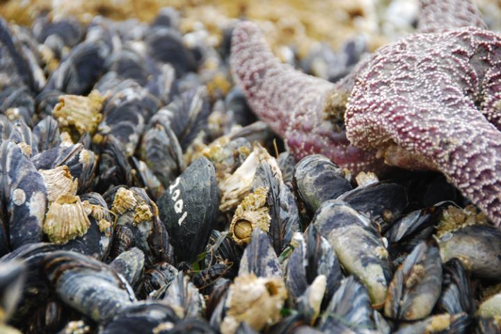
Tiny robots have been helping researchers study how climate change affects biodiversity. Developed by Northeastern University scientist Brian Helmuth, the “robomussels” have the shape, size, and color of actual mussels, with miniature built-in sensors that track temperatures inside the mussel beds.

Caption: This is a robomussel, seen among living mussels and other sea creatures. Credit: Allison Matzelle

Caption: This is a robomussel, seen among living mussels and other sea creatures. Credit: Allison Matzelle

An Oct. 12, 2016 Northeastern University news release (also on EurekAlert), which originated the news item, describes a project some 20 years in the making,

For the past 18 years, every 10 to 15 minutes, Helmuth and a global research team of 48 scientists have used robomussels to track internal body temperature, which is determined by the temperature of the surrounding air or water, and the amount of solar radiation the devices absorb. They place the robots inside mussel beds in oceans around the globe and record temperatures. The researchers have built a database of nearly two decades worth of data enabling scientists to pinpoint areas of unusual warming, intervene to help curb damage to vital marine ecosystems, and develop strategies that could prevent extinction of certain species.

Housed at Northeastern’s Marine Science Center in Nahant, Massachusetts, this largest-ever database is not only a remarkable way to track the effects of climate change, the findings can also reveal emerging hotspots so policymakers and scientists can step in and relieve stressors such as erosion and water acidification before it’s too late.

“They look exactly like mussels but they have little green blinking lights in them,” says Helmuth. “You basically pluck out a mussel and then glue the device to the rock right inside the mussel bed. They enable us to link our field observations with the physiological impact of global climate change on these ecologically and economically important animals.”

For ecological forecasters such as Helmuth, mussels act as a barometer of climate change. That’s because they rely on external sources of heat such as air temperature and sun exposure for their body heat and thrive, or not, depending on those conditions. Using fieldwork along with mathematical and computational models, Helmuth forecasts the patterns of growth, reproduction, and survival of mussels in intertidal zones.

Over the years, he and his colleagues have found surprises: “Our expectations of where to look for the effects of climate change in nature are more complex than anticipated,” says Helmuth. For example, in an earlier paper in the journal Science, his team found that hotspots existed not only at the southern end of the species’ distribution, in this case, southern California; they also existed at sites up north, in Oregon and Washington state.

“These datasets tell us when and where to look for the effects of climate change,” he says. “Without them we could miss early warning signs of trouble.”

The robomussels’ near-continuous measurements serve as an early warning system. “If we start to see sites where the animals are regularly getting to temperatures that are right below what kills them, we know that any slight increase is likely to send them over the edge, and we can act,” says Helmuth.

It’s not only the mussels that may be pulled back from the brink. The advance notice could inform everything from maintaining the biodiversity of coastal systems to determining the best–and worst–places to locate mussel farms.

“Losing mussel beds is essentially like clearing a forest,” says Helmuth. “If they go, everything that’s living in them will go. They are a major food supply for many species, including lobsters and crabs. They also function as filters along near-shore waters, clearing huge amounts of particulates. So losing them can affect everything from the growth of species we care about because we want to eat them to water clarity to biodiversity of all the tiny animals that live on the insides of the beds.”

Here’s a link to and a citation for the paper,

Long-term, high frequency in situ measurements of intertidal mussel bed temperatures using biomimetic sensors by Brian Helmuth, Francis Choi, Gerardo Zardi.  Scientific Data 3, Article number: 160087 (2016) doi:10.1038/sdata.2016.87 Published online: 11 October 2016

This paper is open access.

Nanotechnology, tobacco plants, and the Ebola virus

Before presenting information about the current Ebola crisis and issues with vaccines and curatives, here’s a description of the disease from its Wikipedia entry,

Ebola virus disease (EVD) or Ebola hemorrhagic fever (EHF) is a disease of humans and other primates caused by an ebola virus. Symptoms start two days to three weeks after contracting the virus, with a fever, sore throat, muscle pain, and headaches. Typically nausea, vomiting, and diarrhea follow, along with decreased functioning of the liver and kidneys. Around this time, affected people may begin to bleed both within the body and externally. [1]

As for the current crisis in countries situated on the west coast of the African continent, there’s this from an Aug. 14, 2014 news item on ScienceDaily,

The outbreak of Ebola virus disease that has claimed more than 1,000 lives in West Africa this year poses a serious, ongoing threat to that region: the spread to capital cities and Nigeria — Africa’s most populous nation — presents new challenges for healthcare professionals. The situation has garnered significant attention and fear around the world, but proven public health measures and sharpened clinical vigilance will contain the epidemic and thwart a global spread, according to a new commentary by Anthony S. Fauci, M.D., director of the National Institute of Allergy and Infectious Diseases (NIAID), part of the National Institutes of Health.

Dr. Fauci’s Aug. 13, 2014 commentary (open access) in the New England Journal of Medicine provides more detail (Note: A link has been removed),

An outbreak of Ebola virus disease (EVD) has jolted West Africa, claiming more than 1000 lives since the virus emerged in Guinea in early 2014 (see figure) Ebola Virus Cases and Deaths in West Africa (Guinea, Liberia, Nigeria, and Sierra Leone), as of August 11, 2014 (Panel A), and Over Time (Panel B).). The rapidly increasing numbers of cases in the African countries of Guinea, Liberia, and Sierra Leone have had public health authorities on high alert throughout the spring and summer. More recent events including the spread of EVD to Nigeria (Africa’s most populous country) and the recent evacuation to the United States of two American health care workers with EVD have captivated the world’s attention and concern. Health professionals and the general public are struggling to comprehend these unfolding dynamics and to separate misinformation and speculation from truth.

In early 2014, EVD emerged in a remote region of Guinea near its borders with Sierra Leone and Liberia. Since then, the epidemic has grown dramatically, fueled by several factors. First, Guinea, Sierra Leone, and Liberia are resource-poor countries already coping with major health challenges, such as malaria and other endemic diseases, some of which may be confused with EVD. Next, their borders are porous, and movement between countries is constant. Health care infrastructure is inadequate, and health workers and essential supplies including personal protective equipment are scarce. Traditional practices, such as bathing of corpses before burial, have facilitated transmission. The epidemic has spread to cities, which complicates tracing of contacts. Finally, decades of conflict have left the populations distrustful of governing officials and authority figures such as health professionals. Add to these problems a rapidly spreading virus with a high mortality rate, and the scope of the challenge becomes clear.

Although the regional threat of Ebola in West Africa looms large, the chance that the virus will establish a foothold in the United States or another high-resource country remains extremely small. Although global air transit could, and most likely will, allow an infected, asymptomatic person to board a plane and unknowingly carry Ebola virus to a higher-income country, containment should be readily achievable. Hospitals in such countries generally have excellent capacity to isolate persons with suspected cases and to care for them safely should they become ill. Public health authorities have the resources and training necessary to trace and monitor contacts. Protocols exist for the appropriate handling of corpses and disposal of biohazardous materials. In addition, characteristics of the virus itself limit its spread. Numerous studies indicate that direct contact with infected bodily fluids — usually feces, vomit, or blood — is necessary for transmission and that the virus is not transmitted from person to person through the air or by casual contact. Isolation procedures have been clearly outlined by the Centers for Disease Control and Prevention (CDC). A high index of suspicion, proper infection-control practices, and epidemiologic investigations should quickly limit the spread of the virus.

Fauci’s article makes it clear that public concerns are rising in the US and I imagine that’s true of Canada too and many other parts of the world, not to mention the countries currently experiencing the EVD outbreak. In the midst of all this comes a US Food and Drug Administration (FDA) warning as per an Aug. 15, 2014 news item (originated by Reuters reporter Toni Clarke) on Nanowerk,

The U.S. Food and Drug Administration said on Thursday [Aug. 14, 2014] it has become aware of products being sold online that fraudulently claim to prevent or treat Ebola.

The FDA’s warning comes on the heels of comments by Nigeria’s top health official, Onyebuchi Chukwu, who reportedly said earlier Thursday [Aug. 14, 2014] that eight Ebola patients in Lagos, the country’s capital, will receive an experimental treatment containing nano-silver.

Erica Jefferson, a spokeswoman for the FDA, said she could not provide any information about the product referenced by the Nigerians.

The Aug. 14,  2014 FDA warning reads in part,

The U.S. Food and Drug Administration is advising consumers to be aware of products sold online claiming to prevent or treat the Ebola virus. Since the outbreak of the Ebola virus in West Africa, the FDA has seen and received consumer complaints about a variety of products claiming to either prevent the Ebola virus or treat the infection.

There are currently no FDA-approved vaccines or drugs to prevent or treat Ebola. Although there are experimental Ebola vaccines and treatments under development, these investigational products are in the early stages of product development, have not yet been fully tested for safety or effectiveness, and the supply is very limited. There are no approved vaccines, drugs, or investigational products specifically for Ebola available for purchase on the Internet. By law, dietary supplements cannot claim to prevent or cure disease.

As per the FDA’s reference to experimental vaccines, an Aug. 6, 2014 article by Caroline Chen, Mark Niquette, Mark Langreth, and Marie French for Bloomberg describes the ZMapp vaccine/treatment (Note: Links have been removed),

On a small plot of land incongruously tucked amid a Kentucky industrial park sit five weather-beaten greenhouses. At the site, tobacco plants contain one of the most promising hopes for developing an effective treatment for the deadly Ebola virus.

The plants contain designer antibodies developed by San Diego-based Mapp Biopharmaceutical Inc. and are grown in Kentucky by a unit of Reynolds American Inc. Two stricken U.S. health workers received an experimental treatment containing the antibodies in Liberia last week. Since receiving doses of the drug, both patients’ conditions have improved.

Tobacco plant-derived medicines, which are also being developed by a company whose investors include Philip Morris International Inc., are part of a handful of cutting edge plant-based treatments that are in the works for everything from pandemic flu to rabies using plants such as lettuce, carrots and even duckweed. While the technique has existed for years, the treatments have only recently begun to reach the marketplace.

Researchers try to identify the best antibodies in the lab, before testing them on mice, then eventually on monkeys. Mapp’s experimental drug, dubbed ZMapp, has three antibodies, which work together to alert the immune system and neutralize the Ebola virus, she [Erica Ollman Saphire, a molecular biologist at the Scripps Research Institute,] said.

This is where the tobacco comes in: the plants are used as hosts to grow large amounts of the antibodies. Genes for the desired antibodies are fused to genes for a natural tobacco virus, Charles Arntzen, a plant biotechnology expert at Arizona State University, said in an Aug. 4 [2014] telephone interview.

The tobacco plants are then infected with this new artificial virus, and antibodies are grown inside the plant. Eventually, the tobacco is ground up and the antibody is extracted, Arntzen said.

The process of growing antibodies in mammals risks transferring viruses that could infect humans, whereas “plants are so far removed, so if they had some sort of plant virus we wouldn’t get sick because viruses are host-specific,” said Qiang Chen, a plant biologist at Arizona State University in Tempe, Arizona, in a telephone interview.

There is a Canadian (?) company working on a tobacco-based vaccines including one for EVD but as the Bloomberg writers note the project is highly secret,

Another tobacco giant-backed company working on biotech drugs grown in tobacco plants is Medicago Inc. in Quebec City, which is owned by Mitsubishi Tanabe Pharma Corp. and Philip Morris. [emphasis mine]

Medicago is working on testing a vaccine for pandemic influenza and has a production greenhouse facility in North Carolina, said Jean-Luc Martre, senior director for government affairs at Medicago. Medicago is planning a final stage trial of the pandemic flu vaccine for next year, he said in a telephone interview.

The plant method is flexible and capable of making antibodies and vaccines for numerous types of viruses, said Martre. In addition to influenza, the company’s website says it is in early stages of testing products for rabies and rotavirus.

Medicago ‘‘is currently closely working with partners for the production of an Ebola antibody as well as other antibodies that are of interest for bio-defense,” he said in an e-mail. He would not disclose who the partners were. [emphasis mine]

I have checked both the English and French language versions of Medicago’s website and cannot find any information about their work on ebola. (The Bloomberg article provides a good overview of the ebola situation and more. I recommend reading it and/or the Aug. 15, 2014 posting on CTV [Canadian Television Network] which originated from an Associated Press article by Malcolm Ritter).

Moving on to more research and ebola, Dexter Johnson in an Aug. 14, 2014 posting (on his Nanoclast blog on the IEEE [Institute of Electrical and Electronics Engineers] website,) describes some work from Northeastern University (US), Note: Links have been removed,

With the Ebola virus death toll now topping 1000 and even the much publicized experimental treatment ZMapp failing to save the life of a Spanish missionary priest who was treated with it, it is clear that scientists need to explore new ways of fighting the deadly disease. For researchers at Northeastern University in Boston, one possibility may be using nanotechnology.

“It has been very hard to develop a vaccine or treatment for Ebola or similar viruses because they mutate so quickly,” said Thomas Webster, the chair of Northeastern’s chemical engineering department, in a press release. “In nanotechnology we turned our attention to developing nanoparticles that could be attached chemically to the viruses and stop them from spreading.”

Webster, along with many researchers in the nanotechnology community, have been trying to use gold nanoparticles, in combination with near-infrared light, to kill cancer cells with heat. The hope is that the same approach could be used to kill the Ebola virus.

There is also an Aug. 6, 2014 Northeastern University news release by Joe O’Connell describing the technique being used by Webster’s team,

… According to Web­ster, gold nanopar­ti­cles are cur­rently being used to treat cancer. Infrared waves, he explained, heat up the gold nanopar­ti­cles, which, in turn, attack and destroy every­thing from viruses to cancer cells, but not healthy cells.

Rec­og­nizing that a larger sur­face area would lead to a quicker heat-​​up time, Webster’s team cre­ated gold nanos­tars. “The star has a lot more sur­face area, so it can heat up much faster than a sphere can,” Web­ster said. “And that greater sur­face area allows it to attack more viruses once they absorb to the par­ti­cles.” The problem the researchers face, how­ever, is making sure the hot gold nanopar­ti­cles attack the virus or cancer cells rather than the healthy cells.

At this point, there don’t seem to be any curative measures generally available although some are available experimentally in very small quantities.

Snow reveals the truth about crystalline growth

A Jan. 24, 2014 news item on Nanowerk has a beautiful and timely (given the snowy, frigid weather in Eastern Canada and the US) opening for a story about crystals and metallic nanorods,

This time of year it’s not hard to imagine the world buried under a smooth blanket of snow. A picnic table on a flat lawn eventually vanishes as trillions of snowflakes collect around it, a crystalline sheet obscuring the normall – visible peaks and valleys of our summertime world.

This is basically how scientists understand the classical theory of crystalline growth. Height steps gradually disappear as atoms of a given material—be it snow or copper or aluminum—collect on a surface and then tumble down to lower heights to fill in the gaps. The only problem with this theory is that it totally falls apart when applied to extremely small situations—i.e., the nanoscale.

The Jan. 23, 2014 Northeastern University news release by Angela Herring, which originated the news item, goes on to provide some context and describe this work concerning nanorods,

Hanchen Huang, pro­fessor and chair of the Depart­ment of Mechan­ical and Indus­trial Engi­neering [Northeastern University located in Massachusetts, US], has spent the last 10 years revising the clas­sical theory of crystal growth that accounts for his obser­va­tions of nanorod crys­tals. His work has gar­nered the con­tinued sup­port of the U.S, Depart­ment of Energy’s Basic Energy Sci­ence Core Program.

Nanorods are minis­cule fibers grown per­pen­dic­ular to a sub­strate, each one about 100,000 times thinner than a human hair. Sur­face steps, or the minor vari­a­tions in the ver­tical land­scape of that sub­strate, deter­mine how the rods will grow.

“Even if some sur­face steps are closer and others more apart at the start, with time the clas­sical theory pre­dicts they become more equal­ized,” Huang said. “But we found that the clas­sical theory missed a pos­i­tive feed­back mechanism.”

This mech­a­nism, he explained, causes the steps to “cluster,” making it more dif­fi­cult for atoms to fall from a higher step to a lower one. So, instead of filling in the height gaps of a vari­able sur­face, atoms in a nanorod crystal localize to the highest levels.

“The taller region gets taller,” Huang said. “It’s like, if you ever play bas­ket­ball, you know the taller guys will get more rebounds.” That’s basi­cally what hap­pens with nanorod growth.

Huang’s theory, which was pub­lished in the journal Phys­ical Review Let­ters this year, rep­re­sents the first time anyone has pro­vided a the­o­ret­ical frame­work for under­standing nanorod crystal growth. “Lots of money has been spent over the past decades on nanoscience and nan­otech­nology,” Huang said. “But we can only turn that into real-​​world appli­ca­tions if we under­stand the science.”

Indeed, his con­tri­bu­tion to under­standing the sci­ence allowed him and his col­leagues to pre­dict the smallest pos­sible size for copper nanorods and then suc­cess­fully syn­the­size them. Not only are they the smallest nanorods ever pro­duced, but with Huang’s theory he can con­fi­dently say they are the smallest nanorods pos­sible using phys­ical vapor deposition.

Here’s a link to and a citation for the paper,

Smallest Metallic Nanorods Using Physical Vapor Deposition by Xiaobin Niu, Stephen P. Stagon, Hanchen Huang, J. Kevin Baldwin, and Amit Misra. Phys. Rev. Lett. 110 (no. 13), 136102 (2013) [5 pages] DoI:
10.1103/PhysRevLett.110.136102

This paper is behind a paywall.

Nature imitates art at Northeastern University (US)

It’s an intriguing mental exercise trying to flip the tables on nature as an inspiration for art to start discussing ‘artmimetics’ as they seem to be doing at Northeastern University (Boston, Massachusetts, US), according to a Dec. 11, 2013 news item on Azonano,

There are exam­ples of art imi­tating nature all around us—whether it’s Monet’s pastel Water Lilies or Chihuly’s glass­blown Seaforms, the human con­cep­tion of nat­ural phe­nomena daz­zles but does not often surprise.

Yet when asso­ciate pro­fessor of physics Latika Menon peered under the elec­tron micro­scope last fall, she dis­cov­ered the exact oppo­site. Instead of art imi­tating nature, she found nature imi­tating art.

The Dec. 10, 2013 Northeastern University news release by Angela Herring, which ‘inspired’ the news item, describes how Menon and her colleagues came to reverse the inspirational direction,

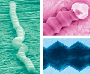
Menon grew up in the eastern region of India and was vaguely familiar with a cul­tural dance from the western state of Rajasthan known as the Bhavai pot dance. Nimble dancers sway their hips as a tall stack of wide-​​bellied pots bal­ances gin­gerly atop their heads. Back in the lab at North­eastern, Menon’s team recently cre­ated  gal­lium nitride nanowires, which bore a striking resem­blance to that stack of pots.

What’s more, a post­doc­toral research asso­ciate in Menon’s lab, Eugen Panaitescu, jumped on the band­wagon with a cul­tural art ref­er­ence of his own. Panaitescu, who hails from Romania, also saw his country’s famous End­less Column reflected in the nanowires. Ded­i­cated to the fallen Romanian heroes of World War I, Con­stantin Brancusi’s 96-​​foot-​​tall mono­lith is con­structed of 17 three-​​dimensional rhom­buses, peri­od­i­cally wavering from a wider cir­cum­fer­ence to a nar­rower one.

The news release goes on to explain more about applications using gallium nitride and why Menon’s insight may prove useful in developing new uses for gallium nitride nanowires,

… Gal­lium nitride is used across a range of tech­nolo­gies, including most ubiq­ui­tously in light emit­ting diodes. The mate­rial also holds great poten­tial for solar cell arrays, mag­netic semi­con­duc­tors, high-​​frequency com­mu­ni­ca­tion devices, and many other things. But these advanced appli­ca­tions are restricted by our lim­ited ability to con­trol the material’s growth on the nanoscale.

The very thing that makes Menon’s nanowires beau­tiful rep­re­sents a break­through in her ability to process them for these novel uses. She deposited onto a sil­icon sub­strate small droplets of liquid gold metal, which act as cat­a­lysts to grab gaseous gal­lium nitride from the atmos­phere of the exper­i­mental system. The net forces between the tiny gold droplet, the solid sub­strate, and the gas cause the nanowire to grow in a par­tic­ular direc­tion, she explained. Depending on the size of the gold cat­a­lyst, she can create wires that exhibit peri­odic serrations.

“It first tries to grow out­ward, but that gives the gold a larger sur­face area,” she said. “So now the wire gets pulled in the inward direc­tion, and then the gold gets a smaller sur­face area, so it grows out­ward again.” This inward and out­ward growth repeated itself again and again to create a peri­odic struc­ture nearly 6 mil­lion times smaller than the end­less column and is sig­nif­i­cantly more promising for its use in advanced devices.

“That there is very little imple­men­ta­tion of nanowire tech­nology in elec­tronics or optical devices is due to the fact that it’s very hard to con­trol their shape and dimen­sions,” said Menon. But now that she has a very simple way of con­trol­ling growth, the next step is to con­trol the size of the cat­alytic droplet with which she starts.

Another advan­tage of Menon’s approach is using what Panaitescu called “macro­scopic tech­niques” to create nanoscale mate­rials, thus making it scal­able and inex­pen­sive. “We just con­trol a few para­me­ters and then leave it, let it do it’s nat­ural thing,” explained Menon.

Here’s an image the researchers have supplied to illustrate their insights and their work,

Depending on the size of the gold cat­a­lyst used to make them, Latika Menon’s nanowires will exhibit peri­odic grooves that resemble common motifs in art. Images cour­tesy of Latika Menon. - See more at: http://www.northeastern.edu/news/2013/12/menon-nanowires/#sthash.LkgJU4es.dpuf

Depending on the size of the gold cat­a­lyst used to make them, Latika Menon’s nanowires will exhibit peri­odic grooves that resemble common motifs in art. Images cour­tesy of Latika Menon. – See more at: http://www.northeastern.edu/news/2013/12/menon-nanowires/#sthash.LkgJU4es.dpuf

I’m not sure I can connect the  imagery in this pot dance video (it does show some pretty astonishing feats of balance) with any of the images from Menon’s lab but sometimes the source of an inspiration is not readily accessible to those who are not amongst the inspired or perhaps there other versions of the dance that make it more obvious to an untrained eye,

Here’s an image of the other artistic inspiration, Constantin Brancusi’s Endless Column found on Dr. Cătălina Köpetz’s (University of Maryland) webpage featuring Brancusi’s work along with this quote from him “Create like a god, comand like a king, work like a slave.”

The Endless Column, Târgu Jiu, România  [downlaoded from http://terpconnect.umd.edu/~ckopetz/brancusi.htm]

The Endless Column,
Târgu Jiu, România [downlaoded from http://terpconnect.umd.edu/~ckopetz/brancusi.htm]

Interestingly, Dr. Köpetz is a social psychologist working in the university’s Center for Addictions, Personality, and Emotion Research.

For anyone who’d like to read more about Menon’s work, here’s a link to a webpage featuring a PDF selection of her papers and a citation for her latest paper on the work described in the news release,

Vapor–liquid–solid growth of serrated GaN nanowires: shape selection driven by kinetic frustration by Zheng Ma, Dillon McDowell, Eugen Panaitescu, Albert V. Davydov, Moneesh Upmanyu and Latika Menon, Physics Faculty Publications (2013)

Compound semiconducting nanowires are promising building blocks for several nanoelectronic devices yet the inability to…

The paper is open access although you will have to click a few times to retrieve it.

Inhale the drugs for Parkinson’s disease

The news out of Northeastern University’s Dr. Barbara Waszczak’s lab is exciting but it’s a single high point in a larger narrative.  First, here’s the high point described in the Apr. 24, 2013 news item on Azonano,

Researchers at Northeastern University in Boston have developed a gene therapy approach that may one day stop Parkinson’s disease (PD) in it tracks, preventing disease progression and reversing its symptoms. The novelty of the approach lies in the nasal route of administration and nanoparticles containing a gene capable of rescuing dying neurons in the brain.

The Apr. 21, 2013 news release on EurekAlert, which originated the news item, provides some information about Parkinson’s disease,

Parkinson’s is a devastating neurodegenerative disorder caused by the death of dopamine neurons in a key motor area of the brain, the substantia nigra (SN). Loss of these neurons leads to the characteristic tremor and slowed movements of PD, which get increasingly worse with time. Currently, more than 1% of the population over age 60 has PD and approximately 60,000 Americans are newly diagnosed every year. The available drugs on the market for PD mimic or replace the lost dopamine but do not get to the heart of the problem, which is the progressive loss of the dopamine neurons.

Here’s how the disease got its name, from the Wikipedia essay: Parkinson’s disease (Note: Links have been removed),

The disease is named after the English doctor James Parkinson, who published the first detailed description in An Essay on the Shaking Palsy in 1817. Several major organizations promote research and improvement of quality of life of those with the disease and their families. Public awareness campaigns include Parkinson’s disease day (on the birthday of James Parkinson, April 11) and the use of a red tulip as the symbol of the disease. People with parkinsonism who have increased the public’s awareness include Michael J. Fox and Muhammad Ali.

Now for some information about the background work leading up to this new, exciting, high point (from the news release on EurekAlert),

The focus of Dr. Barbara Waszczak’s lab at Northeastern University in Boston is to find a way to harvest the potential of glial cell line-derived neurotrophic factor (GDNF) as a treatment for PD. GDNF is a protein known to nourish dopamine neurons by activating survival and growth-promoting pathways inside the cells. Not surprisingly, GDNF is able to protect dopamine neurons from injury and restore the function of damaged and dying neurons in many animal models of PD. However, the action of GDNF is limited by its inability to cross the blood-brain barrier (BBB), thus requiring direct surgical injection into the brain. To circumvent this problem, Waszczak’s lab is investigating intranasal delivery as a way to bypass the BBB. Their previous work showed that intranasal delivery of GDNF protects dopamine neurons from damage by the neurotoxin, 6-hydroxydopamine (6-OHDA), a standard rat model of PD.

According to the Michael J. Fox Foundation, this research work dates from 2007 (at least), from the Intranasal Delivery of GDNF for Parkinson’s Disease: Next Steps grant page,

FINAL OUTCOME

The results of this Drug Delivery 2008 project confirm and extend the conclusions reached under a previous 2007 Rapid Response Innovation Award. The research team has demonstrated that intranasal administration of GDNF has neuroprotective efficacy in a preclinical model of Parkinson’s disease, that the protein gets into the brain and reaches target structures (the striatum and substantia nigra) within an hour of nasal administration, and that the nasal route causes no apparent toxicity in the nose. Longer term efficacy and toxicology studies will be necessary in other relevant preclinical models before testing can be initiated in humans.

The results of this work strongly supports pursuit of intranasal administration as a promising approach for harvesting the therapeutic potential of GDNF. Such an approach could ultimately provide an effective, non-invasive means of delivering GDNF to the brain for the treatment of Parkinson’s disease.

Here’s the 2013 innovation on intranasal delivery of GDNF therapy (from the news release on EurekAlert),

Taking this work a step further, Brendan Harmon, working in Waszczak’s lab, has adapted the intranasal approach so that cells in the brain can continuously produce GDNF. His work utilized nanoparticles, developed by Copernicus Therapeutics, Inc., which are able to transfect brain cells with an expression plasmid carrying the gene for GDNF (pGDNF). When given intranasally to rats, these pGDNF nanoparticles increase GDNF production throughout the brain for long periods, avoiding the need for frequent re-dosing. Now, in new research presented on April 20 at 12:30 pm during Experimental Biology 2013 in Boston, MA, Harmon reports that intranasal administration of Copernicus’ pGDNF nanoparticles results in GDNF expression sufficient to protect SN dopamine neurons in the 6-OHDA model of PD.

Waszczak and Harmon believe that intranasal delivery of Copernicus’ nanoparticles may provide an effective and non-invasive means of GDNF gene therapy for PD, and an avenue for transporting other gene therapy vectors to the brain. This work, which was funded in part by the Michael J. Fox Foundation for Parkinson’s Research and Northeastern University, has the potential to greatly expand treatment options for PD and many other central nervous system disorders.

For the curious, there’s more about Copernicus Therapeutics at the company website.

Congratulations to Harmon and Waszczak! I imagine the next step will be human clinical trials.