Tag Archives: Northwestern University

“The Heart’s Knowledge: Science and Empathy in the Art of Dario Robleto” from Jan. 27 to July 9, 2023 at The Block Museum of Art (Northwestern University, Chicago, Illinois)

I’m sorry to be late. Thankfully this show extends into July 2023, so, there’s still plenty of time to get to Chicago’s The Block Museum of Art (at Northwestern University) art/science exhibition. I found this on the museum’s exhibition page for “The Heart’s Knowledge: Science and Empathy in the Art of Dario Robleto” show,

What do we owe to the memories of one another’s hearts?

For American artist Dario Robleto (b. 1972), artists and scientists share a common aspiration: to increase the sensitivity of their observations. Throughout the history of scientific invention, instruments like the cardiograph and the telescope have extended the reach of perception from the tiniest stirrings of the human body to the farthest reaches of space. In his prints, sculptures, and video and sound installations, Robleto contemplates the emotional significance of these technologies, bringing us closer to the latent traces of life buried in the scientific record.

The Hearts Knowledge concentrates on the most recent decade of Robleto’s creative practice, a period of deepening engagement with histories of medicine, biomedical engineering, sound recording, and space exploration. The exhibition organizes the artist’s conceptually ambitious, elegantly wrought artworks as a series of multisensory encounters between art and science.  Each work seeks to attune viewers to the material traces of life at scales ranging from the intimate to the universal, returning always to the question: Does empathy extend beyond the boundaries of time and space?

Banner image for “The Heart’s Knowledge: Science and Empathy in the Art of Dario Robleto” exhibition page. Courtesy of The Block Museum of Art (Northwestern University) and artist, Dario Robleto

Here’s more from a January 27, 2023 Northwestern University news release (received via email),

Exhibition searches for meaning at the limits of science and perception

“The Heart’s Knowledge: Science and Empathy in the Art of Dario Robleto” is on view Jan. 27 to July 9 [2023] at The Block Museum of Art

  • Works are informed by dialogue with Northwestern Engineering researchers during five-year residency  
  • Exhibition a tribute to NASA Golden Record creator, ‘whose heart has left the solar system’
  • Opening conversation with the artist will take place at 2 p.m. on [Saturday] Feb. 4 [2023]

American artist Dario Robleto (b. 1972) believes artists and scientists share a common aspiration: to increase the sensitivity of their observations.

From understanding the human body’s pulses and brainwaves to viewing the faintest glimmers of light from the edge of the observable universe, groundbreaking science pushes the limits of perception. Similarly, the perceptive work of artists can extend the boundaries of empathy and understanding.

Since 2018, Robleto served as an artist-at-large at the McCormick School of Engineering. This unique partnership between The Block Museum and McCormick gave the artist an open “hall pass” to learn from, collaborate with and question scientists, engineers and experts from across the University.

Robleto’s five-year residency concludes with the exhibition “The Heart’s Knowledge: Science and Empathy in the Art of Dario Robleto.” Co-presented by The Block Museum and McCormick, the exhibition is on view from Jan. 27 to July 9 [2023] at The Block Museum, 40 Arts Circle Drive on Northwestern’s Evanston campus. [emphasis mine]

A free opening conversation with the artist will take place at 2 p.m. on Saturday, Feb. 4 [2023], in Norris University Center’s McCormick Auditorium, 1999 Campus Drive in Evanston.

About the Exhibition: “The Heart’s Knowledge”

Throughout the history of scientific invention, instruments like the cardiograph and the telescope have extended the reach of perception from the tiniest stirrings of the human body to the farthest reaches of space.

Robleto’s prints, sculptures and video and sound installations contemplate the emotional significance of these technologies, bringing viewers closer to the latent traces of life buried in the scientific record.

“The Heart’s Knowledge” represents a decade of Robleto’s creative practice, from 2012 to 2022, a period marked by a deepening engagement with science, including astronomy, synthetic biology and exobiology, and a widening embrace of new materials and creative forms, from 3D-printed objects to film.

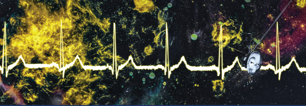
Robleto dedicates the exhibition to Ann Druyan, the creative director of NASA’s Golden Record for the Voyager 1 and 2 projects. The record includes Druyan’s brainwaves and heartbeats, recorded as she reflected on her secret love for famed astronomer and future husband Carl Sagan. The act of sneaking “love on board the Voyager” inspired Robleto to compose a love letter to the only human whose “heart has left the solar system.”

Robleto sees Druyan’s act to include her emotions on the record as the central inspiration of his work. “I consider it the greatest work of subversive, avant-garde art not yet given its due,” Robleto said. “The Golden Record and Ann’s radical act brought us all together to think about what it means to be human — to one another and to unknown beings on other worlds.”

The exhibition organizes the artist’s conceptually ambitious, elegantly wrought artworks as a series of multisensory encounters between art and science. Each asks viewers to seek out the material traces of life in scales ranging from the intimate to the universal, and to question: Does empathy extend beyond the boundaries of time and space?

“Whether he’s addressing the most minute phenomena of the body or the horizons of the known universe, Robleto binds the rigor of scientific inquiry with artistic expression,” said exhibition curator Michael Metzger, The Block’s Pick-Laudati Curator of Media Arts.

“Straining at the bounds of observation, Robleto discovers unity at the limits; the common endeavor of art and science to achieve a form of knowledge that language alone cannot speak,” Metzger said.

The exhibition includes three sections:

Heartbeats
Rooted in the artist’s longstanding fascination with the clinical and cultural history of the human heart, “Heartbeats” draws inspiration from 19th-century pioneers of cardiography, whose instruments graphically measured heart activity for the first time, leaving behind poignant records of human subjectivity. In “The First Time, the Heart (A Portrait of Life 1854-1913)” (2017), Robleto transforms early measurements of heartbeats into photolithographs executed on paper hand-sooted with candle flames. For the installation “The Pulse Armed with a Pen (An Unknown History of the Human Heartbeat)” (2014), Robleto collaborated with sound historian Patrick Feaster to digitally resurrect these heartbeats in audio form, giving visitors access to intimate pulses of life recorded before the invention of sound playback.

Wavelengths
Robleto has recently embraced digital video to create works that narrate transformational episodes in the recording and study of wave phenomena. “Wavelengths” comprises two hour-long immersive video installations. “The Boundary of Life is Quietly Crossed” (2019) is inspired by NASA’s Voyager Golden Record, a gold-plated phonographic disc launched into space onboard the Voyager I and II space probes in 1977. In “The Aorta of an Archivist” (2020-2021), Robleto investigates three breakthroughs in the history of recording: the first recording of a choral performance made with an Edison wax cylinder, the first heartbeat captured while listening to music and the first effort to transcribe the brain wave activity of a dreaming subject.

Horizons
In the final section, “Horizons,” Robleto evokes the spirit of the Hubble telescope and the search for extraterrestrial life, gazing out at the boundaries of the observable universe. Inspired by his time as an artist-in-residence at the SETI Institute (Search for Extraterrestrial Intelligence) and as artistic consultant to the Breakthrough Initiatives, his intricate sculptures, such as “Small Crafts on Sisyphean Seas” (2018), give shape to the speculative search for intelligent life in the universe. Other works like “The Computer of Jupiter” (2019) are framed as “gifts for extraterrestrials” offering an alternative view of the best way to begin a dialogue with alien intelligences.

The Artist-at-Large Program at Northwestern

Lisa Corrin, the Ellen Philips Katz Executive Director of The Block Museum of Art, and Julio M. Ottino, dean of McCormick School of Engineering, envisioned the possibilities of this unconventional partnership between scientist and artist when they launched the artist-at-large initiative together. The work is part of an ongoing Art + Engineering initiative and a part of the whole-brain engineering philosophy at Northwestern Engineering.

“Here, a university’s school of engineering and its art museum come together in the shared belief that transformative innovation can happen at the intersections of usually distinct academic disciplines and modes of creativity and inquiry,” Corrin said. “We had faith that something meaningful would emerge organically if we merely provided structures in which informal interactions might take place.”

“We wanted to model for young engineers the value of embracing uncertainty as part of the journey that leads to innovation and opens pathways within the imagination — as artists do,” Ottino said. “We are grateful to Dario Robleto for accepting our invitation to come to Northwestern and to enter the unknown with us. He has taught us that our shared future resides in our capacity for compassion and for empathy, the ethos at the heart of his work that holds the most promise for those at the forefront of science in the interest of humankind.”

More information about Robleto’s residency can be found in the article “Dario is our Socrates” on the Block Museum website and by viewing the Northwestern Engineering video “Artist-at-Large Program: Dario Robleto.”

Exhibition Events

“The Heart’s Knowledge” will include six months of events and dialogues that will illuminate the intersections in Robleto’s practice. All events are free and open to the public. For current program information, visit The Block Museum website.

Program highlights for February and March include:

Science, Art and the Search for Meaning: Opening Conversation with Dario Robleto
Saturday, Feb. 4 [2023], 2 p.m.
Norris University Center, McCormick Auditorium
1999 Campus Drive

The Block Museum hosts a discussion that reaches across boundaries to examine the shared pursuit that binds artists and scientists. The conversation features artist Dario Robleto; Jennifer Roberts, professor of the humanities at Harvard University; Lucianne Walkowicz, astronomer and co-founder of the JustSpace Alliance; and Michael Metzger, Pick-Laudati Curator of Media Arts and curator of “The Heart’s Knowledge.”

“X: The Man with the X-Ray Eyes” (1963)
Friday, Feb. 10 [2023], 7 p.m.
Block Cinema
40 Arts Circle Drive

A Science on Screen program with Catherine Belling, associate professor of medical education at Northwestern University Feinberg School of Medicine.

“First Man” (2018)
Saturday Feb. 18 [2023], 1 p.m.
Block Cinema
40 Arts Circle Drive

A Science on Screen program featuring history researcher Jordan Bimm of the University of Chicago, who will discuss the military origins of “space medicine.”

Exhibition Conversation: Interstellar Aesthetics and Acts of Translation in Art and Science
Wednesday, Feb. 22 [2023], 6 p.m.
Block Museum

Joining artist Dario Robleto in conversation are Elizabeth Kessler, exhibition publication contributor and a lecturer in American Studies at Stanford University, and Shane Larson, research professor of physics and astronomy and associate director of CIERA (Center for Interdisciplinary Exploration and Research in Astrophysics) at Northwestern.

Gallery Talk: Stillness, Wonder and Gifts for Extraterrestrials
Thursday, Feb. 23 [2023], 12:30 p.m.
Block Museum

Elizabeth Kessler of Stanford University will discuss Robleto’s “gifts for extraterrestrials” series.

Online Conversation: Ann Druyan, The Golden Record and the Memory of Our Hearts
Wednesday, March 8 [2023], 6 p.m. [ET]
Block Cinema

Ann Druyan, creative director for NASA’s Voyager Interstellar Messaging Project and writer and producer of the PBS television series “Cosmos,” joins Robleto and art historian Jennifer Roberts for a conversation about the Golden Record and the heart’s memory.

Exhibition Publication

In conjunction with the exhibition, The Block Museum of Art and the McCormick School of Engineering are proud to announce the publication of “The Heart’s Knowledge: Science and Empathy in the Art of Dario Robleto,” (Artbook, D.A.P., 2023).

The publication is edited by Michael Metzger with contributions by Metzger, Robert M. Brain, Daniel K. L. Chua, Patrick Feaster, Stefan Helmreich, Elizabeth A. Kessler ,Julius B. Lucks, Elizabeth Kathleen Mitchell, Alexander Rehding, Jennifer L. Roberts, Claire Isabel Webb and Dario Robleto.

About Dario Robleto

Dario Robleto was born in San Antonio, Texas, in 1972 and received his BFA from the University of Texas at San Antonio in 1997. He lives and works in Houston, Texas. The artist has had numerous solo exhibitions since 1997, most recently at the Spencer Museum of Art, Lawrence, Kansas (2021); the Radcliffe Institute for Advanced Study at Harvard University (2019); the McNay Museum, San Antonio, Texas (2018); Menil Collection, Houston, Texas (2014); the Baltimore Museum of Art (2014); the New Orleans Museum of Art (2012); and the Museum of Contemporary Art, Denver (2011).

He is currently working on his first book, “Life Signs: The Tender Science of the Pulsewave,” co-authored with art historian Jennifer Roberts, the Elizabeth Cary Agassiz Professor of the Humanities at Harvard (University of Chicago Press).

Exhibition Credits

“The Heart’s Knowledge: Science and Empathy in the Art of Dario Robleto” exhibition is made possible through a partnership with the Robert R. McCormick School of Engineering and Applied Science at Northwestern University. Major support also was provided by the National Endowment for the Arts. Additional support is contributed by the Dorothy J. Speidel Fund; the Bernstein Family Contemporary Art Fund; the Barry and Mary Ann MacLean Fund for Art and Engineering; the Illinois Arts Council Agency; and the Alumnae of Northwestern University. The exhibition publication is made possible in part by the Sandra L. Riggs Publications Fund.

Should you be in the Chicago area and interested in the exhibit, you can find all the information for your visit here.

Implantable living pharmacy

I stumbled across a very interesting US Defense Advanced Research Projects Agency (DARPA) project (from an August 30, 2021 posting on Northwestern University’s Rivnay Lab [a laboratory for organic bioelectronics] blog),

Our lab has received a cooperative agreement with DARPA to develop a wireless, fully implantable ‘living pharmacy’ device that could help regulate human sleep patterns. The project is through DARPA’s BTO (biotechnology office)’s Advanced Acclimation and Protection Tool for Environmental Readiness (ADAPTER) program, meant to address physical challenges of travel, such as jetlag and fatigue.

The device, called NTRAIN (Normalizing Timing of Rhythms Across Internal Networks of Circadian Clocks), would control the body’s circadian clock, reducing the time it takes for a person to recover from disrupted sleep/wake cycles by as much as half the usual time.

The project spans 5 institutions including Northwestern, Rice University, Carnegie Mellon, University of Minnesota, and Blackrock Neurotech.

Prior to the Aug. 30, 2021 posting, Amanda Morris wrote a May 13, 2021 article for Northwestern NOW (university magazine), which provides more details about the project, Note: A link has been removed,

The first phase of the highly interdisciplinary program will focus on developing the implant. The second phase, contingent on the first, will validate the device. If that milestone is met, then researchers will test the device in human trials, as part of the third phase. The full funding corresponds to $33 million over four-and-a-half years. 

Nicknamed the “living pharmacy,” the device could be a powerful tool for military personnel, who frequently travel across multiple time zones, and shift workers including first responders, who vacillate between overnight and daytime shifts.

Combining synthetic biology with bioelectronics, the team will engineer cells to produce the same peptides that the body makes to regulate sleep cycles, precisely adjusting timing and dose with bioelectronic controls. When the engineered cells are exposed to light, they will generate precisely dosed peptide therapies. 

“This control system allows us to deliver a peptide of interest on demand, directly into the bloodstream,” said Northwestern’s Jonathan Rivnay, principal investigator of the project. “No need to carry drugs, no need to inject therapeutics and — depending on how long we can make the device last — no need to refill the device. It’s like an implantable pharmacy on a chip that never runs out.” 

Beyond controlling circadian rhythms, the researchers believe this technology could be modified to release other types of therapies with precise timing and dosing for potentially treating pain and disease. The DARPA program also will help researchers better understand sleep/wake cycles, in general.

“The experiments carried out in these studies will enable new insights into how internal circadian organization is maintained,” said Turek [Fred W. Turek], who co-leads the sleep team with Vitaterna [Martha Hotz Vitaterna]. “These insights will lead to new therapeutic approaches for sleep disorders as well as many other physiological and mental disorders, including those associated with aging where there is often a spontaneous breakdown in temporal organization.” 

For those who like to dig even deeper, Dieynaba Young’s June 17, 2021 article for Smithsonian Magazine (GetPocket.com link to article) provides greater context and greater satisfaction, Note: Links have been removed,

In 1926, Fritz Kahn completed Man as Industrial Palace, the preeminent lithograph in his five-volume publication The Life of Man. The illustration shows a human body bustling with tiny factory workers. They cheerily operate a brain filled with switchboards, circuits and manometers. Below their feet, an ingenious network of pipes, chutes and conveyer belts make up the blood circulatory system. The image epitomizes a central motif in Kahn’s oeuvre: the parallel between human physiology and manufacturing, or the human body as a marvel of engineering.

An apparatus in the embryonic stage of development at the time of this writing in June of 2021—the so-called “implantable living pharmacy”—could have easily originated in Kahn’s fervid imagination. The concept is being developed by the Defense Advanced Research Projects Agency (DARPA) in conjunction with several universities, notably Northwestern and Rice. Researchers envision a miniaturized factory, tucked inside a microchip, that will manufacture pharmaceuticals from inside the body. The drugs will then be delivered to precise targets at the command of a mobile application. …

The implantable living pharmacy, which is still in the “proof of concept” stage of development, is actually envisioned as two separate devices—a microchip implant and an armband. The implant will contain a layer of living synthetic cells, along with a sensor that measures temperature, a short-range wireless transmitter and a photo detector. The cells are sourced from a human donor and reengineered to perform specific functions. They’ll be mass produced in the lab, and slathered onto a layer of tiny LED lights.

The microchip will be set with a unique identification number and encryption key, then implanted under the skin in an outpatient procedure. The chip will be controlled by a battery-powered hub attached to an armband. That hub will receive signals transmitted from a mobile app.

If a soldier wishes to reset their internal clock, they’ll simply grab their phone, log onto the app and enter their upcoming itinerary—say, a flight departing at 5:30 a.m. from Arlington, Virginia, and arriving 16 hours later at Fort Buckner in Okinawa, Japan. Using short-range wireless communications, the hub will receive the signal and activate the LED lights inside the chip. The lights will shine on the synthetic cells, stimulating them to generate two compounds that are naturally produced in the body. The compounds will be released directly into the bloodstream, heading towards targeted locations, such as a tiny, centrally-located structure in the brain called the suprachiasmatic nucleus (SCN) that serves as master pacemaker of the circadian rhythm. Whatever the target location, the flow of biomolecules will alter the natural clock. When the solider arrives in Okinawa, their body will be perfectly in tune with local time.

The synthetic cells will be kept isolated from the host’s immune system by a membrane constructed of novel biomaterials, allowing only nutrients and oxygen in and only the compounds out. Should anything go wrong, they would swallow a pill that would kill the cells inside the chip only, leaving the rest of their body unaffected.

If you have the time, I recommend reading Young’s June 17, 2021 Smithsonian Magazine article (GetPocket.com link to article) in its entirety. Young goes on to discuss, hacking, malware, and ethical/societal issues and more.

There is an animation of Kahn’s original poster in a June 23, 2011 posting on openculture.com (also found on Vimeo; Der Mensch als Industriepalast [Man as Industrial Palace])

Credits: Idea & Animation: Henning M. Lederer / led-r-r.net; Sound-Design: David Indge; and original poster art: Fritz Kahn.

Shaving the ‘hairs’ off nanocrystals for more efficient electronics

A March 24, 2022 news item on phys.org announced research into nanoscale crystals and how they might be integrated into electronic devices, Note: A link has been removed,

You can carry an entire computer in your pocket today because the technological building blocks have been getting smaller and smaller since the 1950s. But in order to create future generations of electronics—such as more powerful phones, more efficient solar cells, or even quantum computers—scientists will need to come up with entirely new technology at the tiniest scales.

One area of interest is nanocrystals. These tiny crystals can assemble themselves into many configurations, but scientists have had trouble figuring out how to make them talk to each other.  

A new study introduces a breakthrough in making nanocrystals function together electronically. Published March 25 [2022] in Science, the research may open the doors to future devices with new abilities. 

A March 25, 2022 University of Chicago news release (also on EurekAlert but published on March 24, 2022), which originated the news item, expands on the possibilities the research makes possible, Note: Links have been removed,

“We call these super atomic building blocks, because they can grant new abilities—for example, letting cameras see in the infrared range,” said University of Chicago Prof. Dmitri Talapin, the corresponding author of the paper. “But until now, it has been very difficult to both assemble them into structures and have them talk to each other. Now for the first time, we don’t have to choose. This is a transformative improvement.”  

In their paper, the scientists lay out design rules which should allow for the creation of many different types of materials, said Josh Portner, a Ph.D. student in chemistry and one of the first authors of the study. 

A tiny problem

Scientists can grow nanocrystals out of many different materials: metals, semiconductors, and magnets will each yield different properties. But the trouble was that whenever they tried to assemble these nanocrystals together into arrays, the new supercrystals would grow with long “hairs” around them. 

These hairs made it difficult for electrons to jump from one nanocrystal to another. Electrons are the messengers of electronic communication; their ability to move easily along is a key part of any electronic device. 

The researchers needed a method to reduce the hairs around each nanocrystal, so they could pack them in more tightly and reduce the gaps in between. “When these gaps are smaller by just a factor of three, the probability for electrons to jump across is about a billion times higher,” said Talapin, the Ernest DeWitt Burton Distinguished Service Professor of Chemistry and Molecular Engineering at UChicago and a senior scientist at Argonne National Laboratory. “It changes very strongly with distance.”

To shave off the hairs, they sought to understand what was going on at the atomic level. For this, they needed the aid of powerful X-rays at the Center for Nanoscale Materials at Argonne and the Stanford Synchrotron Radiation Lightsource at SLAC National Accelerator Laboratory, as well as powerful simulations and models of the chemistry and physics at play. All these allowed them to understand what was happening at the surface—and find the key to harnessing their production.

Part of the process to grow supercrystals is done in solution—that is, in liquid. It turns out that as the crystals grow, they undergo an unusual transformation in which gas, liquid and solid phases all coexist. By precisely controlling the chemistry of that stage, they could create crystals with harder, slimmer exteriors which could be packed in together much more closely. “Understanding their phase behavior was a massive leap forward for us,” said Portner. 

The full range of applications remains unclear, but the scientists can think of multiple areas where the technique could lead. “For example, perhaps each crystal could be a qubit in a quantum computer; coupling qubits into arrays is one of the fundamental challenges of quantum technology right now,” said Talapin. 

Portner is also interested in exploring the unusual intermediate state of matter seen during supercrystal growth: “Triple phase coexistence like this is rare enough that it’s intriguing to think about how to take advantage of this chemistry and build new materials.”

The study included scientists with the University of Chicago, Technische Universität Dresden, Northwestern University, Arizona State University, SLAC, Lawrence Berkeley National Laboratory, and the University of California, Berkeley.

Here’s a link to and a citation for the paper,

Self-assembly of nanocrystals into strongly electronically coupled all-inorganic supercrystals by Igor Coropceanu, Eric M. Janke, Joshua Portner, Danny Haubold, Trung Dac Nguyen, Avishek Das, Christian P. N. Tanner, James K. Utterback, Samuel W. Teitelbaum¸ Margaret H. Hudson, Nivedina A. Sarma, Alex M. Hinkle, Christopher J. Tassone, Alexander Eychmüller, David T. Limmer, Monica Olvera de la Cruz, Naomi S. Ginsberg and Dmitri V. Talapin. Science • 24 Mar 2022 • Vol 375, Issue 6587 • pp. 1422-1426 • DOI: 10.1126/science.abm6753

This paper is behind a paywall.

Will you be my friend? Yes, after we activate our ultraminiature, wireless, battery-free, fully implantable devices

Perhaps I’m the only one who’s disconcerted?

Here’s the research (in text form) as to why we’re watching these scampering, momentary mouse friends, from a May 10, 2021 Northwestern University news release (also on EurekAlert) by Amanda Morris,

Northwestern University researchers are building social bonds with beams of light.

For the first time ever, Northwestern engineers and neurobiologists have wirelessly programmed — and then deprogrammed — mice to socially interact with one another in real time. The advancement is thanks to a first-of-its-kind ultraminiature, wireless, battery-free and fully implantable device that uses light to activate neurons.

This study is the first optogenetics (a method for controlling neurons with light) paper exploring social interactions within groups of animals, which was previously impossible with current technologies.

The research was published May 10 [2021] in the journal Nature Neuroscience.

The thin, flexible, wireless nature of the implant allows the mice to look normal and behave normally in realistic environments, enabling researchers to observe them under natural conditions. Previous research using optogenetics required fiberoptic wires, which restrained mouse movements and caused them to become entangled during social interactions or in complex environments.

“With previous technologies, we were unable to observe multiple animals socially interacting in complex environments because they were tethered,” said Northwestern neurobiologist Yevgenia Kozorovitskiy, who designed the experiment. “The fibers would break or the animals would become entangled. In order to ask more complex questions about animal behavior in realistic environments, we needed this innovative wireless technology. It’s tremendous to get away from the tethers.”

“This paper represents the first time we’ve been able to achieve wireless, battery-free implants for optogenetics with full, independent digital control over multiple devices simultaneously in a given environment,” said Northwestern bioelectronics pioneer John A. Rogers, who led the technology development. “Brain activity in an isolated animal is interesting, but going beyond research on individuals to studies of complex, socially interacting groups is one of the most important and exciting frontiers in neuroscience. We now have the technology to investigate how bonds form and break between individuals in these groups and to examine how social hierarchies arise from these interactions.”

Kozorovitskiy is the Soretta and Henry Shapiro Research Professor of Molecular Biology and associate professor of neurobiology in Northwestern’s Weinberg College of Arts and Sciences. She also is a member of the Chemistry of Life Processes Institute. Rogers is the Louis Simpson and Kimberly Querrey Professor of Materials Science and Engineering, Biomedical Engineering and Neurological Surgery in the McCormick School of Engineering and Northwestern University Feinberg School of Medicine and the director of the Querrey Simpson Institute for Bioelectronics.

Kozorovitskiy and Rogers led the work with Yonggang Huang, the Jan and Marcia Achenbach Professor in Mechanical Engineering at McCormick, and Zhaoqian Xie, a professor of engineering mechanics at Dalian University of Technology in China. The paper’s co-first authors are Yiyuan Yang, Mingzheng Wu and Abraham Vázquez-Guardado — all at Northwestern.

Promise and problems of optogenetics

Because the human brain is a system of nearly 100 billion intertwined neurons, it’s extremely difficult to probe single — or even groups of — neurons. Introduced in animal models around 2005, optogenetics offers control of specific, genetically targeted neurons in order to probe them in unprecedented detail to study their connectivity or neurotransmitter release. Researchers first modify neurons in living mice to express a modified gene from light-sensitive algae. Then they can use external light to specifically control and monitor brain activity. Because of the genetic engineering involved, the method is not yet approved in humans.

“It sounds like sci-fi, but it’s an incredibly useful technique,” Kozorovitskiy said. “Optogenetics could someday soon be used to fix blindness or reverse paralysis.”

Previous optogenetics studies, however, were limited by the available technology to deliver light. Although researchers could easily probe one animal in isolation, it was challenging to simultaneously control neural activity in flexible patterns within groups of animals interacting socially. Fiberoptic wires typically emerged from an animal’s head, connecting to an external light source. Then a software program could be used to turn the light off and on, while monitoring the animal’s behavior.

“As they move around, the fibers tugged in different ways,” Rogers said. “As expected, these effects changed the animal’s patterns of motion. One, therefore, has to wonder: What behavior are you actually studying? Are you studying natural behaviors or behaviors associated with a physical constraint?”

Wireless control in real time

A world-renowned leader in wireless, wearable technology, Rogers and his team developed a tiny, wireless device that gently rests on the skull’s outer surface but beneath the skin and fur of a small animal. The half-millimeter-thick device connects to a fine, flexible filamentary probe with LEDs on the tip, which extend down into the brain through a tiny cranial defect.

The miniature device leverages near-field communication protocols, the same technology used in smartphones for electronic payments. Researchers wirelessly operate the light in real time with a user interface on a computer. An antenna surrounding the animals’ enclosure delivers power to the wireless device, thereby eliminating the need for a bulky, heavy battery.

Activating social connections

To establish proof of principle for Rogers’ technology, Kozorovitskiy and colleagues designed an experiment to explore an optogenetics approach to remote-control social interactions among pairs or groups of mice.

When mice were physically near one another in an enclosed environment, Kozorovitskiy’s team wirelessly synchronously activated a set of neurons in a brain region related to higher order executive function, causing them to increase the frequency and duration of social interactions. Desynchronizing the stimulation promptly decreased social interactions in the same pair of mice. In a group setting, researchers could bias an arbitrarily chosen pair to interact more than others.

“We didn’t actually think this would work,” Kozorovitskiy said. “To our knowledge, this is the first direct evaluation of a major long-standing hypothesis about neural synchrony in social behavior.”

Here’s a citation and a link to the paper,

Wireless multilateral devices for optogenetic studies of individual and social behaviors by Yiyuan Yang, Mingzheng Wu, Amy J. Wegener, Jose G. Grajales-Reyes, Yujun Deng, Taoyi Wang, Raudel Avila, Justin A. Moreno, Samuel Minkowicz, Vasin Dumrongprechachan, Jungyup Lee, Shuangyang Zhang, Alex A. Legaria, Yuhang Ma, Sunita Mehta, Daniel Franklin, Layne Hartman, Wubin Bai, Mengdi Han, Hangbo Zhao, Wei Lu, Yongjoon Yu, Xing Sheng, Anthony Banks, Xinge Yu, Zoe R. Donaldson, Robert W. Gereau IV, Cameron H. Good, Zhaoqian Xie, Yonggang Huang, Yevgenia Kozorovitskiy and John A. Rogers. Nature Neuroscience (2021)
DOI: https://doi.org/10.1038/s41593-021-00849-x Published 10 May 2021

This paper is behind a paywall.

This latest research seems to be the continuation of research featured here in a July 16, 2019 posting: “Controlling neurons with light: no batteries or wires needed.”

Synaptic transistor better then memristor when it comes to brainlike learning for computers

An April 30, 2021 news item on Nanowerk announced research from a joint team at Northwestern University (located in Chicago, Illinois, US) and University of Hong Kong of researchers in the field of neuromorphic (brainlike) computing,

Researchers have developed a brain-like computing device that is capable of learning by association.

Similar to how famed physiologist Ivan Pavlov conditioned dogs to associate a bell with food, researchers at Northwestern University and the University of Hong Kong successfully conditioned their circuit to associate light with pressure.

The device’s secret lies within its novel organic, electrochemical “synaptic transistors,” which simultaneously process and store information just like the human brain. The researchers demonstrated that the transistor can mimic the short-term and long-term plasticity of synapses in the human brain, building on memories to learn over time.

With its brain-like ability, the novel transistor and circuit could potentially overcome the limitations of traditional computing, including their energy-sapping hardware and limited ability to perform multiple tasks at the same time. The brain-like device also has higher fault tolerance, continuing to operate smoothly even when some components fail.

“Although the modern computer is outstanding, the human brain can easily outperform it in some complex and unstructured tasks, such as pattern recognition, motor control and multisensory integration,” said Northwestern’s Jonathan Rivnay, a senior author of the study. “This is thanks to the plasticity of the synapse, which is the basic building block of the brain’s computational power. These synapses enable the brain to work in a highly parallel, fault tolerant and energy-efficient manner. In our work, we demonstrate an organic, plastic transistor that mimics key functions of a biological synapse.”

Rivnay is an assistant professor of biomedical engineering at Northwestern’s McCormick School of Engineering. He co-led the study with Paddy Chan, an associate professor of mechanical engineering at the University of Hong Kong. Xudong Ji, a postdoctoral researcher in Rivnay’s group, is the paper’s first author.

Caption: By connecting single synaptic transistors into a neuromorphic circuit, researchers demonstrated that their device could simulate associative learning. Credit: Northwestern University

An April 30, 2021 Northwestern University news release (also on EurekAlert), which originated the news item, includes a good explanation about brainlike computing and information about how synaptic transistors work along with some suggestions for future applications,

Conventional, digital computing systems have separate processing and storage units, causing data-intensive tasks to consume large amounts of energy. Inspired by the combined computing and storage process in the human brain, researchers, in recent years, have sought to develop computers that operate more like the human brain, with arrays of devices that function like a network of neurons.

“The way our current computer systems work is that memory and logic are physically separated,” Ji said. “You perform computation and send that information to a memory unit. Then every time you want to retrieve that information, you have to recall it. If we can bring those two separate functions together, we can save space and save on energy costs.”

Currently, the memory resistor, or “memristor,” is the most well-developed technology that can perform combined processing and memory function, but memristors suffer from energy-costly switching and less biocompatibility. These drawbacks led researchers to the synaptic transistor — especially the organic electrochemical synaptic transistor, which operates with low voltages, continuously tunable memory and high compatibility for biological applications. Still, challenges exist.

“Even high-performing organic electrochemical synaptic transistors require the write operation to be decoupled from the read operation,” Rivnay said. “So if you want to retain memory, you have to disconnect it from the write process, which can further complicate integration into circuits or systems.”

How the synaptic transistor works

To overcome these challenges, the Northwestern and University of Hong Kong team optimized a conductive, plastic material within the organic, electrochemical transistor that can trap ions. In the brain, a synapse is a structure through which a neuron can transmit signals to another neuron, using small molecules called neurotransmitters. In the synaptic transistor, ions behave similarly to neurotransmitters, sending signals between terminals to form an artificial synapse. By retaining stored data from trapped ions, the transistor remembers previous activities, developing long-term plasticity.

The researchers demonstrated their device’s synaptic behavior by connecting single synaptic transistors into a neuromorphic circuit to simulate associative learning. They integrated pressure and light sensors into the circuit and trained the circuit to associate the two unrelated physical inputs (pressure and light) with one another.

Perhaps the most famous example of associative learning is Pavlov’s dog, which naturally drooled when it encountered food. After conditioning the dog to associate a bell ring with food, the dog also began drooling when it heard the sound of a bell. For the neuromorphic circuit, the researchers activated a voltage by applying pressure with a finger press. To condition the circuit to associate light with pressure, the researchers first applied pulsed light from an LED lightbulb and then immediately applied pressure. In this scenario, the pressure is the food and the light is the bell. The device’s corresponding sensors detected both inputs.

After one training cycle, the circuit made an initial connection between light and pressure. After five training cycles, the circuit significantly associated light with pressure. Light, alone, was able to trigger a signal, or “unconditioned response.”

Future applications

Because the synaptic circuit is made of soft polymers, like a plastic, it can be readily fabricated on flexible sheets and easily integrated into soft, wearable electronics, smart robotics and implantable devices that directly interface with living tissue and even the brain [emphasis mine].

“While our application is a proof of concept, our proposed circuit can be further extended to include more sensory inputs and integrated with other electronics to enable on-site, low-power computation,” Rivnay said. “Because it is compatible with biological environments, the device can directly interface with living tissue, which is critical for next-generation bioelectronics.”

Here’s a link to and a citation for the paper,

Mimicking associative learning using an ion-trapping non-volatile synaptic organic electrochemical transistor by Xudong Ji, Bryan D. Paulsen, Gary K. K. Chik, Ruiheng Wu, Yuyang Yin, Paddy K. L. Chan & Jonathan Rivnay . Nature Communications volume 12, Article number: 2480 (2021) DOI: https://doi.org/10.1038/s41467-021-22680-5 Published: 30 April 2021

This paper is open access.

“… devices that directly interface with living tissue and even the brain,” would I be the only one thinking about cyborgs?

Concrete collapse and research into durability

I have two items about concrete buildings, one concerns the June 24, 2021 collapse of a 12-storey condominium building in Surfside, close to Miami Beach in Florida. There are at least 20 people dead and, I believe, over 120 are still unaccounted for (July 2, 2021 Associated Press news item on Canadian Broadcasting Corporation news online website).

Miami collapse

Nate Berg’s June 25, 2021 article for Fast Company provides an instructive overview of the building collapse (Note: A link has been removed),

Why the building collapsed is not yet known [emphasis mine]. David Darwin is a professor of civil engineering at the University of Kansas and an expert in reinforced concrete structures, and he says the eventual investigation of the Surfside collapse will explore all the potential causes, ranging from movement in the foundation before the collapse, corrosion in the debris, and excessive cracking in the part of the building that remains standing. “There are all sorts of potential causes of failure,” Darwin says. “At this point, speculation is not helpful for anybody.”

Sometimes I can access the entire article, and at other times, only a few paragraphs; I hope you get access to all of it as it provides a lot of information.

The Surfside news puts this research from Northwestern University (Chicago, Illinois) into much sharper relief than might otherwise be the case. (Further on I have some information about the difference between cement and concrete and how cement leads to concrete.)

Smart cement for more durable roads and cities

Coincidentally, just days before the Miami Beach building collapse, a June 21, 2021 Northwestern University news release (also on EurekAlert), announced research into improving water and fracture resistance in cement,

Forces of nature have been outsmarting the materials we use to build our infrastructure since we started producing them. Ice and snow turn major roads into rubble every year; foundations of houses crack and crumble, in spite of sturdy construction. In addition to the tons of waste produced by broken bits of concrete, each lane-mile of road costs the U.S. approximately $24,000 per year to keep it in good repair.

Engineers tackling this issue with smart materials typically enhance the function of materials by increasing the amount of carbon, but doing so makes materials lose some mechanical performance. By introducing nanoparticles into ordinary cement, Northwestern University researchers have formed a smarter, more durable and highly functional cement.

The research was published today (June 21 [2021]) in the journal Philosophical Transactions of the Royal Society A.

With cement being the most widely consumed material globally and the cement industry accounting for 8% of human-caused greenhouse gas emissions, civil and environmental engineering professor Ange-Therese Akono turned to nanoreinforced cement to look for a solution. Akono, the lead author on the study and an assistant professor in the McCormick School of Engineering, said nanomaterials reduce the carbon footprint of cement composites, but until now, little was known about its impact on fracture behavior.

“The role of nanoparticles in this application has not been understood before now, so this is a major breakthrough,” Akono said. “As a fracture mechanics expert by training, I wanted to understand how to change cement production to enhance the fracture response.”

Traditional fracture testing, in which a series of light beams is cast onto a large block of material, involves lots of time and materials and seldom leads to the discovery of new materials.

By using an innovative method called scratch testing, Akono’s lab efficiently formed predictions on the material’s properties in a fraction of the time. The method tests fracture response by applying a conical probe with increasing vertical force against the surface of microscopic bits of cement. Akono, who developed the novel method during her Ph.D. work, said it requires less material and accelerates the discovery of new ones.

“I was able to look at many different materials at the same time,” Akono said. “My method is applied directly at the micrometer and nanometer scales, which saves a considerable amount of time. And then based on this, we can understand how materials behave, how they crack and ultimately predict their resistance to fracture.”

Predictions formed through scratch tests also allow engineers to make changes to materials that enhance their performance at the larger scale. In the paper, graphene nanoplatelets, a material rapidly gaining popularity in forming smart materials, were used to improve the resistance to fracture of ordinary cement. Incorporating a small amount of the nanomaterial also was shown to improve water transport properties including pore structure and water penetration resistance, with reported relative decreases of 76% and 78%, respectively.

Implications of the study span many fields, including building construction, road maintenance, sensor and generator optimization and structural health monitoring.

By 2050, the United Nations predicts two-thirds of the world population will be concentrated in cities. Given the trend toward urbanization, cement production is expected to skyrocket.

Introducing green concrete that employs lighter, higher-performing cement will reduce its overall carbon footprint by extending maintenance schedules and reducing waste.

Alternately, smart materials allow cities to meet the needs of growing populations in terms of connectivity, energy and multifunctionality. Carbon-based nanomaterials including graphene nanoplatelets are already being considered in the design of smart cement-based sensors for structural health monitoring.

Akono said she’s excited for both follow-ups to the paper in her own lab and the ways her research will influence others. She’s already working on proposals that look into using construction waste to form new concrete and is considering “taking the paper further” by increasing the fraction of nanomaterial that cement contains.

“I want to look at other properties like understanding the long-term performance,” Akono said. “For instance, if you have a building made of carbon-based nanomaterials, how can you predict the resistance in 10, 20 even 40 years?”

The study, “Fracture toughness of one- and two-dimensional nanoreinforced cement via scratch testing,” was supported by the National Science Foundation Division of Civil, Mechanical and Manufacturing Innovation (award number 18929101).

Akono will give a talk on the paper at The Royal Society’s October [2021] meeting, “A Cracking Approach to Inventing Tough New Materials: Fracture Stranger Than Friction,” which will highlight major advances in fracture mechanics from the past century.

I don’t often include these kinds of photos (one or more of the researchers posing (sometimes holding something) for the camera but I love the professor’s first name, Ange-Therese (which means angel in French, I don’t know if she ever uses the French spelling for Thérèse),

Caption: Professor Ange-Therese Akono holds a sample of her smart cement. Credit: Northwestern University

Here’s a link to and a citation for the paper,

Fracture toughness of one- and two-dimensional nanoreinforced cement via scratch testing by Ange-Therese Akono. Philosophical Transactions of the Royal Society A: Mathematical, Physical & Engineering Sciences 2021 379 (2203): 20200288 DOI: 10.1098/rsta.2020.0288 Published June 21, 2021

This paper appears to be open access.

Cement vs. concrete

Andrew Logan’s April 3, 2020 article for MIT (Massachusetts Institute of Technology) News is a very readable explanation of how cement and concrete differ and how they are related,

There’s a lot the average person doesn’t know about concrete. For example, it’s porous; it’s the world’s most-used material after water; and, perhaps most fundamentally, it’s not cement.

Though many use “cement” and “concrete” interchangeably, they actually refer to two different — but related — materials: Concrete is a composite made from several materials, one of which is cement. [emphasis mine]

Cement production begins with limestone, a sedimentary rock. Once quarried, it is mixed with a silica source, such as industrial byproducts slag or fly ash, and gets fired in a kiln at 2,700 degrees Fahrenheit. What comes out of the kiln is called clinker. Cement plants grind clinker down to an extremely fine powder and mix in a few additives. The final result is cement.

“Cement is then brought to sites where it is mixed with water, where it becomes cement paste,” explains Professor Franz-Josef Ulm, faculty director of the MIT Concrete Sustainability Hub (CSHub). “If you add sand to that paste it becomes mortar. And if you add to the mortar large aggregates — stones of a diameter of up to an inch — it becomes concrete.”

Final thoughts

I offer my sympathies to the folks affected by the building collapse and my hopes that research will lead the way to more durable cement and, ultimately, concrete buildings.

Cortical spheroids (like mini-brains) could unlock (larger) brain’s mysteries

A March 19, 2021 Northwestern University news release on EurekAlert announces the creation of a device designed to monitor brain organoids (for anyone unfamiliar with brain organoids there’s more information after the news),

A team of scientists, led by researchers at Northwestern University, Shirley Ryan AbilityLab and the University of Illinois at Chicago (UIC), has developed novel technology promising to increase understanding of how brains develop, and offer answers on repairing brains in the wake of neurotrauma and neurodegenerative diseases.

Their research is the first to combine the most sophisticated 3-D bioelectronic systems with highly advanced 3-D human neural cultures. The goal is to enable precise studies of how human brain circuits develop and repair themselves in vitro. The study is the cover story for the March 19 [March 17, 2021 according to the citation] issue of Science Advances.

The cortical spheroids used in the study, akin to “mini-brains,” were derived from human-induced pluripotent stem cells. Leveraging a 3-D neural interface system that the team developed, scientists were able to create a “mini laboratory in a dish” specifically tailored to study the mini-brains and collect different types of data simultaneously. Scientists incorporated electrodes to record electrical activity. They added tiny heating elements to either keep the brain cultures warm or, in some cases, intentionally overheated the cultures to stress them. They also incorporated tiny probes — such as oxygen sensors and small LED lights — to perform optogenetic experiments. For instance, they introduced genes into the cells that allowed them to control the neural activity using different-colored light pulses.

This platform then enabled scientists to perform complex studies of human tissue without directly involving humans or performing invasive testing. In theory, any person could donate a limited number of their cells (e.g., blood sample, skin biopsy). Scientists can then reprogram these cells to produce a tiny brain spheroid that shares the person’s genetic identity. The authors believe that, by combining this technology with a personalized medicine approach using human stem cell-derived brain cultures, they will be able to glean insights faster and generate better, novel interventions.

“The advances spurred by this research will offer a new frontier in the way we study and understand the brain,” said Shirley Ryan AbilityLab’s Dr. Colin Franz, co-lead author on the paper who led the testing of the cortical spheroids. “Now that the 3-D platform has been developed and validated, we will be able to perform more targeted studies on our patients recovering from neurological injury or battling a neurodegenerative disease.”

Yoonseok Park, postdoctoral fellow at Northwestern University and co-lead author, added, “This is just the beginning of an entirely new class of miniaturized, 3-D bioelectronic systems that we can construct to expand the capacity of the regenerative medicine field. For example, our next generation of device will support the formation of even more complex neural circuits from brain to muscle, and increasingly dynamic tissues like a beating heart.”

Current electrode arrays for tissue cultures are 2-D, flat and unable to match the complex structural designs found throughout nature, such as those found in the human brain. Moreover, even when a system is 3-D, it is extremely challenging to incorporate more than one type of material into a small 3-D structure. With this advance, however, an entire class of 3-D bioelectronics devices has been tailored for the field of regenerative medicine.

“Now, with our small, soft 3-D electronics, the capacity to build devices that mimic the complex biological shapes found in the human body is finally possible, providing a much more holistic understanding of a culture,” said Northwestern’s John Rogers, who led the technology development using technology similar to that found in phones and computers. “We no longer have to compromise function to achieve the optimal form for interfacing with our biology.”

As a next step, scientists will use the devices to better understand neurological disease, test drugs and therapies that have clinical potential, and compare different patient-derived cell models. This understanding will then enable a better grasp of individual differences that may account for the wide variation of outcomes seen in neurological rehabilitation.

“As scientists, our goal is to make laboratory research as clinically relevant as possible,” said Kristen Cotton, research assistant in Dr. Franz’s lab. “This 3-D platform opens the door to new experiments, discovery and scientific advances in regenerative neurorehabilitation medicine that have never been possible.”

Caption: Three dimensional multifunctional neural interfaces for cortical spheroids and engineered assembloids Credit: Northwestern University

As for what brain ogranoids might be, Carl Zimmer in an Aug. 29, 2019 article for the New York Times provides an explanation,

Organoids Are Not Brains. How Are They Making Brain Waves?

Two hundred and fifty miles over Alysson Muotri’s head, a thousand tiny spheres of brain cells were sailing through space.

The clusters, called brain organoids, had been grown a few weeks earlier in the biologist’s lab here at the University of California, San Diego. He and his colleagues altered human skin cells into stem cells, then coaxed them to develop as brain cells do in an embryo.

The organoids grew into balls about the size of a pinhead, each containing hundreds of thousands of cells in a variety of types, each type producing the same chemicals and electrical signals as those cells do in our own brains.

In July, NASA packed the organoids aboard a rocket and sent them to the International Space Station to see how they develop in zero gravity.

Now the organoids were stowed inside a metal box, fed by bags of nutritious broth. “I think they are replicating like crazy at this stage, and so we’re going to have bigger organoids,” Dr. Muotri said in a recent interview in his office overlooking the Pacific.

What, exactly, are they growing into? That’s a question that has scientists and philosophers alike scratching their heads.

On Thursday, Dr. Muotri and his colleagues reported that they  have recorded simple brain waves in these organoids. In mature human brains, such waves are produced by widespread networks of neurons firing in synchrony. Particular wave patterns are linked to particular forms of brain activity, like retrieving memories and dreaming.

As the organoids mature, the researchers also found, the waves change in ways that resemble the changes in the developing brains of premature babies.

“It’s pretty amazing,” said Giorgia Quadrato, a neurobiologist at the University of Southern California who was not involved in the new study. “No one really knew if that was possible.”

But Dr. Quadrato stressed it was important not to read too much into the parallels. What she, Dr. Muotri and other brain organoid experts build are clusters of replicating brain cells, not actual brains.

If you have the time, I recommend reading Zimmer’s article in its entirety. Perhaps not coincidentally, Zimmer has an excerpt titled “Lab-Grown Brain Organoids Aren’t Alive. But They’re Not Not Alive, Either.” published in Slate.com,

From Life’s Edge: The Search For What It Means To Be Alive by Carl Zimmer, published by Dutton, an imprint of Penguin Publishing Group, a division of Penguin Random House, LLC. Copyright © 2021 by Carl Zimmer.

Cleber Trujillo led me to a windowless room banked with refrigerators, incubators, and microscopes. He extended his blue-gloved hands to either side and nearly touched the walls. “This is where we spend half our day,” he said.

In that room Trujillo and a team of graduate students raised a special kind of life. He opened an incubator and picked out a clear plastic box. Raising it above his head, he had me look up at it through its base. Inside the box were six circular wells, each the width of a cookie and filled with what looked like watered-down grape juice. In each well 100 pale globes floated, each the size of a housefly head.

Getting back to the research about monitoring brain organoids, here’s a link to and a citation for the paper about cortical spheroids,

Three-dimensional, multifunctional neural interfaces for cortical spheroids and engineered assembloids by Yoonseok Park, Colin K. Franz, Hanjun Ryu, Haiwen Luan, Kristen Y. Cotton, Jong Uk Kim, Ted S. Chung, Shiwei Zhao, Abraham Vazquez-Guardado, Da Som Yang, Kan Li, Raudel Avila, Jack K. Phillips, Maria J. Quezada, Hokyung Jang, Sung Soo Kwak, Sang Min Won, Kyeongha Kwon, Hyoyoung Jeong, Amay J. Bandodkar, Mengdi Han, Hangbo Zhao, Gabrielle R. Osher, Heling Wang, KunHyuck Lee, Yihui Zhang, Yonggang Huang, John D. Finan and John A. Rogers. Science Advances 17 Mar 2021: Vol. 7, no. 12, eabf9153 DOI: 10.1126/sciadv.abf9153

This paper appears to be open access.

According to a March 22, 2021 posting on the Shirley Riley AbilityLab website, the paper is featured on the front cover of Science Advances (vol. 7 no. 12).

Transplanting healthy neurons could be possible with walking molecules and 3D printing

A February 23, 2021 news item on ScienceDaily announces work which may lead to healing brain injuries and diseases,

Imagine if surgeons could transplant healthy neurons into patients living with neurodegenerative diseases or brain and spinal cord injuries. And imagine if they could “grow” these neurons in the laboratory from a patient’s own cells using a synthetic, highly bioactive material that is suitable for 3D printing.

By discovering a new printable biomaterial that can mimic properties of brain tissue, Northwestern University researchers are now closer to developing a platform capable of treating these conditions using regenerative medicine.

A February 22, 2021 Northwestern University news release (also received by email and available on EurekAlert) by Lila Reynolds, which originated the news item, delves further into self-assembling ‘walking’ molecules and the nanofibers resulting in a new material designed to promote the growth of healthy neurons,

A key ingredient to the discovery is the ability to control the self-assembly processes of molecules within the material, enabling the researchers to modify the structure and functions of the systems from the nanoscale to the scale of visible features. The laboratory of Samuel I. Stupp published a 2018 paper in the journal Science which showed that materials can be designed with highly dynamic molecules programmed to migrate over long distances and self-organize to form larger, “superstructured” bundles of nanofibers.

Now, a research group led by Stupp has demonstrated that these superstructures can enhance neuron growth, an important finding that could have implications for cell transplantation strategies for neurodegenerative diseases such as Parkinson’s and Alzheimer’s disease, as well as spinal cord injury.

“This is the first example where we’ve been able to take the phenomenon of molecular reshuffling we reported in 2018 and harness it for an application in regenerative medicine,” said Stupp, the lead author on the study and the director of Northwestern’s Simpson Querrey Institute. “We can also use constructs of the new biomaterial to help discover therapies and understand pathologies.

Walking molecules and 3D printing

The new material is created by mixing two liquids that quickly become rigid as a result of interactions known in chemistry as host-guest complexes that mimic key-lock interactions among proteins, and also as the result of the concentration of these interactions in micron-scale regions through a long scale migration of “walking molecules.”

The agile molecules cover a distance thousands of times larger than themselves in order to band together into large superstructures. At the microscopic scale, this migration causes a transformation in structure from what looks like an uncooked chunk of ramen noodles into ropelike bundles.

“Typical biomaterials used in medicine like polymer hydrogels don’t have the capabilities to allow molecules to self-assemble and move around within these assemblies,” said Tristan Clemons, a research associate in the Stupp lab and co-first author of the paper with Alexandra Edelbrock, a former graduate student in the group. “This phenomenon is unique to the systems we have developed here.”

Furthermore, as the dynamic molecules move to form superstructures, large pores open that allow cells to penetrate and interact with bioactive signals that can be integrated into the biomaterials.

Interestingly, the mechanical forces of 3D printing disrupt the host-guest interactions in the superstructures and cause the material to flow, but it can rapidly solidify into any macroscopic shape because the interactions are restored spontaneously by self-assembly. This also enables the 3D printing of structures with distinct layers that harbor different types of neural cells in order to study their interactions.

Signaling neuronal growth

The superstructure and bioactive properties of the material could have vast implications for tissue regeneration. Neurons are stimulated by a protein in the central nervous system known as brain-derived neurotrophic factor (BDNF), which helps neurons survive by promoting synaptic connections and allowing neurons to be more plastic. BDNF could be a valuable therapy for patients with neurodegenerative diseases and injuries in the spinal cord but these proteins degrade quickly in the body and are expensive to produce.

One of the molecules in the new material integrates a mimic of this protein that activates its receptor known as Trkb, and the team found that neurons actively penetrate the large pores and populate the new biomaterial when the mimetic signal is present. This could also create an environment in which neurons differentiated from patient-derived stem cells mature before transplantation.

Now that the team has applied a proof of concept to neurons, Stupp believes he could now break into other areas of regenerative medicine by applying different chemical sequences to the material. Simple chemical changes in the biomaterials would allow them to provide signals for a wide range of tissues.

“Cartilage and heart tissue are very difficult to regenerate after injury or heart attacks, and the platform could be used to prepare these tissues in vitro from patient-derived cells,” Stupp said. “These tissues could then be transplanted to help restore lost functions. Beyond these interventions, the materials could be used to build organoids to discover therapies or even directly implanted into tissues for regeneration since they are biodegradable.”

Here’s a link to and a citation for the paper,

Superstructured Biomaterials Formed by Exchange Dynamics and Host–Guest Interactions in Supramolecular Polymers by Alexandra N. Edelbrock, Tristan D. Clemons, Stacey M. Chin, Joshua J. W. Roan, Eric P. Bruckner, Zaida Álvarez, Jack F. Edelbrock, Kristen S. Wek, Samuel I. Stupp. Advanced Science DOI: https://doi.org/10.1002/advs.202004042 First published: 22 February 2021

This paper is open access.

3D-printed graphene sensors for highly sensitive food freshness detection

I love the opening line (lede). From a June 29, 2020 news item on Nanowerk,

Researchers dipped their new, printed sensors into tuna broth and watched the readings.

It turned out the sensors – printed with high-resolution aerosol jet printers on a flexible polymer film and tuned to test for histamine, an allergen and indicator of spoiled fish and meat – can detect histamine down to 3.41 parts per million.

The U.S. Food and Drug Administration has set histamine guidelines of 50 parts per million in fish, making the sensors more than sensitive enough to track food freshness and safety.

I find using 3D-printing techniques to produce graphene, a 2-d material, intriguing. Apparently, the technique is cheaper and offers an advantage as it allows for greater precision than other techniques (inkjet printing, chemical vapour depostion [CVD], etc.)

Here’s more detail from a June 25, 2020 Iowa State University news release (also on EurekAlert but published June 29, 2020), which originated the news item,

Making the sensor technology possible is graphene, a supermaterial that’s a carbon honeycomb just an atom thick and known for its strength, electrical conductivity, flexibility and biocompatibility. Making graphene practical on a disposable food-safety sensor is a low-cost, aerosol-jet-printing technology that’s precise enough to create the high-resolution electrodes necessary for electrochemical sensors to detect small molecules such as histamine.

“This fine resolution is important,” said Jonathan Claussen, an associate professor of mechanical engineering at Iowa State University and one of the leaders of the research project. “The closer we can print these electrode fingers, in general, the higher the sensitivity of these biosensors.”

Claussen and the other project leaders – Carmen Gomes, an associate professor of mechanical engineering at Iowa State; and Mark Hersam, the Walter P. Murphy Professor of Materials Science and Engineering at Northwestern University in Evanston, Illinois – have recently reported their sensor discovery in a paper published online by the journal 2D Materials. (…)

The paper describes how graphene electrodes were aerosol jet printed on a flexible polymer and then converted to histamine sensors by chemically binding histamine antibodies to the graphene. The antibodies specifically bind histamine molecules.

The histamine blocks electron transfer and increases electrical resistance, Gomes said. That change in resistance can be measured and recorded by the sensor.

“This histamine sensor is not only for fish,” Gomes said. “Bacteria in food produce histamine. So it can be a good indicator of the shelf life of food.”

The researchers believe the concept will work to detect other kinds of molecules, too.

“Beyond the histamine case study presented here, the (aerosol jet printing) and functionalization process can likely be generalized to a diverse range of sensing applications including environmental toxin detection, foodborne pathogen detection, wearable health monitoring, and health diagnostics,” they wrote in their research paper.

For example, by switching the antibodies bonded to the printed sensors, they could detect salmonella bacteria, or cancers or animal diseases such as avian influenza, the researchers wrote.

Claussen, Hersam and other collaborators (…) have demonstrated broader application of the technology by modifying the aerosol-jet-printed sensors to detect cytokines, or markers of inflammation. The sensors, as reported in a recent paper published by ACS Applied Materials & Interfaces, can monitor immune system function in cattle and detect deadly and contagious paratuberculosis at early stages.

Claussen, who has been working with printed graphene for years, said the sensors have another characteristic that makes them very useful: They don’t cost a lot of money and can be scaled up for mass production.

“Any food sensor has to be really cheap,” Gomes said. “You have to test a lot of food samples and you can’t add a lot of cost.”

Claussen and Gomes know something about the food industry and how it tests for food safety. Claussen is chief scientific officer and Gomes is chief research officer for NanoSpy Inc., a startup company based in the Iowa State University Research Park that sells biosensors to food processing companies.

They said the company is in the process of licensing this new histamine and cytokine sensor technology.

It, after all, is what they’re looking for in a commercial sensor. “This,” Claussen said, “is a cheap, scalable, biosensor platform.”

Here’s a link to and a citation for the two papers mentioned in the news release,

Aerosol-jet-printed graphene electrochemical histamine sensors for food safety monitoring by Kshama Parate, Cícero C Pola, Sonal V Rangnekar, Deyny L Mendivelso-Perez, Emily A Smith, Mark C Hersam, Carmen L Gomes and Jonathan C Claussen. 2D Materials, Volume 7, Number 3 DOI https://doi.org/10.1088/2053-1583/ab8919 Published 10 June 2020 • © 2020 IOP Publishing Ltd

Aerosol-Jet-Printed Graphene Immunosensor for Label-Free Cytokine Monitoring in Serum by Kshama Parate, Sonal V. Rangnekar, Dapeng Jing, Deyny L. Mendivelso-Perez, Shaowei Ding, Ethan B. Secor, Emily A. Smith, Jesse M. Hostetter, Mark C. Hersam, and Jonathan C. Claussen. ACS Appl. Mater. Interfaces 2020, 12, 7, 8592–8603 DOI: https://doi.org/10.1021/acsami.9b22183 Publication Date: February 10, 2020 Copyright © 2020 American Chemical Society

Both papers are behind paywalls.

You can find the NanoSpy website here.

Shining a light on flurocarbon bonds and robotic ‘soft’ matter research

Both of these news bits are concerned with light for one reason or another.

Rice University (Texas, US) and breaking fluorocarbon bonds

The secret to breaking fluorocarbon bonds is light according to a June 22, 2020 news item on Nanowerk,

Rice University engineers have created a light-powered catalyst that can break the strong chemical bonds in fluorocarbons, a group of synthetic materials that includes persistent environmental pollutants.

A June 22, 2020 Rice University news release (also on EurekAlert), which originated the news item, describes the work in greater detail,

In a study published this month in Nature Catalysis, Rice nanophotonics pioneer Naomi Halas and collaborators at the University of California, Santa Barbara (UCSB) and Princeton University showed that tiny spheres of aluminum dotted with specks of palladium could break carbon-fluorine (C-F) bonds via a catalytic process known as hydrodefluorination in which a fluorine atom is replaced by an atom of hydrogen.

The strength and stability of C-F bonds are behind some of the 20th century’s most recognizable chemical brands, including Teflon, Freon and Scotchgard. But the strength of those bonds can be problematic when fluorocarbons get into the air, soil and water. Chlorofluorocarbons, or CFCs, for example, were banned by international treaty in the 1980s after they were found to be destroying Earth’s protective ozone layer, and other fluorocarbons were on the list of “forever chemicals” targeted by a 2001 treaty.

“The hardest part about remediating any of the fluorine-containing compounds is breaking the C-F bond; it requires a lot of energy,” said Halas, an engineer and chemist whose Laboratory for Nanophotonics (LANP) specializes in creating and studying nanoparticles that interact with light.

Over the past five years, Halas and colleagues have pioneered methods for making “antenna-reactor” catalysts that spur or speed up chemical reactions. While catalysts are widely used in industry, they are typically used in energy-intensive processes that require high temperature, high pressure or both. For example, a mesh of catalytic material is inserted into a high-pressure vessel at a chemical plant, and natural gas or another fossil fuel is burned to heat the gas or liquid that’s flowed through the mesh. LANP’s antenna-reactors dramatically improve energy efficiency by capturing light energy and inserting it directly at the point of the catalytic reaction.

In the Nature Catalysis study, the energy-capturing antenna is an aluminum particle smaller than a living cell, and the reactors are islands of palladium scattered across the aluminum surface. The energy-saving feature of antenna-reactor catalysts is perhaps best illustrated by another of Halas’ previous successes: solar steam. In 2012, her team showed its energy-harvesting particles could instantly vaporize water molecules near their surface, meaning Halas and colleagues could make steam without boiling water. To drive home the point, they showed they could make steam from ice-cold water.

The antenna-reactor catalyst design allows Halas’ team to mix and match metals that are best suited for capturing light and catalyzing reactions in a particular context. The work is part of the green chemistry movement toward cleaner, more efficient chemical processes, and LANP has previously demonstrated catalysts for producing ethylene and syngas and for splitting ammonia to produce hydrogen fuel.

Study lead author Hossein Robatjazi, a Beckman Postdoctoral Fellow at UCSB who earned his Ph.D. from Rice in 2019, conducted the bulk of the research during his graduate studies in Halas’ lab. He said the project also shows the importance of interdisciplinary collaboration.

“I finished the experiments last year, but our experimental results had some interesting features, changes to the reaction kinetics under illumination, that raised an important but interesting question: What role does light play to promote the C-F breaking chemistry?” he said.

The answers came after Robatjazi arrived for his postdoctoral experience at UCSB. He was tasked with developing a microkinetics model, and a combination of insights from the model and from theoretical calculations performed by collaborators at Princeton helped explain the puzzling results.

“With this model, we used the perspective from surface science in traditional catalysis to uniquely link the experimental results to changes to the reaction pathway and reactivity under the light,” he said.

The demonstration experiments on fluoromethane could be just the beginning for the C-F breaking catalyst.

“This general reaction may be useful for remediating many other types of fluorinated molecules,” Halas said.

Caption: An artist’s illustration of the light-activated antenna-reactor catalyst Rice University engineers designed to break carbon-fluorine bonds in fluorocarbons. The aluminum portion of the particle (white and pink) captures energy from light (green), activating islands of palladium catalysts (red). In the inset, fluoromethane molecules (top) comprised of one carbon atom (black), three hydrogen atoms (grey) and one fluorine atom (light blue) react with deuterium (yellow) molecules near the palladium surface (black), cleaving the carbon-fluorine bond to produce deuterium fluoride (right) and monodeuterated methane (bottom). Credit: H. Robatjazi/Rice University

Here’s a link to and a citation for the paper,

Plasmon-driven carbon–fluorine (C(sp3)–F) bond activation with mechanistic insights into hot-carrier-mediated pathways by Hossein Robatjazi, Junwei Lucas Bao, Ming Zhang, Linan Zhou, Phillip Christopher, Emily A. Carter, Peter Nordlander & Naomi J. Halas. Nature Catalysis (2020) DOI: https://doi.org/10.1038/s41929-020-0466-5 Published: 08 June 2020

This paper is behind a paywall.

Northwestern University (Illinois, US) brings soft robots to ‘life’

This June 22, 2020 news item on ScienceDaily reveals how scientists are getting soft robots to mimic living creatures,

Northwestern University researchers have developed a family of soft materials that imitates living creatures.

When hit with light, the film-thin materials come alive — bending, rotating and even crawling on surfaces.

A June 22, 2020 Northwestern University news release (also on EurekAlert) by Amanda Morris, which originated the news item, delves further into the details,

Called “robotic soft matter by the Northwestern team,” the materials move without complex hardware, hydraulics or electricity. The researchers believe the lifelike materials could carry out many tasks, with potential applications in energy, environmental remediation and advanced medicine.

“We live in an era in which increasingly smarter devices are constantly being developed to help us manage our everyday lives,” said Northwestern’s Samuel I. Stupp, who led the experimental studies. “The next frontier is in the development of new science that will bring inert materials to life for our benefit — by designing them to acquire capabilities of living creatures.”

The research will be published on June 22 [2020] in the journal Nature Materials.

Stupp is the Board of Trustees Professor of Materials Science and Engineering, Chemistry, Medicine and Biomedical Engineering at Northwestern and director of the Simpson Querrey Institute He has appointments in the McCormick School of Engineering, Weinberg College of Arts and Sciences and Feinberg School of Medicine. George Schatz, the Charles E. and Emma H. Morrison Professor of Chemistry in Weinberg, led computer simulations of the materials’ lifelike behaviors. Postdoctoral fellow Chuang Li and graduate student Aysenur Iscen, from the Stupp and Schatz laboratories, respectively, are co-first authors of the paper.

Although the moving material seems miraculous, sophisticated science is at play. Its structure comprises nanoscale peptide assemblies that drain water molecules out of the material. An expert in materials chemistry, Stupp linked the peptide arrays to polymer networks designed to be chemically responsive to blue light.

When light hits the material, the network chemically shifts from hydrophilic (attracts water) to hydrophobic (resists water). As the material expels the water through its peptide “pipes,” it contracts — and comes to life. When the light is turned off, water re-enters the material, which expands as it reverts to a hydrophilic structure.

This is reminiscent of the reversible contraction of muscles, which inspired Stupp and his team to design the new materials.

“From biological systems, we learned that the magic of muscles is based on the connection between assemblies of small proteins and giant protein polymers that expand and contract,” Stupp said. “Muscles do this using a chemical fuel rather than light to generate mechanical energy.”

For Northwestern’s bio-inspired material, localized light can trigger directional motion. In other words, bending can occur in different directions, depending on where the light is located. And changing the direction of the light also can force the object to turn as it crawls on a surface.

Stupp and his team believe there are endless possible applications for this new family of materials. With the ability to be designed in different shapes, the materials could play a role in a variety of tasks, ranging from environmental clean-up to brain surgery.

“These materials could augment the function of soft robots needed to pick up fragile objects and then release them in a precise location,” he said. “In medicine, for example, soft materials with ‘living’ characteristics could bend or change shape to retrieve blood clots in the brain after a stroke. They also could swim to clean water supplies and sea water or even undertake healing tasks to repair defects in batteries, membranes and chemical reactors.”

Fascinating, eh? No batteries, no power source, just light to power movement. For the curious, here’s a link to and a citation for the paper,

Supramolecular–covalent hybrid polymers for light-activated mechanical actuation by Chuang Li, Aysenur Iscen, Hiroaki Sai, Kohei Sato, Nicholas A. Sather, Stacey M. Chin, Zaida Álvarez, Liam C. Palmer, George C. Schatz & Samuel I. Stupp. Nature Materials (2020) DOI: https://doi.org/10.1038/s41563-020-0707-7 Published: 22 June 2020

This paper is behind a paywall.