Tag Archives: ocean

Canadian and Guadeloupean oysters: exposure to nanoplastics and arsenic

A May 27, 2021 news item on phys.org describes research into oysters and nanoplastics,

Oysters’ exposure to plastics is concerning, particularly because these materials can accumulate and release metals which are then absorbed by the mollusks. According to a recent study published in the journal Chemosphere, the combined presence of nanoplastics and arsenic affects the biological functions of oysters. This study was conducted by the Institut national de la recherche scientifique (INRS) in Québec City and the French National Centre for Scientific Research (CNRS) at the University of Bordeaux in France

A May 27, 2021 INRS news release (French language version here and an English language version on EurekAlert), which originated the news item, provides fascinating details,

The international research team chose to study arsenic, since it is one of the most common metals absorbed by the plastic debris collected from the beaches of Guadeloupe. “Oysters easily accumulate metals from the environment into their tissues. We therefore wanted to test whether the combined exposure to nanoplastics and arsenic would increase the bioaccumulation of this contaminant,” reported Marc Lebordais, the Master’s student in charge of the research.

The scientists proved that the bioaccumulation of arsenic does not increase when nanoplastics are also present. However, it remained higher in the gills of the Canadian Crassostrea virginica oyster [emphasis mine] than in the Isognomon alatus oyster, found in Guadeloupe. These results are the first to highlight the diverging sensitivity of different species. [emphasis mine]

Gene deregulation

In addition to bioaccumulation, the team also observed an overexpression of genes responsible for cell death and the number of mitochondria–a cell’s energy centres–in C. virginica. In I. alatus, the expression of these same genes was less significant.

“Evaluating the expression of genes involved in important functions, such as cell death and detoxification, gives us information on the toxicity of nanoplastics and arsenic on a cellular level,” explained the young researcher, who is co-directed by Professors Valérie Langlois of INRS and Magalie Baudrimont of the University of Bordeaux.

The food chain

The next step, after characterizing the presence of nanoplastics and arsenic in oysters, would be to study how these contaminants are transferred through the food chain.

“Analytical tools are currently being developed to quantify the presence of nanoplastics in biological tissues,” said Marc Lebordais. “Understanding the amount of nanoplastics in farmed oysters currently boils down to a technical issue.” ?

Here’s a link to and a citation for the paper,

Molecular impacts of dietary exposure to nanoplastics combined with arsenic in Canadian oysters (Crassostrea virginica) and bioaccumulation comparison with Caribbean oysters (Isognomon alatus) by Marc Lebordais, Juan Manuel Gutierrez-Villagomez, Julien Gigault, Magalie Baudrimont, and Valérie Langlois. Chemosphere Volume 277, August 2021, 130331 DOI: https://doi.org/10.1016/j.chemosphere.2021.130331 First published online 19 March 2021.

This paper is open access.

Space debris, water, and DIY biology, science events in Canada (Jan. 22 – 23, 2020)

There is a lot happening in the next day or two. I have two Vancouver (Canada) science events and an online event, which can be attended from anywhere.

Space debris on January 23, 2020 in Vancouver

I was surprised to learn about space debris (it was described as a floating junkyard in space) in 1992. It seems things have not gotten better. Here’s more from the Cosmic Nights: Space Debris event page on the H.R. MacMillan Space Centre website,

Cosmic Nights: Space Debris

….

There are tens of thousands of pieces of man-made debris, or “space junk,” orbiting the Earth that threaten satellites and other spacecraft. With the increase of space exploration and no debris removal processes in place that number is sure to increase.

Learn more about the impact space debris will have on current and future missions, space law, and the impact human activity, both scientific, and commercial are having on space as we discuss what it will take to make space exploration more sustainable. Physics professors Dr. Aaron Rosengren, and Dr. Aaron Boley will be joining us to share their expertise on the subject.

Tickets available for 7:30pm or 9:00pm planetarium star theatre shows.
________________

7:30 ticket holder schedule:
6:30 – check-in
7:00 – “Pooping in Space” (GroundStation Canada Theatre)
7:30 – 8:30 “Go Boldly and Sustainably” show (Planetarium Star Theatre)
9:00 – 9:30 “Space Debris” lecture

9:00 ticket holder schedule:
6:30 – check-in
7:00 – 9:00 (runs every 30 mins) “Pooping in Space” show (GroundStation Canada Theatre)
8:00 – 8:30 “Space Debris” lecture
9:00 – 10:00 “Go Boldly and Sustainably” show (Planetarium Star Theatre)
The bar will be open from 6:30 – 10:00pm in the Cosmic Courtyard.

Only planetarium shows are ticketed, all other activities are optional.

7:00pm, 7:30pm, 8:00pm, 8:30pm – “Pooping in Space” – GroundStation Canada Theatre
The ultimate waste! What happens when you have to “GO” in space? In this live show you’ll see how astronauts handle this on the ISS, look at some new innovations space suit design for future missions, and we’ll have some fun astronaut trivia.

7:30pm and 9:00pm – “Go Boldly and Sustainably” – Planetarium Star Theatre
As humans venture into a solar system, where no one can own anything, it is becoming increasingly important to create policies to control for waste and promote sustainability. But who will enact these policies? Will it be our governments or private companies? Our astronomer Rachel Wang, and special guest Dr. Aaron Boley will explore these concepts under the dome in the Planetarium Star Theatre. For the 7:30 show SFU’s Paul Meyer will be making an appearance to talk about the key aspects of space security diplomacy and how it relates to the space debris challenge.

Dr. Aaron Boley is an Assistant Professor in the Physics and Astronomy department at UBC whose research program uses theory and observations to explore a wide range of processes in the formation of planets, from the birth of planet-forming discs to the long-term evolution of planetary systems.

Paul Meyer is Fellow in International Security and Adjunct Professor of International Studies at Simon Fraser University and a founding member of the Outer Space Institute. Prior to his assuming his current positions in 2011, Mr. Meyer had a 35-year career with the Canadian Foreign Service, including serving as Canada’s Ambassador to the United Nations and to the Conference on Disarmament in Geneva (2003-2007). He teaches a course on diplomacy at SFU’s School for International Studies and writes on issues of nuclear non-proliferation and disarmament, outer space security and international cyber security.

8:00pm and 9:00pm – “Space Junk: Our Quest to Conquer the Space Environment Problem” lecture by Dr. Aaron Rosengren

At the end of 2019, after nearly two decades, the U.S. government issued updated orbital debris mitigation guidelines, but the revision fell short of the sweeping changes many in the space debris research community expected. The updated guidelines sets new quantitative limits on events that can create debris and updates the classes of orbits to be used for the retirement of satellites, even allowing for the new exotic idea of passive disposal through gravitational resonances (similar phenomena have left their mark on the asteroid belt between Mars and Jupiter). The revised guidelines, however, do not make major changes, and leave intact the 25-year time frame for end-of-life disposal of low-Earth orbit satellites, a period many now believe to be far too long with the ever increasing orbital traffic in near-Earth space. In this talk, I will discuss various approaches to cleaning up or containing space junk, such as a recent exciting activity in Australia to use laser photo pressure to nudge inactive debris to safe orbits.

Dr. Aaron J. Rosengren is an Assistant Professor in the College of Engineering at the University of Arizona and Member of the Interdisciplinary Graduate Program in Applied Mathematics. Prior to joining UA in 2017, he spent one year at the Aristotle University of Thessaloniki in Greece working in the Department of Physics, as part of the European Union H2020 Project ReDSHIFT. He has also served as a member of the EU Asteroid and Space Debris Network, Stardust, working for two years at the Institute of Applied Physics Nello Carrara of the Italian National Research Council. His research interests include space situational awareness, orbital debris, celestial mechanics, and planetary science. Aaron is currently part of the Space Situational Awareness (SSA)-Arizona initiative at the University of Arizona, a member of the Outer Space Institute (OSI) for the sustainable development of Space at the University of British Columbia, and a research affiliate of the Center for Orbital Debris Education and Research (CODER) at the University of Maryland.

*Choose between either the 7:30pm or 9:00pm planetarium show when purchasing your ticket.*

This is a 19+ event. All attendees will be required to provide photo ID upon entry.

Date and Time

Thu, 23 January 2020
6:30 PM – 10:00 PM PST

Location

H.R. MacMillan Space Centre
1100 Chestnut Street
Vancouver, BC V6J 3J9

Cosmic Nights is the name for a series of talks about space and astronomy and an opportunity to socialize with your choice of beer or wine for purchase.

Canada-wide 2nd Canadian DIY Biology Summit (live audio and webcast)

This is a January 22, 2020 event accessible Canada-wide. For anyone on Pacific Time, it does mean being ready to check-in at 5 am. The first DIY Biology (‘do-it-yourself’ biology) Summit was held in 2016.

Here’s more about the event from its Open Science Network events page on Meetup,

Organizers of Community Biolabs across Canada are converging on Ottawa this Wednesday for the second Canadian DIY Biology Summit organized by the Public Health Agency of Canada (PHAC). OSN [Open Science Network] President & Co-Founder, Scott Pownall, has been invited to talk about the Future of DIY/Community Biology in Canada.

The agenda was just released. Times are East Standard Time.
https://www.opensciencenet.org/wp-content/uploads/2020/01/2020-2nd-Canadian-DYI-Biology-Summit-Agenda.pdf

You can join in remotely via WebEx or audio conferencing.

WebEx Link: https://gts-ee.webex.com/webappng/sites/gts-ee/meeting/info/1144bc57660846349f15cf6e80a6a35f

A few points of clarification: DIYbio YVR has been renamed Open Science Network on Meetup and, should you wish to attend the summit virtually, there is information about passwords and codes on the agenda, which presumably will help you to get access.

Nerd Nite v. 49: Waterslides, Oil Tankers, and Predator-Prey Relationships on January 22, 2020 in Vancouver

Here’s more about Nerd Nite Vancouver v.49 from its event posting,

When you were young, did you spend your summers zooming down waterslides? We remember days where our calves ached from climbing stairs, and sore bums from well… you know. And, if you were like us, you also stared at those slides and thought “How are these things made? And, is it going to disassemble while I’m on it?”. Today, we spend more of our summer days staring out at the oil tankers lining the shore, or watching seagulls dive down to retrieve waste left behind by tourists on Granville Island, but we maintain that curiousity about the things around us! So, splash into a New Year with us to learn about all three: waterslides, oil tankers, and predator-prey relationships.

Hosted by: Kaylee Byers and Michael Unger

Where: The Fox Cabaret

When: Wednesday January 22nd; Doors @ 7, show starts @ 7:30

Tickets: Eventbrite

Poster by: Armin Mortazavi

Music by: DJ Burger

1. Ecology

Zachary Sherker 

Zachary is completing an MSc at UBC investigating freshwater and estuarine predation on juvenile salmon during their out-migration from natal rivers and works as a part-time contract biologist in the lower mainland. Prior to coming out west, Zach completed an interdisciplinary BSc in Aquatic Resources and Biology at St. F.X. University in Antigonish, N.S. During his undergraduate degree, Zach ran field and lab experiments to explore predator-induced phenotypic plasticity in intertidal blue mussels exposed to the waterborne cues of a drilling predator snail. He also conducted biological surveys on lobster fishing boats and worked as a fisheries observer for the offshore commercial snow crab fleet.

2. Waterslides

Shane Jensen

Shane is a professional mechanical engineer whose career transitioned from submarine designer to waterslide tester. He is currently a product manager for waterslides at WhiteWater West.

3. Oil Tankers 101

Kayla Glynn 

Kayla is an ocean enthusiast. She earned her Masters in Marine Management at Dalhousie University, studying compensation for environmental damage caused by ship-source oil spills. Passionate about sharing her knowledge of the ocean with others, Kayla’s shifted her focus to the realm of science communication to help more people foster a deeper relationship with science and the ocean. Kayla now works as a producer at The Story Collider, a non-profit dedicated to sharing true, personal stories about science, where she hosts live storytelling events and leads workshops on behalf of the organization. Follow her at @kaylamayglynn and catch her live on the Story Collider stage on February 11th, 2020!

There you have it.

In six hours billions of plastic nanoparticles accumulate in marine organisms

For the sake of comparison, I wish they’d thought to include an image of a giant scallop that hadn’t been used in the research (I have an ‘unplastic’ giant scallop image at the end of this posting),

Caption: These are some of the scallops used as part of the current research. Credit: University of Plymouth

But, they did do this,

A scan showing nanoplastic particles accumulated within the scallop’s gills (GI), kidney (K), gonad (GO), intestine (I), hepatopancreas (HP) and muscle (M). Credit: University of Plymouth [downloaded from https://phys.org/news/2018-12-billions-nanoplastics-accumulate-marine-hours.html]

A December 3, 2018 news item on phys.org announces the research,

A ground-breaking study has shown it takes a matter of hours for billions of minute plastic nanoparticles to become embedded throughout the major organs of a marine organism.

The research, led by the University of Plymouth, examined the uptake of nanoparticles by a commercially important mollusc, the great scallop (Pecten maximus).

After six hours exposure in the laboratory, billions of particles measuring 250nm (around 0.00025mm) had accumulated within the scallop’s intestines.

However, considerably more even smaller particles measuring 20nm (0.00002mm) had become dispersed throughout the body including the kidney, gill, muscle and other organs.

A December 3, 2018 University of Plymouth press release (also on EurekAlert), which originated the news item, adds more detail,

The study is the first to quantify the uptake of nanoparticles at predicted environmentally relevant conditions, with previous research having been conducted at far higher concentrations than scientists believe are found in our oceans.

Dr Maya Al Sid Cheikh, Postdoctoral Research Fellow at the University of Plymouth, led the study. She said: “For this experiment, we needed to develop an entirely novel scientific approach. We made nanoparticles of plastic in our laboratories and incorporated a label so that we could trace the particles in the body of the scallop at environmentally relevant concentrations. The results of the study show for the first time that nanoparticles can be rapidly taken up by a marine organism, and that in just a few hours they become distributed across most of the major organs.”

Professor Richard Thompson OBE, Head of the University’s International Marine Litter Research Unit, added: “This is a ground breaking study, in terms of both the scientific approach and the findings. We only exposed the scallops to nanoparticles for a few hours and, despite them being transferred to clean conditions, traces were still present several weeks later. Understanding the dynamics of nanoparticle uptake and release, as well as their distribution in body tissues, is essential if we are to understand any potential effects on organisms. A key next step will be to use this approach to guide research investigating any potential effects of nanoparticles and in particular to consider the consequences of longer term exposures.”

Accepted for publication in the Environmental Science and Technology journal, the study also involved scientists from the Charles River Laboratories in Elphinstone, Scotland; the Institute Maurice la Montagne in Canada; and Heriot-Watt University.

It was conducted as part of RealRiskNano, a £1.1million project funded by the Natural Environment Research Council (NERC). Led by Heriot-Watt and Plymouth, it is exploring the effects which microscopic plastic particles can have on the marine environment.

In this study, the scallops were exposed to quantities of carbon-radiolabeled nanopolystyrene and after six hours, autoradiography was used to show the number of particles present in organs and tissue.

It was also used to demonstrate that the 20nm particles were no longer detectable after 14 days, whereas 250nm particles took 48 days to disappear.

Ted Henry, Professor of Environmental Toxicology at Heriot-Watt University, said: “Understanding whether plastic particles are absorbed across biological membranes and accumulate within internal organs is critical for assessing the risk these particles pose to both organism and human health. The novel use of radiolabelled plastic particles pioneered in Plymouth provides the most compelling evidence to date on the level of absorption of plastic particles in a marine organism.”

Here’s a link to and a citation for the paper,

Uptake, Whole-Body Distribution, and Depuration of Nanoplastics by the Scallop Pecten maximus at Environmentally Realistic Concentrations by Maya Al-Sid-Cheikh, Steve J. Rowland, Karen Stevenson, Claude Rouleau, Theodore B. Henry, and Richard C. Thompson. Environ. Sci. Technol., Article ASAP DOI: 10.1021/acs.est.8b05266 Publication Date (Web): November 20, 2018

Copyright © 2018 American Chemical Society

This paper is behind a paywall.

‘Unplastic giant scallop’

The sea scallop (Placopecten magellanicus) has over 100 blue eyes along the edge of its mantle, with which it senses light intensity. This mollusk has the ability to scoot away from potential danger by flapping the two parts of its shell, like a swimming castenet. Credit: Dann Blackwood, USGS – http://www.sanctuaries.nos.noaa.gov/pgallery/pgstellwagen/living/living_17.html Public Domain

Stunning, isn’t it?

Are living bacteria providing camouflage for crustaceans?

When you have no place to hide, you can develop some unique methods to avoid detection according to an Oct. 27, 2016 news item on ScienceDaily,

Crustaceans that thrive in the vastness of the open ocean have no place to hide from their predators. Consequently, many creatures that live at depths where sunlight fades to darkness have developed transparent bodies to be less visible when spotted against the twilight by upward-looking predators. But they also face predators with bioluminescent searchlights that should cause the clear animals to flash brightly, just like shining a flashlight across a window pane.

Well, it turns out the midwater crustaceans have camouflage for that too.

An Oct. 27, 2016 Duke University news release on EurekAlert, which originated the news item, expands on the theme,

A new study from Duke University and the Smithsonian Institution has found that these midwater hyperiid amphipods are covered with anti-reflective coatings on their legs and bodies that can dampen the reflection of light by 250-fold in some cases and prevent it from bouncing back to a hungry lantern fish’s eye.

Weirder still, these coatings appear to be made of living bacteria.

When viewed under an electron microscope, the optical coating appears as a sheet of fairly uniform beads, smaller than the wavelength of light. “This coating of little spheres reduces reflections the same way putting a shag carpet on the walls of a recording studio would soften echoes,” said study leader Laura Bagge, a Ph.D. candidate at Duke working with biologist Sönke Johnsen.

The spheres range from 50 to 300 nanometers in diameter on different species of amphipod, but a sphere of 110 nm would be optimal, resulting in up to a 250-fold reduction in reflectance, Bagge calculated. “But every size of these bumps helps.”

Adding to the impression that the spheres might be bacteria, they are sometimes connected with a net of filaments like a biofilm. Each of the seven amphipod species Bagge looked at appears to have its own species of symbiotic optical bacteria. But that’s not a sure thing yet.

“They have all the features of bacteria, but to be 100 percent sure, we’re going to have to perform an in-depth sequencing project,” Bagge said. That project is already underway.

If the spheres are bacteria, they’re very small ones. But it’s not hard to imagine the natural selection — having your host spotted and eaten — that would drive the microbes to an optimal size, said research zoologist Karen Osborn of the Smithsonian National Museum of Natural History, who provided some of the species for this study.

If the optical coating is alive, the researchers will have to figure out how this symbiotic relationship got started in the first place.

Crustaceans molt to grow, shedding the old shell and perhaps its attendant anti-reflective bacteria. But Osborn thinks it would be pretty easy to re-seed the animal’s new shell. “In that whole process, they’re touching the old carapace.” There’s also a species of hyperiid, Phronima, that raises its young in a little floating nest hollowed out of the body of a salp. In that case, the kids could adopt mom’s anti-reflective bacteria pretty easily, Osborn said.

Another amphipod species, Cystisoma, also extrudes brush-like structures on the exoskeleton of its legs which are just the right size and shape to serve the same purpose as the antireflective spheres. At up to six inches in length, Cystisoma has a serious need for stealth.

“They’re remarkably transparent,” Osborn said. “Mostly you see them because you don’t see them. When you pull up a trawl bucket packed full of plankton, you see an empty spot – why is nothing there? You reach in and pull out a Cystisoma. It’s a firm cellophane bag, essentially.”

“We care about this for the basic biology,” Bagge said. But the discovery of living anti-reflective coatings may have technological applications as well. Reflection-reducing “nipple arrays” are being used in the design of glass windows and have also been found in the eyes of moths, apparently to help them see better at night.

Here’s a link to and a citation for the paper,

Nanostructures and Monolayers of Spheres Reduce Surface Reflections in Hyperiid Amphipods by Laura E. Bagge, Karen J. Osborn, Sönke Johnsen. Current Biology DOI: http://dx.doi.org/10.1016/j.cub.2016.09.033 Publication stage: In Press Corrected Proof

This paper is behind a paywall.

‘Robomussels’ for climate change

These ‘robomussels’ are not voting but they are being used to monitor mussel bed habitats according to an Oct. 17, 2016 news item on ScienceDaily,

Tiny robots have been helping researchers study how climate change affects biodiversity. Developed by Northeastern University scientist Brian Helmuth, the “robomussels” have the shape, size, and color of actual mussels, with miniature built-in sensors that track temperatures inside the mussel beds.

Caption: This is a robomussel, seen among living mussels and other sea creatures. Credit: Allison Matzelle

Caption: This is a robomussel, seen among living mussels and other sea creatures. Credit: Allison Matzelle

An Oct. 12, 2016 Northeastern University news release (also on EurekAlert), which originated the news item, describes a project some 20 years in the making,

For the past 18 years, every 10 to 15 minutes, Helmuth and a global research team of 48 scientists have used robomussels to track internal body temperature, which is determined by the temperature of the surrounding air or water, and the amount of solar radiation the devices absorb. They place the robots inside mussel beds in oceans around the globe and record temperatures. The researchers have built a database of nearly two decades worth of data enabling scientists to pinpoint areas of unusual warming, intervene to help curb damage to vital marine ecosystems, and develop strategies that could prevent extinction of certain species.

Housed at Northeastern’s Marine Science Center in Nahant, Massachusetts, this largest-ever database is not only a remarkable way to track the effects of climate change, the findings can also reveal emerging hotspots so policymakers and scientists can step in and relieve stressors such as erosion and water acidification before it’s too late.

“They look exactly like mussels but they have little green blinking lights in them,” says Helmuth. “You basically pluck out a mussel and then glue the device to the rock right inside the mussel bed. They enable us to link our field observations with the physiological impact of global climate change on these ecologically and economically important animals.”

For ecological forecasters such as Helmuth, mussels act as a barometer of climate change. That’s because they rely on external sources of heat such as air temperature and sun exposure for their body heat and thrive, or not, depending on those conditions. Using fieldwork along with mathematical and computational models, Helmuth forecasts the patterns of growth, reproduction, and survival of mussels in intertidal zones.

Over the years, he and his colleagues have found surprises: “Our expectations of where to look for the effects of climate change in nature are more complex than anticipated,” says Helmuth. For example, in an earlier paper in the journal Science, his team found that hotspots existed not only at the southern end of the species’ distribution, in this case, southern California; they also existed at sites up north, in Oregon and Washington state.

“These datasets tell us when and where to look for the effects of climate change,” he says. “Without them we could miss early warning signs of trouble.”

The robomussels’ near-continuous measurements serve as an early warning system. “If we start to see sites where the animals are regularly getting to temperatures that are right below what kills them, we know that any slight increase is likely to send them over the edge, and we can act,” says Helmuth.

It’s not only the mussels that may be pulled back from the brink. The advance notice could inform everything from maintaining the biodiversity of coastal systems to determining the best–and worst–places to locate mussel farms.

“Losing mussel beds is essentially like clearing a forest,” says Helmuth. “If they go, everything that’s living in them will go. They are a major food supply for many species, including lobsters and crabs. They also function as filters along near-shore waters, clearing huge amounts of particulates. So losing them can affect everything from the growth of species we care about because we want to eat them to water clarity to biodiversity of all the tiny animals that live on the insides of the beds.”

Here’s a link to and a citation for the paper,

Long-term, high frequency in situ measurements of intertidal mussel bed temperatures using biomimetic sensors by Brian Helmuth, Francis Choi, Gerardo Zardi.  Scientific Data 3, Article number: 160087 (2016) doi:10.1038/sdata.2016.87 Published online: 11 October 2016

This paper is open access.

4th USA Science & Engineering Festival on April 16 and 17, 2016

Thanks to David Bruggeman and a March 23, 2016 posting on his Pasco Phronesis blog for reminding me of this event (Note: Links have been removed),

The Fourth USA Science and Engineering Festival will return [if memory serves this festival takes place every two years] to Washington D.C. next month.  The big Expo is April 16th and 17th [2016] at the Walter Washington Convention Center and open to all.  There is a Sneak Peek taking place on the 15th, which is open to school groups (including homeschooled children) and military families.  Registration is required and now open.

Here’s more from the USA Science and Engineering Festival’s homepage,

Attendees will have access to over 3000 hands-on, interactive activities offered by more than 1000 leading science organizations from around the nation. Imagine chatting with Albert Einstein, flying a simulated jet fighter, being a crime scene investigator, building an underwater robot, taking a vacation in space or watching a science magician. …

The 2016 sneak peek mentioned in the excerpt from David’s post is shown here in a video of 2014 festival sneak peek event,

There aren’t many details about the 2016 programme but I did find this in a March 16, 2016 posting on the USA Science & Engineering festival blog,

Tracking Sharks with Chris Fischer on Stage at the Festival

OCEARCH is a recognized world leader in generating critical scientific data related to tracking (telemetry) and biological studies of keystone marine species such as great white sharks, in conjunction with conservation outreach and education at a measurable global scale. In a collaborative environment established by Founding Chairman and Expedition Leader Chris Fischer, OCEARCH shares real-time data through OCEARCH’s Global Shark Tracker, inspires current and future generations of explorers, scientists, and stewards of the ocean, and enables leading researchers and institutions to generate previously unattainable data. OCEARCH has completed 22 expeditions as of September 2015; by 2016, a total of 26 will be completed.

Meet Chris Fischer as he speaks about his expeditions and efforts to track white sharks and other ocean giants at the USA Science & Engineering Festival. …

The festival itself is free.

You may want to read David’s March 23, 2016 posting in its entirety as he highlights different aspects of the festival.

Nano and a Unified Microbiome Initiative (UMI)

A Jan. 6, 2015 news item on Nanowerk features a proposal by US scientists for a Unified Microbiome Initiative (UMI),

In October [2015], an interdisciplinary group of scientists proposed forming a Unified Microbiome Initiative (UMI) to explore the world of microorganisms that are central to life on Earth and yet largely remain a mystery.

An article in the journal ACS Nano (“Tools for the Microbiome: Nano and Beyond”) describes the tools scientists will need to understand how microbes interact with each other and with us.

A Jan. 6, 2016 American Chemical Society (ACS) news release, which originated the news item, expands on the theme,

Microbes live just about everywhere: in the oceans, in the soil, in the atmosphere, in forests and in and on our bodies. Research has demonstrated that their influence ranges widely and profoundly, from affecting human health to the climate. But scientists don’t have the necessary tools to characterize communities of microbes, called microbiomes, and how they function. Rob Knight, Jeff F. Miller, Paul S. Weiss and colleagues detail what these technological needs are.

The researchers are seeking the development of advanced tools in bioinformatics, high-resolution imaging, and the sequencing of microbial macromolecules and metabolites. They say that such technology would enable scientists to gain a deeper understanding of microbiomes. Armed with new knowledge, they could then tackle related medical and other challenges with greater agility than what is possible today.

Here’s a link to and a citation for the paper,

Tools for the Microbiome: Nano and Beyond by Julie S. Biteen, Paul C. Blainey, Zoe G. Cardon, Miyoung Chun, George M. Church, Pieter C. Dorrestein, Scott E. Fraser, Jack A. Gilbert, Janet K. Jansson, Rob Knight, Jeff F. Miller, Aydogan Ozcan, Kimberly A. Prather, Stephen R. Quake, Edward G. Ruby, Pamela A. Silver, Sharif Taha, Ger van den Engh, Paul S. Weiss, Gerard C. L. Wong, Aaron T. Wright, and Thomas D. Young. ACS Nano, Article ASAP DOI: 10.1021/acsnano.5b07826 Publication Date (Web): December 22, 2015

Copyright © 2015 American Chemical Society

This is an open access paper.

I sped through very quickly and found a couple of references to ‘nano’,

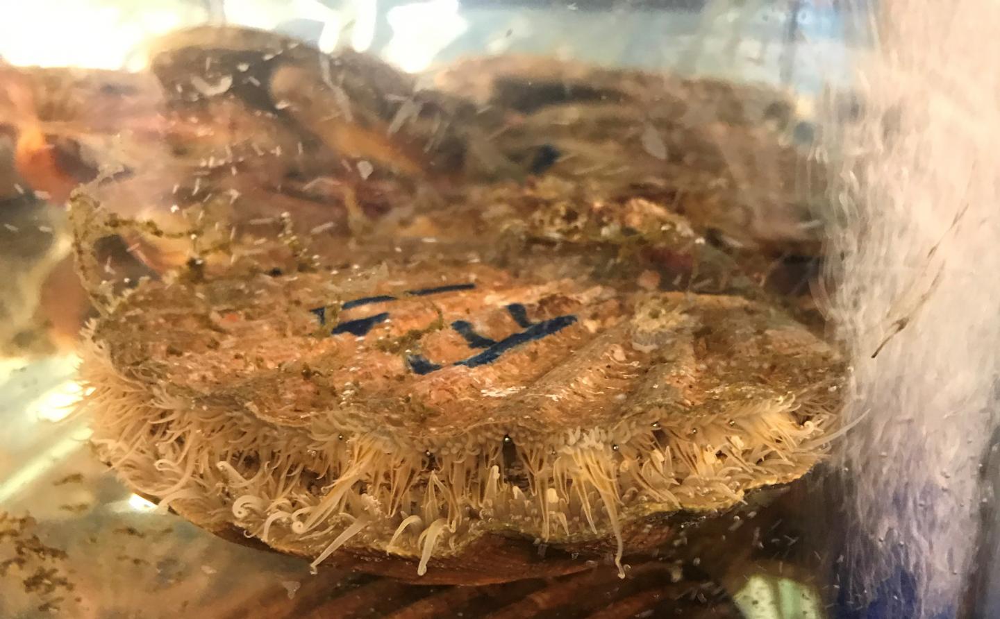
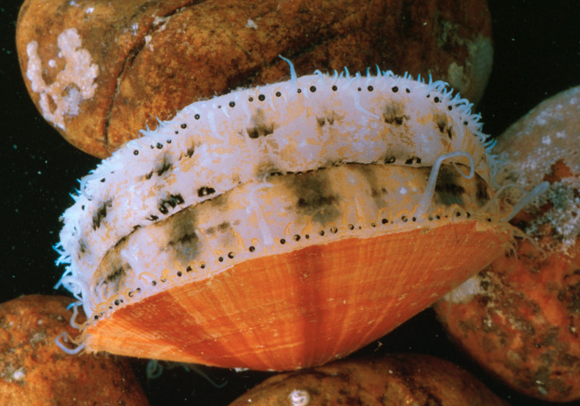
Ocean Microbiomes and Nanobiomes

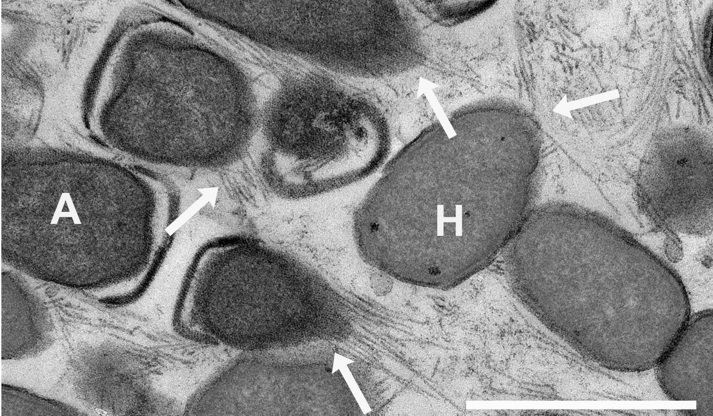
Life in the oceans is supported by a community of extremely small organisms that can be called a “nanobiome.” These nanoplankton particles, many of which measure less than 0.001× the volume of a white blood cell, harvest solar and chemical energy and channel essential elements into the food chain. A deep network of larger life forms (humans included) depends on these tiny microbes for its energy and chemical building blocks.

The importance of the oceanic nanobiome has only recently begun to be fully appreciated. Two dominant forms, Synechococcus and Prochlorococcus, were not discovered until the 1980s and 1990s.(32-34) Prochloroccus has now been demonstrated to be so abundant that it may account for as much as 10% of the world’s living organic carbon. The organism divides on a diel cycle while maintaining constant numbers, suggesting that about 5% of the world’s biomass flows through this species on a daily basis.(35-37)

Metagenomic studies show that many other less abundant life forms must exist but elude direct observation because they can neither be isolated nor grown in culture.

The small sizes of these organisms (and their genomes) indicate that they are highly specialized and optimized. Metagenome data indicate a large metabolic heterogeneity within the nanobiome. Rather than combining all life functions into a single organism, the nanobiome works as a network of specialists that can only exist as a community, therein explaining their resistance to being cultured. The detailed composition of the network is the result of interactions between the organisms themselves and the local physical and chemical environment. There is thus far little insight into how these networks are formed and how they maintain steady-state conditions in the turbulent natural ocean environment.

Rather than combining all life functions into a single organism, the nanobiome works as a network of specialists that can only exist as a community

The serendipitous discovery of Prochlorococcus happened by applying flow cytometry (developed as a medical technique for counting blood cells) to seawater.(34) With these medical instruments, the faint signals from nanoplankton can only be seen with great difficulty against noisy backgrounds. Currently, a small team is adapting flow cytometric technology to improve the capabilities for analyzing individual nanoplankton particles. The latest generation of flow cytometers enables researchers to count and to make quantitative observations of most of the small life forms (including some viruses) that comprise the nanobiome. To our knowledge, there are only two well-equipped mobile flow cytometry laboratories that are regularly taken to sea for real-time observations of the nanobiome. The laboratories include equipment for (meta)genome analysis and equipment to correlate the observations with the local physical parameters and (nutrient) chemistry in the ocean. Ultimately, integration of these measurements will be essential for understanding the complexity of the oceanic microbiome.

The ocean is tremendously undersampled. Ship time is costly and limited. Ultimately, inexpensive, automated, mobile biome observatories will require methods that integrate microbiome and nanobiome measurements, with (meta-) genomics analyses, with local geophysical and geochemical parameters.(38-42) To appreciate how the individual components of the ocean biome are related and work together, a more complete picture must be established.

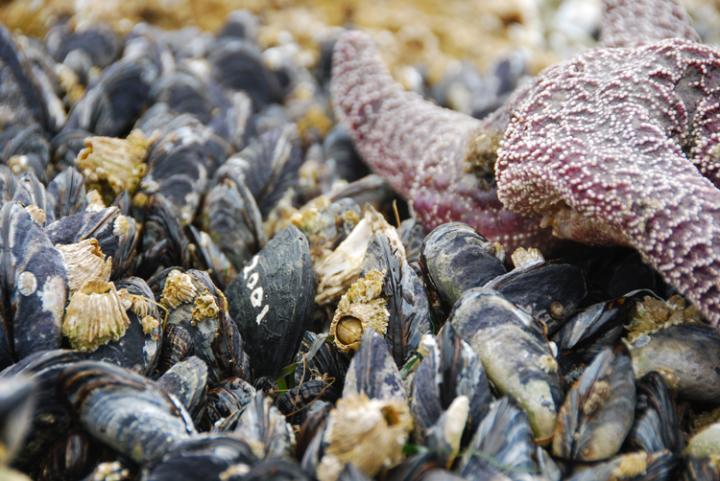
The marine environment consists of stratified zones, each with a unique, characteristic biome.(43) The sunlit waters near the surface are mixed by wind action. Deeper waters may be mixed only occasionally by passing storms. The dark deepest layers are stabilized by temperature/salinity density gradients. Organic material from the photosynthetically active surface descends into the deep zone, where it decomposes into nutrients that are mixed with compounds that are released by volcanic and seismic action. These nutrients diffuse upward to replenish the depleted surface waters. The biome is stratified accordingly, sometimes with sudden transitions on small scales. Photo-autotrophs dominate near the surface. Chemo-heterotrophs populate the deep. The makeup of the microbial assemblages is dictated by the local nutrient and oxygen concentrations. The spatiotemporal interplay of these systems is highly relevant to such issues as the carbon budget of the planet but remains little understood.

And then, there was this,

Nanoscience and Nanotechnology Opportunities

The great advantage of nanoscience and nanotechnology in studying microbiomes is that the nanoscale is the scale of function in biology. It is this convergence of scales at which we can “see” and at which we can fabricate that heralds the contributions that can be made by developing new nanoscale analysis tools.(159-168) Microbiomes operate from the nanoscale up to much larger scales, even kilometers, so crossing these scales will pose significant challenges to the field, in terms of measurement, stimulation/response, informatics, and ultimately understanding.

Some progress has been made in creating model systems(143-145, 169-173) that can be used to develop tools and methods. In these cases, the tools can be brought to bear on more complex and real systems. Just as nanoscience began with the ability to image atoms and progressed to the ability to manipulate structures both directly and through guided interactions,(162, 163, 174-176) it has now become possible to control structure, materials, and chemical functionality from the submolecular to the centimeter scales simultaneously. Whereas substrates and surface functionalization have often been tailored to be resistant to bioadhesion, deliberate placement of chemical patterns can also be used for the growth and patterning of systems, such as biofilms, to be put into contact with nanoscale probes.(177-180) Such methods in combination with the tools of other fields (vide infra) will provide the means to probe and to understand microbiomes.

Key tools for the microbiome will need to be miniaturized and made parallel. These developments will leverage decades of work in nanotechnology in the areas of nanofabrication,(181) imaging systems,(182, 183) lab-on-a-chip systems,(184) control of biological interfaces,(185) and more. Commercialized and commoditized tools, such as smart phone cameras, can also be adapted for use (vide infra). By guiding the development and parallelization of these tools, increasingly complex microbiomes will be opened for study.(167)

Imaging and sensing, in general, have been enjoying a Renaissance over the past decades, and there are various powerful measurement techniques that are currently available, making the Microbiome Initiative timely and exciting from the broad perspective of advanced analysis techniques. Recent advances in various -omics technologies, electron microscopy, optical microscopy/nanoscopy and spectroscopy, cytometry, mass spectroscopy, atomic force microscopy, nuclear imaging, and other techniques, create unique opportunities for researchers to investigate a wide range of questions related to microbiome interactions, function, and diversity. We anticipate that some of these advanced imaging, spectroscopy, and sensing techniques, coupled with big data analytics, will be used to create multimodal and integrated smart systems that can shed light onto some of the most important needs in microbiome research, including (1) analyzing microbial interactions specifically and sensitively at the relevant spatial and temporal scales; (2) determining and analyzing the diversity covered by the microbial genome, transcriptome, proteome, and metabolome; (3) managing and manipulating microbiomes to probe their function, evaluating the impact of interventions and ultimately harnessing their activities; and (4) helping us identify and track microbial dark matter (referring to 99% of micro-organisms that cannot be cultured).

In this broad quest for creating next-generation imaging and sensing instrumentation to address the needs and challenges of microbiome-related research activities comprehensively, there are important issues that need to be considered, as discussed below.

The piece is extensive and quite interesting, if you have the time.

What is the effect of nanoscale plastic on marine life?

A Nov.27, 2015 news item on Nanowerk announces a new UK (United Kingdom) research project designed to answer the question: what impact could nanoscale plastic particles  have on the marine environment?,

As England brings in pricing on plastic carrier bags, and Scotland reveals that similar changes a little over a year ago have reduced the use of such bags by 80%, new research led by Heriot-Watt University in conjunction with Plymouth University will look at the effect which even the most microscopic plastic particles can have on the marine environment.

While images of large ‘islands’ of plastic rubbish or of large marine animals killed or injured by the effects of such discards have brought home some of the obvious negative effects of plastics in the marine environment, it is known that there is more discarded plastic out there than we can account for, and much of it will have degraded into small or even microscopic particles.

It is the effect of these latter, known as nano-plastics, which will be studied under a £1.1m research project, largely funded by NERC [UK Natural Environment Research Council] and run by Heriot-Watt and Plymouth Universities.

A Nov. 25, 2015 Herriot-Watt University press release, which originated the news item, provides more details,

The project, RealRiskNano, will look at the risks these tiny plastic particles pose to the food web including filter-feeding organisms like mussels, clams and sediment dwelling organisms. It will focus on providing information to improve environmental risk assessment for nanoplastics, based on real-world exposure scenarios replicated in the laboratory.

Team leader Dr Theodore Henry, Associate Professor of Toxicology at Heriot-Watt’s School of Life Sciences, said that the study will build on previous research on nano-material toxicology, but will provide information which the earlier studies did not include.

“Pieces of plastic of all sizes have been found in even the most remote marine environments. It’s relatively easy to see some of the results: turtles killed by easting plastic bags which they take for jelly fish, or large marine mammals drowned when caught in discarded ropes and netting.

“But when plastics fragment into microscopic particles, what then? It’s easy to imagine that they simply disappear, but we know that nano-particles pose their own distinct threats purely because of their size. They’re small enough to be transported throughout the environment with unknown effects on organisms including toxicity and interference with processes of the digestive system.

An important component of the project, to be investigated by Dr Tony Gutierrez at Heriot-Watt, will be the study of interactions between microorganisms and the nanoplastics to reveal how these interactions affect their fate and toxicology.

The aim, said Dr Henry, is to provide the information which is needed to effect real change.“We simply don’t know what effects these nano-plastic particles may pose to the marine environment, to filter-feeders and on to fish, and through the RealRiskNano project we aim to provide this urgently needed information to the people whose job it is to assess risk to the marine ecosystem and decide what steps need to be taken to mitigate it.”

You can find the RealRiskNano website here.

A nanoscale bacteria power grid

It’s not often you see the word ‘spectacular’ when reading a science news item but it can be found in an Oct. 21, 2015 news item on ScienceDaily,

Electrical energy from the socket — this convenient type of power supply is apparently used by some microorganisms. Cells can meet their energy needs in the form of electricity through nano-wire connections. Researchers from the Max Planck Institute for Marine Microbiology in Bremen have discovered these possibly smallest power grids in the world when examining cell aggregates of methane degrading microorganisms. They consist of two completely different cell types, which can only jointly degrade methane. Scientists have discovered wire-like connections between the cells, which are relevant in energy exchanges.

It was a spectacular [emphasis mine] scientific finding when researchers discovered electrical wiring between microorganisms using iron as energy source in 2010. Immediately the question came up if electric power exchange is common in other microbially mediated reactions. One of the processes in question was the anaerobic oxidation of methane (AOM) that is responsible for the degradation of the greenhouse gas methane in the seafloor, and therefore has a great relevance for Earth climate. The microorganisms involved have been described for the first time in 2000 by researchers from Bremen and since then have been extensively studied.

This image accompanies the research,

Caption: Electron micrograph of the nanowires shows connecting archaea and sulphate reducing bacteria. The wires stretch out for several micrometres, longer than a single cell. The white bar represents the length of one micrometre. The arrows indicate the nanowires (A=ANME-Archaeen, H=HotSeep-1 partner bacteria). Credit: MPI f. Biophysical Chemistry

Caption: Electron micrograph of the nanowires shows connecting archaea and sulphate reducing bacteria. The wires stretch out for several micrometres, longer than a single cell. The white bar represents the length of one micrometre. The arrows indicate the nanowires (A=ANME-Archaeen, H=HotSeep-1 partner bacteria).
Credit: MPI f. Biophysical Chemistry

A Oct. 21, 2015 Max Planck press release (also on EurekAlert), which originated the news item, provides more information about methane in the ocean, power wires, and electron transporters,

In the ocean, methane is produced from the decay of dead biomass in subsurface sediments. The methane rises upwards to the seafloor, but before reaching the water column it is degraded by special consortia of archaea and bacteria. The archaea take up methane and oxidise it to carbonate. They pass on energy to their partner bacteria, so that the reaction can proceed. The bacteria respire sulphate instead of oxygen to gain energy (sulphate reducers). This may be an ancient metabolism, already relevant billions of years ago when the Earth’s atmosphere was oxygen-free. Yet today it remains unknown how the anaerobic oxidation of methane works biochemically.

Gunter Wegener, who authors the publication together with PhD student Viola Krukenberg, says: “We focused on thermophilic AOM consortia living at 60 degrees Celsius. For the first time we were able to isolate the partner bacteria to grow them alone. Then we systematically compared the physiology of the isolate with that of the AOM culture. We wanted to know which substances can serve as an energy carrier between the archaea and sulphate reducers.” Most compounds were ruled out quickly. At first, hydrogen was considered as energy source. However, the archaea did not produce sufficient hydrogen to explain the growth of sulphate reducers – hence the researchers had to change their strategy.

Direct power wires and electron transporters

One possible alternative was to look for direct connections channelling electrons between the cells. Using electron microscopy on the thermophilic AOM cultures this idea was confirmed. Dietmar Riedel, head of electron microscopy facilities at the Max Planck Institute in Goettingen says: “It was really challenging to visualize the cable-like structures. We embedded aggregates under high pressure using different embedding media. Ultrathin sections of these aggregates were then examined in near-native state using transmission electron microscopy.”

Viola Krukenberg adds: “We found all genes necessary for biosynthesis of the cellular connections called pili. Only when methane is added as energy source these genes are activated and pili are formed between bacteria and archaea.”

With length of several micrometres the wires can exceed the length of the cells by far, but their diameter is only a few nanometres. These wires provide the contact between the closely spaced cells and explain the spatial structure of the consortium, as was shown by a team of researchers led by Victoria Orphan from Caltech.

“Consortia of archaea and bacteria are abundant in nature. Our next step is to see whether other types also show such nanowire-like connections. It is important to understand how methane-degrading microbial consortia work, as they provide important functions in nature”, explains Antje Boetius, leader of the research group at the Institute in Bremen.

Here’s a link to and a citation for the paper,

Intercellular wiring enables electron transfer between methanotrophic archaea and bacteria by Gunter Wegener, Viola Krukenberg, Dietmar Riedel, Halina E. Tegetmeyer, & Antje Boetius. Nature 526, 587–590 (22 October 2015) doi:10.1038/nature15733 Published online 21 October 2015

This paper is behind a paywall.