Tag Archives: Okinawa Institute of Science and Technology Graduate University (OIST)

Dendritic painting: a physics story

A March 4, 2024 news item on phys.org announces research into the physics of using paints and inks in visual art, Note: A link has been removed,

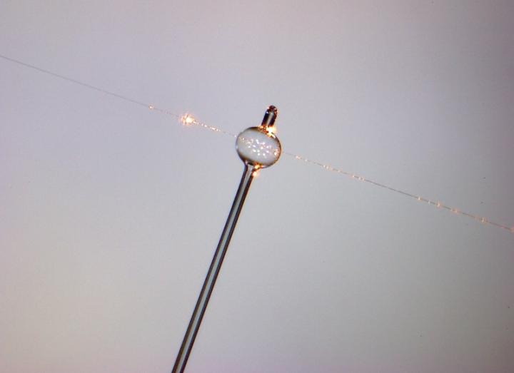
Falling from the tip of a brush suspended in mid-air, an ink droplet touches a painted surface and blossoms into a masterpiece of ever-changing beauty. It weaves a tapestry of intricate, evolving patterns. Some of them resemble branching snowflakes, thunderbolts or neurons, whispering the unique expression of the artist’s vision.

Okinawa Institute of Science and Technology (OIST) researchers set out to analyze the physical principles of this fascinating technique, known as dendritic painting. They took inspiration from the artwork of Japanese media artist, Akiko Nakayama. The work is published in the journal PNAS Nexus.

Caption: Japanese artist Akiko Nakayama manipulates alcohol and inks to create tree-like dendritic patterns during a live painting session. Credit: Photo Credit: Akiko Nakayama

Yes, the ends definitely look tree-like (maybe cedar). A February 29, 2024 Okinawa Institute of Science and Technology (OIST) press release (also on EurekAlert but published March 1, 2024), which originated the news item, goes on to describe the forces at work and provides instructions for creating your own dendritic paintings, Note: Links have been removed,

During her [Akiko Nakayama] live painting performances, she applies colourful droplets of acrylic ink mixed with alcohol atop a flat surface coated with a layer of acrylic paint. Beautiful fractals – tree-like geometrical shapes that repeat at different scales and are often found in nature – appear before the eyes of the audience. This is a captivating art form driven by creativity, but also by the physics of fluid dynamics.

“I have a deep admiration for scientists, such as Ukichiro Nakaya and Torahiko Terada, who made remarkable contributions to both science and art. I was very happy to be contacted by OIST physicist Chan San To. I am envious of his ability ‘to dialogue’ with the dendritic patterns, observing how they change shape in response to different approaches. Hearing this secret conversation was delightful,” explains Nakayama.

“Painters have often employed fluid mechanics to craft unique compositions. We have seen it with David Alfaro Siqueiros, Jackson Pollock, and Naoko Tosa, just to name a few. In our laboratory, we reproduce and study artistic techniques, to understand how the characteristics of the fluids influence the final outcome,” says OIST Professor Eliot Fried of OIST’s Mechanics and Materials Unit, who likes looking at dendritic paintings from artistic and scientific angles.

In dendritic painting, the droplets made of ink and alcohol experience various forces. One of them is surface tension – the force that makes rain droplets spherical in shape, and allows leaves to float on the surface of a pond. In particular, as alcohol evaporates faster than water, it alters the surface tension of the droplet. Fluid molecules tend to be pulled towards the droplet rim, which has higher surface tension compared to its centre. This is called the Marangoni effect and is the same phenomenon responsible for the formation of wine tears – the droplets or streaks of wine that form on the inside of a wine glass after swirling or tilting.

Secondly, the underlying paint layer also plays an important part in this artistic technique. Dr. Chan tested various types of liquids. For fractals to emerge, the liquid must be a fluid that decreases in viscosity under shear strain, meaning it has to behave somewhat like ketchup. It’s common knowledge that it’s hard to get ketchup out of the bottle unless you shake it. This happens because ketchup’s viscosity changes depending on shear strain. When you shake the bottle, the ketchup becomes less viscous, making it easier to pour it onto your dish. How is this applied to dendritic painting?

“In dendritic painting, the expanding ink droplet shears the underlying acrylic paint layer. It is not as strong as the shaking of a ketchup bottle, but it is still a form of shear strain. As with ketchup, the more stress there is, the easier it is for the ink droplets to flow,” explains Dr. Chan.

“We also showed that the physics behind this dendritic painting technique is similar to how liquid travels in a porous medium, such as soil. If you were to look at the mix of acrylic paint under the microscope, you would see a network of microscopic structures made of polymer molecules and pigments. The ink droplet tends to find its way through this underlying network, travelling through paths of least resistance, that leads to the dendritic pattern,” adds Prof. Fried.

Each dendritic print is one-of-a-kind, but there are at least two key aspects that artists can take into consideration to control the outcome of dendritic painting. The first and most important factor is the thickness of the paint layer spread on the surface. Dr. Chan observed that well-refined fractals appear with paint layer thinner than a half millimetre.

The second factor to experiment with is the concentration of diluting medium and paint in this paint layer. Dr. Chan obtained the most detailed fractals using three parts diluting medium and one part paint, or two parts diluting medium and one part paint. If the concentration of paint is higher, the droplet cannot spread well. Conversely, if the concentration of paint is lower, fuzzy edges will form. 

This is not the first science-meets-art project that members of the Mechanics and Materials Unit have embarked on. For example, they designed and installed a mobile sculpture on the OIST campus. The sculpture exemplifies a family of mechanical devices, called Möbius kaleidocycles, invented in the Unit, which may offer guidelines for designing chemical compounds with novel electronic properties.

Currently, Dr. Chan is also developing novel methods of analysing how the complexity of a sketch or painting evolves during its creation. He and Prof. Fried are optimistic that these methods might be applied to uncover hidden structures in experimentally captured or numerically generated images of flowing fluids.

“Why should we confine science to just technological progress?” wonders Dr. Chan. “I like exploring its potential to drive artistic innovation as well. I do digital art, but I really admire traditional artists. I sincerely invite them to experiment with various materials and reach out to us if they’re interested in collaborating and exploring the physics hidden within their artwork.”

Instructions to create dendritic painting at home

Everybody can have fun creating dendritic paintings. The materials needed include a non-absorbent surface (glass, synthetic paper, ceramics, etc.), a brush, a hairbrush, rubbing alcohol (iso-propyl alcohol), acrylic ink, acrylic paint and pouring medium.

  1. Dilute one part of acrylic paint to two or three parts of  pouring medium, or test other ratios to see how the result changes
  2. Apply this to the non-absorbent surface uniformly using a hairbrush. OIST physicists have found out that the thickness of the paint affects the result. For the best fractals, a layer of paint thinner than half millimetre is recommended.
  3. Mix rubbing alcohol with acrylic ink. The density of the ink may differ for different brands: have a try mixing alcohol and ink in different ratios
  4. When the white paint is still wet (hasn’t dried yet), apply a droplet of the ink with alcohol mix using a brush or another tool, such as a bamboo stick or a toothpick.
  5. Enjoy your masterpiece as it develops before your eyes. 

Here’s a link to and a citation for the paper,

Marangoni spreading on liquid substrates in new media art by San To Chan and Eliot Fried. PNAS Nexus, Volume 3, Issue 2, February 2024, pgae059 DOI: https://doi.org/10.1093/pnasnexus/pgae059 Published: 08 February 2024

This paper is open access.

Nanoparticle detection with whispers and bubbles

Caption: A magnified photograph of a glass Whispering Gallery Resonator. The bubble is extremely small, less than the width of a human hair. Credit: OIST (Okinawa Institute of Science and Technology Graduate University)

It was the reference to a whispering gallery which attracted my attention; a July 11, 2018 news item on Nanowerk is where I found it,

Technology created by researchers at the Okinawa Institute of Science and Technology Graduate University (OIST) [Japan] is literally shedding light on some of the smallest particles to detect their presence – and it’s made from tiny glass bubbles.

The technology has its roots in a peculiar physical phenomenon known as the “whispering gallery,” described by physicist Lord Rayleigh (John William Strutt) in 1878 and named after an acoustic effect inside the dome of St Paul’s Cathedral in London. Whispers made at one side of the circular gallery could be heard clearly at the opposite side. It happens because sound waves travel along the walls of the dome to the other side, and this effect can be replicated by light in a tiny glass sphere just a hair’s breadth wide called a Whispering Gallery Resonator (WGR).

A July 11, 2018 OIST press release by Andrew Scott (also on EurekAlert), provides more details,

When light is shined into the sphere, it bounces around and around the inner surface, creating an optical carousel. Photons bouncing along the interior of the tiny sphere can end up travelling for long distances, sometimes as far as 100 meters. But each time a photon bounces off the sphere’s surface, a small amount of light escapes. This leaking light creates a sort of aura around the sphere, known as an evanescent light field. When nanoparticles come within range of this field, they distort its wavelength, effectively changing its color. Monitoring these color changes allows scientists to use the WGRs as a sensor; previous research groups have used them to detect individual virus particles in solution, for example. But at OIST’s Light-Matter Interactions Unit, scientists saw they could improve on previous work and create even more sensitive designs. The study is published in Optica.

Today, Dr. Jonathan Ward is using WGRs to detect minute particles more efficiently than ever before. The WGRs they have made are hollow glass bubbles rather than balls, explains Dr. Ward. “We heated a small glass tube with a laser and had air blown down it – it’s a lot like traditional glass blowing”. Blowing the air down the heated glass tube creates a spherical chamber that can support the sensitive light field. The most noticeable difference between a blown glass ornament and these precision instruments is the scale: the glass bubbles can be as small as 100 microns- a fraction of a millimeter in width. Their size makes them fragile to handle, but also malleable.

Working from theoretical models, Dr. Ward showed that they could increase the size of the light field by using a thin spherical shell (a bubble, in other words) instead of a solid sphere. A bigger field would increase the range in which particles can be detected, increasing the efficacy of the sensor. “We knew we had the techniques and the materials to fabricate the resonator”, said Dr. Ward. “Next we had to demonstrate that it could outperform the current types used for particle detection”.

To prove their concept, the team came up with a relatively simple test. The new bubble design was filled with a liquid solution containing tiny particles of polystyrene, and light was shined along a glass filament to generate a light field in its liquid interior. As particles passed within range of the light field, they produced noticeable shifts in the wavelength that were much more pronounced than those seen with a standard spherical WGR.

With a more effective tool now at their disposal, the next challenge for the team is to find applications for it. Learning what changes different materials make to the light field would allow Dr Ward to identify and target them, and even control their activity.

Despite their fragility, these new versions of WGRs are easy to manufacture and can be safely transported in custom made cases. That means these sensors could be used in a wide verity of fields, such as testing for toxic molecules in water to detect pollution, or detecting blood borne viruses in extremely rural areas where healthcare may be limited.

For Dr. Ward however, there’s always room from improvement: “We’re always pushing to get even more sensitivity and find the smallest particle this sensor can detect. We want to push our detection to the physical limits.”

Here’s a link to and a citation for the paper,

Nanoparticle sensing beyond evanescent field interaction with a quasi-droplet microcavity by Jonathan M. Ward, Yong Yang, Fuchuan Lei, Xiao-Chong Yu, Yun-Feng Xiao, and Síle Nic Chormaic. Optica Vol. 5, Issue 6, pp. 674-677 (2018) https://doi.org/10.1364/OPTICA.5.000674

This paper is open access.

Molecular electronics: one atom at a time

This research on developing molecular electronics comes from Okinawa (Japan) according to a January 17, 2018 news item on Nanowerk,

Electronic devices are getting smaller and smaller. Early computers filled entire rooms. Today you can hold one in the palm of your hand. Now the field of molecular electronics is taking miniaturization to the next level. Researchers are creating electronic components so tiny they can’t be seen with the naked eye.

Molecular electronics is a branch of nanotechnology that uses single molecules, or nanoscale collections of molecules, as electronic components. The purpose is to create miniature computing devices, replacing bulk materials with molecular blocks.

For instance, metal atoms can be made into nanoscale ‘molecular wires.’ Also known as Extended Metal Atom Chains (EMACs), molecular wires are one-dimensional chains of single metal atoms connected to an organic molecule, called a ligand, that acts as a support. Molecular wire-type compounds have a diverse array of potential uses, from LED lights to catalysts.

Researchers at the Okinawa Institute of Science and Technology Graduate University (OIST) have found a simple way to create copper molecular wires of different lengths by adding or removing copper atoms one by one. “This is the first example of a molecular copper wire being formed in a stepwise, atom-by-atom process,” says Julia Khusnutdinova, head of the OIST Coordination Chemistry and Catalysis Unit. “Our method can be compared to Lego construction in which you add one brick at a time,” she says.

A January 16, 2018 OIST press release (also on EurekAlert but with a January 17, 2018 date) by Sophie Protheroe, which originated the news item, adds detail,

Molecular wires can vary in length, with different lengths having different molecular properties and practical applications.  At present, the longest EMAC reported in the literature is based on nickel and it contains 11 metal atoms in a single linear chain.

The structure of the longest EMAC reported in the literature, confirmed by X-ray crystallography. It contains 11 nickel atoms arranged in a linear chain.

Creating molecular wires of different lengths is difficult because it requires a specific ligand to be synthesized each time. The ligand, which can be seen as an ‘insulator’ by analogy to the macro world, helps the wires to form by bringing the metal atoms together and aligning them into a linear string. However, creating ligands of different lengths can be an elaborate and complicated process.

The OIST researchers have found a new way to overcome this problem.  “We have created a single dynamic ligand that can be used to synthesize multiple chain lengths,” says Dr. Orestes Rivada-Wheelaghan, first author of the paper.  “This is much more efficient than making a new ligand each time,” he says.

In their paper, published in Angewandte Chemie International Edition, the researchers describe their new stepwise method of creating copper molecular wires.  “The ligand opens up from one end to let a metal atom enter and, when the chain extends, the ligand undergoes a sliding movement along the chain to accommodate more metal atoms,” says Prof. Khusnutdinova.  “This can be likened to a molecular accordion that can be extended and shortened,” says Rivada-Wheelaghan. By adding or removing copper atoms one at a time in this way, the researchers can construct molecular wires of different lengths, varying from 1 to 4 copper atoms.

A cartoon by Dr. Rivada-Wheelaghan shows the simple stepwise process of copper atom chain synthesis using a dynamic ligand. Copper atoms can be added or removed one by one to make chains of different lengths.

This dynamic ligand offers a new way for chemists to synthesize molecules with specific shapes and properties, creating potential for many practical applications in microelectronics and beyond.

“The next step is to develop dynamic ligands that could be used to create molecular wires made from other metals, or a combination of different metals,” says Dr. Rivada-Wheelaghan. “For example, by selectively inserting copper atoms at the termini of the chain, and using a different type of metal at the center of the chain, we could create new compounds with interesting electronic properties,” says Prof. Khusnutdinova.

I particularly enjoy the cartoon. Getting back to business, here’s a link to and a citation for the paper,

Controlled and Reversible Stepwise Growth of Linear Copper(I) Chains Enabled by Dynamic Ligand Scaffolds by Dr. Orestes Rivada-Wheelaghan, Sandra L. Aristizábal, Dr. Joaquín López-Serrano, Dr. Robert R. Fayzullin, and Prof. Julia R. Khusnutdinova. Angewandte Chemie International Edition Version of Record online: 23 NOV 2017 Volume 56, Issue 51, pages 16267–16271, December 18, 2017 DOI: 10.1002/anie.201709167

© 2017 Wiley-VCH Verlag GmbH & Co. KGaA, Weinheim

This paper is behind a paywall.

Ukidama-structured nanoparticles discovered

The researchers discovered a new nanoparticle structure that resemble the ukidama, glass fishing floats, used regularly by Japanese fishermen. The nanoparticle has a core of one element (copper) and is surrounded by a “cage” of another element (silver). The silver does not cover certain areas of the copper core, which is very similar to the rope that surrounds the glass float. Courtesy: Okinawa Institute of Science and Technology (OIST)

The researchers discovered a new nanoparticle structure that resemble the ukidama, glass fishing floats, used regularly by Japanese fishermen. The nanoparticle has a core of one element (copper) and is surrounded by a “cage” of another element (silver). The silver does not cover certain areas of the copper core, which is very similar to the rope that surrounds the glass float. Courtesy: Okinawa Institute of Science and Technology (OIST)

What a beautiful image to illustrate the new ukidama nanoparticle structure! Here’s the announcement in a June 13, 2016 news item on ScienceDaily,

Sometimes it is the tiny things in the world that can make an incredible difference. One of these things is the nanoparticle. Nanoparticles may be small, but they have a variety of important applications in areas such as, medicine, manufacturing, and energy. A team of researchers from Okinawa Institute of Science and Technology Graduate University (OIST) recently discovered a unique copper-silver nanoparticle structure that has a core of one element surrounded by a “cage” of the other element. However, the cage does not cover certain areas of the core, which very much resembles the Japanese glass fishing floats traditionally covered with rope called ukidama.

This previously undiscovered ukidama structure may have properties that can help the team on their mission for optimal nanotechnology. …

A June 13, 2016 OIST press release by Rebecca Holland (also on EurekAlert; the June 12, 2016 publication date discrepancy is likely due to timezone issues), which originated the news item, provides more insight into the research team’s workings,

“The ukidama is a unique structure, which means that it can likely give us unique properties,” said Panagiotis Grammatikopoulos, first author and group leader of the OIST Nanoparticles by Design Unit. “The idea is that now that we know about this structure we may be able to fine tune it to our applications.”

The OIST researchers are continually working to create and design nanoparticles that can be used in biomedical technology. Specifically, the team works to design the optimal nanoparticles for technologies like smart gas sensors that can send information about what is going on inside your body to your smart phone for better diagnoses. Another application is the label free biosensor, a device that can detect chemical substances without the hindrance of fluorescent or radioactive labels. The identification of the ukidama structure is important in this endeavour because having a new structure increases the possibilities for technological advancements.

“The more parameters that we can control the more flexibility we have in our applications and devices,” Prof. Mukhles Sowwan, author and head of OIST’s Nanoparticles by Design Unit said. “Therefore, we need to optimize many properties of these nanoparticles: the size, chemical composition, crystallinity, shape, and structure.”

The discovery of the ukidama structure was found through sputtering copper and silver atoms simultaneously, but independently, through a magnetron-sputtering system at high temperatures. When the atoms began to cool they combined into bi-metallic nanoparticles. During the sputtering process, researchers could control the ratio of silver to copper, with the rate of power with which the atoms were sputtered. They found that the ukidama structure was possible, especially when the copper was the dominant element, since silver atoms have a higher tendency to diffuse on the nanoparticle surface. From their experimental findings, the team was able to create simulations that can clearly show how the ukidama nanoparticles form.

The team is now looking to see if this structure can be recreated in other types of nanoparticles, which could be an even bigger step in the optimization of nanoparticles for biomedical application and nanotechnology.

“We design and optimize nanoparticles for biomedical devices and nanotechnology,” Sowwan said. “Because the ukidama is a new structure, it may have properties that could be utilized in our applications.”

Co-author, Antony Galea, formerly of the Nanoparticles by Design Unit, was responsible for the experimental portion of this study and has since moved to OIST’s Technology and Licensing Section to help research – like this work being done with nanoparticles that can be utilized in applications – move into the market.

“Our aim is to take research created by OIST from the lab to the real world,” Galea said. “This is a way that work done at OIST, such as by the Nanoparticles by Design Unit, can benefit society.”

Here’s a link to and a citation for the paper,

Kinetic trapping through coalescence and the formation of patterned Ag–Cu nanoparticles by Panagiotis Grammatikopoulos, Joseph Kioseoglou, Antony Galea, Jerome Vernieres, Maria Benelmekki, Rosa E. Diaz, Mukhles Sowwan. Nanoscale, 2016; 8 (18): 9780 DOI: 10.1039/C5NR08256K

I believe this paper is behind a paywall.