Tag Archives: Oliver Kempski

Gold nanoparticle tattoo changes medical diagnostics?

The tattoos are in fact implantable sensors. Here’s more from an April 6, 2021 news item on ScienceDaily,

The idea of implantable sensors that continuously transmit information on vital values and concentrations of substances or drugs in the body has fascinated physicians and scientists for a long time. Such sensors enable the constant monitoring of disease progression and therapeutic success. However, until now implantable sensors have not been suitable to remain in the body permanently but had to be replaced after a few days or weeks. On the one hand, there is the problem of implant rejection because the body recognizes the sensor as a foreign object. On the other hand, the sensor’s color which indicates concentration changes has been unstable so far and faded over time. Scientists at Johannes Gutenberg University Mainz (JGU) have developed a novel type of implantable sensor which can be operated in the body for several months. The sensor is based on color-stable gold nanoparticles that are modified with receptors for specific molecules. Embedded into an artificial polymeric tissue, the nanogold is implanted under the skin where it reports changes in drug concentrations by changing its color.

An April 6, 2021 Johannes Gutenberg Universitaet Mainz press release (also on EurekAlert), which originated the news item, provides more detail about the proposed tattoo/implantable sensors,

Implant reports information as an “invisible tattoo”

Professor Carsten Sönnichsen’s research group at JGU has been using gold nanoparticles as sensors to detect tiny amounts of proteins in microscopic flow cells for many years. Gold nanoparticles act as small antennas for light: They strongly absorb and scatter it and, therefore, appear colorful. They react to alterations in their surrounding by changing color. Sönnichsen’s team has exploited this concept for implanted medical sensing.

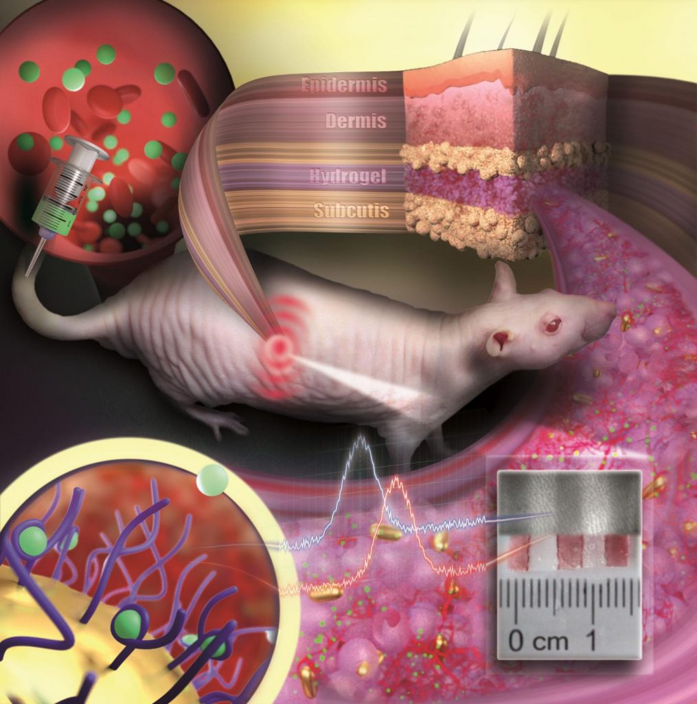
To prevent the tiny particles from swimming away or being degraded by immune cells, they are embedded in a porous hydrogel with a tissue-like consistency. Once implanted under the skin, small blood vessels and cells grow into the pores. The sensor is integrated in the tissue and is not rejected as a foreign body. “Our sensor is like an invisible tattoo, not much bigger than a penny and thinner than one millimeter,” said Professor Carsten Sönnichsen, head of the Nanobiotechnology Group at JGU. Since the gold nanoparticles are infrared, they are not visible to the eye. However, a special kind of measurement device can detect their color noninvasively through the skin.

In their study published in Nano Letters, the JGU researchers implanted their gold nanoparticle sensors under the skin of hairless rats. Color changes in these sensors were monitored following the administration of various doses of an antibiotic. The drug molecules are transported to the sensor via the bloodstream. By binding to specific receptors on the surface of the gold nanoparticles, they induce color change that is dependent on drug concentration. Thanks to the color-stable gold nanoparticles and the tissue-integrating hydrogel, the sensor was found to remain mechanically and optically stable over several months.

Huge potential of gold nanoparticles as long-lasting implantable medical sensors

“We are used to colored objects bleaching over time. Gold nanoparticles, however, do not bleach but keep their color permanently. As they can be easily coated with various different receptors, they are an ideal platform for implantable sensors,” explained Dr. Katharina Kaefer, first author of the study.

The novel concept is generalizable and has the potential to extend the lifetime of implantable sensors. In future, gold nanoparticle-based implantable sensors could be used to observe concentrations of different biomarkers or drugs in the body simultaneously. Such sensors could find application in drug development, medical research, or personalized medicine, such as the management of chronic diseases.

Interdisciplinary team work brought success

Sönnichsen had the idea of using gold nanoparticles as implanted sensors already in 2004 when he started his research in biophysical chemistry as a junior professor in Mainz. However, the project was not realized until ten years later in cooperation with Dr. Thies Schroeder and Dr. Katharina Kaefer, both scientists at JGU. Schroeder was experienced in biological research and laboratory animal science and had already completed several years of research work in the USA. Kaefer was looking for an exciting topic for her doctorate and was particularly interested in the complex and interdisciplinary nature of the project. Initial results led to a stipend awarded to Kaefer by the Max Planck Graduate Center (MPGC) as well as financial support from Stiftung Rheinland-Pfalz für Innovation. “Such a project requires many people with different scientific backgrounds. Step by step we were able to convince more and more people of our idea,” said Sönnichsen happily. Ultimately, it was interdisciplinary teamwork that resulted in the successful development of the first functional implanted sensor with gold nanoparticles.

The researchers have provided an image which illustrates several elements described in the press release,

Caption: Gold nanoparticles embedded in a porous hydrogel can be implanted under the skin and used as medical sensors. The sensor is like an invisible tattoo revealing concentration changes of substances in the blood by color change. Credit: ill./©: Nanobiotechnology Group, JGU Department of Chemistry

Here’s a link to and a citation for the paper,

Implantable Sensors Based on Gold Nanoparticles for Continuous Long-Term Concentration Monitoring in the Body by Katharina Kaefer, Katja Krüger, Felix Schlapp, Hüseyin Uzun, Sirin Celiksoy, Bastian Flietel, Axel Heimann, Thies Schroeder, Oliver Kempski, and Carsten Sönnichsen. Nano Lett. 2021, XXXX, XXX, XXX-XXX DOI: https://doi.org/10.1021/acs.nanolett.1c00887 Publication Date:March 30, 2021 © 2021 The Authors. Published by American Chemical Society

This paper is behind a paywall.

Graphene-based neural probes

I have two news bits (dated almost one month apart) about the use of graphene in neural probes, one from the European Union and the other from Korea.

European Union (EU)

This work is being announced by the European Commission’s (a subset of the EU) Graphene Flagship (one of two mega-funding projects announced in 2013; 1B Euros each over ten years for the Graphene Flagship and the Human Brain Project).

According to a March 27, 2017 news item on ScienceDaily, researchers have developed a graphene-based neural probe that has been tested on rats,

Measuring brain activity with precision is essential to developing further understanding of diseases such as epilepsy and disorders that affect brain function and motor control. Neural probes with high spatial resolution are needed for both recording and stimulating specific functional areas of the brain. Now, researchers from the Graphene Flagship have developed a new device for recording brain activity in high resolution while maintaining excellent signal to noise ratio (SNR). Based on graphene field-effect transistors, the flexible devices open up new possibilities for the development of functional implants and interfaces.

The research, published in 2D Materials, was a collaborative effort involving Flagship partners Technical University of Munich (TU Munich; Germany), Institut d’Investigacions Biomèdiques August Pi i Sunyer (IDIBAPS; Spain), Spanish National Research Council (CSIC; Spain), The Biomedical Research Networking Center in Bioengineering, Biomaterials and Nanomedicine (CIBER-BBN; Spain) and the Catalan Institute of Nanoscience and Nanotechnology (ICN2; Spain).

Caption: Graphene transistors integrated in a flexible neural probe enables electrical signals from neurons to be measured with high accuracy and density. Inset: The tip of the probe contains 16 flexible graphene transistors. Credit: ICN2

A March 27, 2017 Graphene Flagship press release on EurekAlert, which originated the news item, describes the work,  in more detail,

The devices were used to record the large signals generated by pre-epileptic activity in rats, as well as the smaller levels of brain activity during sleep and in response to visual light stimulation. These types of activities lead to much smaller electrical signals, and are at the level of typical brain activity. Neural activity is detected through the highly localised electric fields generated when neurons fire, so densely packed, ultra-small measuring devices is important for accurate brain readings.

The neural probes are placed directly on the surface of the brain, so safety is of paramount importance for the development of graphene-based neural implant devices. Importantly, the researchers determined that the graphene-based probes are non-toxic, and did not induce any significant inflammation.

Devices implanted in the brain as neural prosthesis for therapeutic brain stimulation technologies and interfaces for sensory and motor devices, such as artificial limbs, are an important goal for improving quality of life for patients. This work represents a first step towards the use of graphene in research as well as clinical neural devices, showing that graphene-based technologies can deliver the high resolution and high SNR needed for these applications.

First author Benno Blaschke (TU Munich) said “Graphene is one of the few materials that allows recording in a transistor configuration and simultaneously complies with all other requirements for neural probes such as flexibility, biocompability and chemical stability. Although graphene is ideally suited for flexible electronics, it was a great challenge to transfer our fabrication process from rigid substrates to flexible ones. The next step is to optimize the wafer-scale fabrication process and improve device flexibility and stability.”

Jose Antonio Garrido (ICN2), led the research. He said “Mechanical compliance is an important requirement for safe neural probes and interfaces. Currently, the focus is on ultra-soft materials that can adapt conformally to the brain surface. Graphene neural interfaces have shown already great potential, but we have to improve on the yield and homogeneity of the device production in order to advance towards a real technology. Once we have demonstrated the proof of concept in animal studies, the next goal will be to work towards the first human clinical trial with graphene devices during intraoperative mapping of the brain. This means addressing all regulatory issues associated to medical devices such as safety, biocompatibility, etc.”

Caption: The graphene-based neural probes were used to detect rats’ responses to visual stimulation, as well as neural signals during sleep. Both types of signals are small, and typically difficult to measure. Credit: ICN2

Here’s a link to and a citation for the paper,

Mapping brain activity with flexible graphene micro-transistors by Benno M Blaschke, Núria Tort-Colet, Anton Guimerà-Brunet, Julia Weinert, Lionel Rousseau, Axel Heimann, Simon Drieschner, Oliver Kempski, Rosa Villa, Maria V Sanchez-Vives. 2D Materials, Volume 4, Number 2 DOI https://doi.org/10.1088/2053-1583/aa5eff Published 24 February 2017

© 2017 IOP Publishing Ltd

This paper is behind a paywall.

Korea

While this research from Korea was published more recently, the probe itself has not been subjected to in vivo (animal testing). From an April 19, 2017 news item on ScienceDaily,

Electrodes placed in the brain record neural activity, and can help treat neural diseases like Parkinson’s and epilepsy. Interest is also growing in developing better brain-machine interfaces, in which electrodes can help control prosthetic limbs. Progress in these fields is hindered by limitations in electrodes, which are relatively stiff and can damage soft brain tissue.

Designing smaller, gentler electrodes that still pick up brain signals is a challenge because brain signals are so weak. Typically, the smaller the electrode, the harder it is to detect a signal. However, a team from the Daegu Gyeongbuk Institute of Science & Technology [DGIST} in Korea developed new probes that are small, flexible and read brain signals clearly.

This is a pretty interesting way to illustrate the research,

Caption: Graphene and gold make a better brain probe. Credit: DGIST

An April 19, 2017 DGIST press release (also on EurekAlert), which originated the news item, expands on the theme (Note: A link has been removed),

The probe consists of an electrode, which records the brain signal. The signal travels down an interconnection line to a connector, which transfers the signal to machines measuring and analysing the signals.

The electrode starts with a thin gold base. Attached to the base are tiny zinc oxide nanowires, which are coated in a thin layer of gold, and then a layer of conducting polymer called PEDOT. These combined materials increase the probe’s effective surface area, conducting properties, and strength of the electrode, while still maintaining flexibility and compatibility with soft tissue.

Packing several long, thin nanowires together onto one probe enables the scientists to make a smaller electrode that retains the same effective surface area of a larger, flat electrode. This means the electrode can shrink, but not reduce signal detection. The interconnection line is made of a mix of graphene and gold. Graphene is flexible and gold is an excellent conductor. The researchers tested the probe and found it read rat brain signals very clearly, much better than a standard flat, gold electrode.

“Our graphene and nanowires-based flexible electrode array can be useful for monitoring and recording the functions of the nervous system, or to deliver electrical signals to the brain,” the researchers conclude in their paper recently published in the journal ACS Applied Materials and Interfaces.

The probe requires further clinical tests before widespread commercialization. The researchers are also interested in developing a wireless version to make it more convenient for a variety of applications.

Here’s a link to and a citation for the paper,

Enhancement of Interface Characteristics of Neural Probe Based on Graphene, ZnO Nanowires, and Conducting Polymer PEDOT by Mingyu Ryu, Jae Hoon Yang, Yumi Ahn, Minkyung Sim, Kyung Hwa Lee, Kyungsoo Kim, Taeju Lee, Seung-Jun Yoo, So Yeun Kim, Cheil Moon, Minkyu Je, Ji-Woong Choi, Youngu Lee, and Jae Eun Jang. ACS Appl. Mater. Interfaces, 2017, 9 (12), pp 10577–10586 DOI: 10.1021/acsami.7b02975 Publication Date (Web): March 7, 2017

Copyright © 2017 American Chemical Society

This paper is behind a paywall.