Tag Archives: Olli Ikkala

Novel self-assembly at 102 atoms

A Jan. 13, 2017 news item on ScienceDaily announces a discovery about self-assembly of 102-atom gold nanoclusters,

Self-assembly of matter is one of the fundamental principles of nature, directing the growth of larger ordered and functional systems from smaller building blocks. Self-assembly can be observed in all length scales from molecules to galaxies. Now, researchers at the Nanoscience Centre of the University of Jyväskylä and the HYBER Centre of Excellence of Aalto University in Finland report a novel discovery of self-assembling two- and three-dimensional materials that are formed by tiny gold nanoclusters of just a couple of nanometres in size, each having 102 gold atoms and a surface layer of 44 thiol molecules. The study, conducted with funding from the Academy of Finland and the European Research Council, has been published in Angewandte Chemie.

A Jan. 13, 2017 Academy of Finland press release, which originated the news item, provides more technical information about the work,

The atomic structure of the 102-atom gold nanocluster was first resolved by the group of Roger D Kornberg at Stanford University in 2007 (2). Since then, several further studies of its properties have been conducted in the Jyväskylä Nanoscience Centre, where it has also been used for electron microscopy imaging of virus structures (3). The thiol surface of the nanocluster has a large number of acidic groups that can form directed hydrogen bonds to neighbouring nanoclusters and initiate directed self-assembly.

The self-assembly of gold nanoclusters took place in a water-methanol mixture and produced two distinctly different superstructures that were imaged in a high-resolution electron microscope at Aalto University. In one of the structures, two-dimensional hexagonally ordered layers of gold nanoclusters were stacked together, each layer being just one nanocluster thick. Modifying the synthesis conditions, also three-dimensional spherical, hollow capsid structures were observed, where the thickness of the capsid wall corresponds again to just one nanocluster size (see figure).

While the details of the formation mechanisms of these superstructures warrant further systemic investigations, the initial observations open several new views into synthetically made self-assembling nanomaterials.

“Today, we know of several tens of different types of atomistically precise gold nanoclusters, and I believe they can exhibit a wide variety of self-assembling growth patterns that could produce a range of new meta-materials,” said Academy Professor Hannu Häkkinen, who coordinated the research at the Nanoscience Centre. “In biology, typical examples of self-assembling functional systems are viruses and vesicles. Biological self-assembled structures can also be de-assembled by gentle changes in the surrounding biochemical conditions. It’ll be of great interest to see whether these gold-based materials can be de-assembled and then re-assembled to different structures by changing something in the chemistry of the surrounding solvent.”

“The free-standing two-dimensional nanosheets will bring opportunities towards new-generation functional materials, and the hollow capsids will pave the way for highly lightweight colloidal framework materials,” Postdoctoral Researcher Nonappa (Aalto University) said.

Professor Olli Ikkala of Aalto University said: “In a broader framework, it has remained as a grand challenge to master the self-assemblies through all length scales to tune the functional properties of materials in a rational way. So far, it has been commonly considered sufficient to achieve sufficiently narrow size distributions of the constituent nanoscale structural units to achieve well-defined structures. The present findings suggest a paradigm change to pursue strictly defined nanoscale units for self-assemblies.”

References:

(1)    Nonappa, T. Lahtinen, J.S. Haataja, T.-R. Tero, H. Häkkinen and O. Ikkala, “Template-Free Supracolloidal Self-Assembly of Atomically Precise Gold Nanoclusters: From 2D Colloidal Crystals to Spherical Capsids”, Angewandte Chemie International Edition, published online 23 November 2016, DOI: 10.1002/anie.201609036

(2)    P. Jadzinsky et al., “Structure of a thiol-monolayer protected gold nanoparticle at 1.1Å resolution”, Science 318, 430 (2007)

(3)    V. Marjomäki et al., “Site-specific targeting of enterovirus capsid by functionalized monodispersed gold nanoclusters”, PNAS 111, 1277 (2014)

Here’s the figure mentioned in the news release,
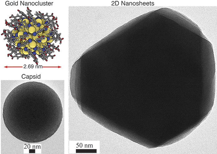
Figure: 2D hexagonal sheet-like and 3D capsid structures based on atomically precise gold nanoclusters as guided by hydrogen bonding between the ligands. The inset in the top left corner shows the atomic structure of one gold nanocluster.

Here’s a link to and a citation for the paper,

Template-Free Supracolloidal Self-Assembly of Atomically Precise Gold Nanoclusters: From 2D Colloidal Crystals to Spherical Capsids by Dr. Nonappa, Dr. Tanja Lahtinen, M. Sc. Johannes. S. Haataja, Dr. Tiia-Riikka Tero, Prof. Hannu Häkkinen, and Prof. Olli Ikkala. Angewandte Chemie International Edition Volume 55, Issue 52, pages 16035–16038, December 23, 2016 Version of Record online: 23 NOV 2016 DOI: 10.1002/anie.201609036

© 2016 Wiley-VCH Verlag GmbH & Co. KGaA, Weinheim

This paper is behind a paywall.

Nanocellulose at the American Chemical Society’s 243rd annual meeting

Nanocellulose seems to be one of the major topics at the ACS’s (Americal Chemical Society) 243rd annual meeting themed Chemistry of Life  in San Diego, California, March 25-29, 2012. From the March 25, 2012 news item on Nanowerk,

… almost two dozen reports in the symposium titled, “Cellulose-Based Biomimetic and Biomedical Materials,” that focused on the use of specially processed cellulose in the design and engineering of materials modeled after biological systems. Cellulose consists of long chains of the sugar glucose linked together into a polymer, a natural plastic–like material. Cellulose gives wood its remarkable strength and is the main component of plant stems, leaves and roots. Traditionally, cellulose’s main commercial uses have been in producing paper and textiles –– cotton being a pure form of cellulose. But development of a highly processed form of cellulose, termed nanocellulose, has expanded those applications and sparked intense scientific research. Nanocellulose consists of the fibrils of nanoscale diameters so small that 50,000 would fit across the width of the period at the end of this sentence.

“We are in the middle of a Golden Age, in which a clearer understanding of the forms and functions of cellulose architectures in biological systems is promoting the evolution of advanced materials,” said Harry Brumer, Ph.D., of Michael Smith Laboratories, University of British Columbia, Vancouver. He was a co-organizer of the symposium with J. Vincent Edwards, Ph.D., a research chemist with the Agricultural Research Service, U.S. Department of Agriculture in New Orleans, Louisiana. “This session on cellulose-based biomimetic and biomedical materials is really very timely due to the sustained and growing interest in the use of cellulose, particularly nanoscale cellulose, in biomaterials.”

One of the presenters has a very charming way of describing the nanocellulose product his team is working on (from the news item),

Olli Ikkala, Ph.D., [Aalto University, Finland] described the new buoyant material, engineered to mimic the water strider’s long, thin feet and made from an “aerogel” composed of the tiny nano-fibrils from the cellulose in plants. Aerogels are so light that some of them are denoted as “solid smoke. [emphasis mine]” The nanocellulose aerogels also have remarkable mechanical properties and are flexible.

There were some 20 presentations in this symposium held under the auspices of the ACS annual meeting. Here’s a few of the presentations (some of these folks have been featured on this blog previously), from the news item,

Native cellulose nanofibers: From biomimetic nanocomposites to functionalized gel spun fibers and functional aerogels Olli Ikkala, Professor, PhD, Aalto University, P.O. Box 5100, Espoo, Finland, FIN-02015, Finland , 358-9-470 23154, olli.ikkala@aalto.fi Native cellulose nanofibers and whiskers attract interest even beyond the traditional cellulose community due to their mechanical properties, availability and sustainability. We describe biomimetic nanocomposites with aligned self-assemblies combining nanocellulose with nanoclays, polymers, block copolymer, or graphene, allowing exciting mechanical properties. Functional ductile and even flexible aerogels are presented, combining superhydrophobicity, superoleophobicity, oil-spill absorption, photocatalytics, optically switchable water absorption, sensing, and antimicrobial properties. Finally mechanically excellent fibers are gel-spun and functionalized for electric, magnetic, optical and drug-release properties.

Evaluation of skin tissue repair materials from bacterial cellulose Lina Fu, Miss, Huazhong University of Science & Technology, College of Life Science & Technology, 1037 Luoyu Road, Wuhan, Hubei, 430074, China , 86-18971560696, runa0325@gmail.com Bacterial cellulose (BC) has been reported as the materials in the tissue engineering fields, such as skin, bone, vascular and cartilage tissue engineering. Exploitation of the skin substitutes and modern wound dressing materials by using BC has attracted much attention. A skin tissue repair materials based on BC have been biosynthesized by Gluconacetobacter xylinus. The nano-composites of BC and chitosan form a cohesive gel structure, and the cell toxicity of the composite is excellent. Unlike other groups, which showed more inflammatory behavior, the inflammatory cells of the BC group were mainly polymorph-nuclear and showed few lymphocytes. The BC skin tissue repair material has an obviously curative effect in promoting the healing of epithelial tissue and reducing inflammation. With its superior mechanical properties, and the excellent biocompatibility, these skin tissue repair materials based on BC have great promise and potential for wound healing and very high clinical value.

….

New materials from nanocrystalline cellulose Mark MacLachlan [mentioned in my Nov. 18, 2010 posting], University of British Columbia, Department of Chemistry, 2036 Main Mall, Vancouver, BC, V6T 1Z1, Canada , 604-822-3070, mmaclach@chem.ubc.ca Nanocrystalline cellulose (NCC) is available from the acid-catalyzed degradation of cellulosic materials. NCC is composed of cylindrical crystallites with diameters of ca. 5-10 nm and large aspect ratios. This form of cellulose has intriguing properties, including its ability to form a chiral nematic structure. By using the chiral nematic organization of NCC as a template, we have been able to create highly porous silica films and carbon films with chiral nematic organization.1,2 These materials are iridescent and their structures mimic the shells of jewel beetles. In this paper, I will describe our recent efforts to use NCC to create new materials with interesting optical properties.

Factors influencing chiral nematic pitch and texture of cellulose nanocrystal films Derek G Gray, McGill University, Department of Chemistry, Pulp and Paper Building, 3420 University Street, Montreal, QC, H3A 2A7, Canada , 1-514-398-6182, derek.gray@mcgill.ca Appropriately stabilized cellulose nanocrystal (NCC) suspensions in water form chiral nematic liquid crystalline phases above some critical concentration. In the absence of added electrolye, the chiral nematic pitch of such suspensions is longer than that of visible light. Films prepared by evaporation from the suspensions also often display the characteristic fingerprint patterns characteristic of long-pitch chiral nematic phases, but the pitch values can be shifted into the visible range by adding small quantities of electrolyte to the evaporating suspension. The factors that control the final pitch have been the subject of some confusion. While still not well understood, it is clear that at high nanocrystal concentrations and in solid films, the pitch is not simply a reversible function of nanocrystal concentration. We examine some of the factors that control the pitch and liquid crystal texture during the drying of chiral nematic NCC films.

….

Bioprinting of 3D porous nanocellulose scaffolds for tissue engineering and organ regeneration Paul Gatenholm, Professor, [mentioned in my March 19, 2012 posting] Wallenberg Wood Science Center, Chalmers, Department of Chemical and Biological Engineering, Kemigarden 4, Goteborg, V. Gotaland, SE41296, Sweden , 46317723407, paul.gatenholm@chalmers.se Nanocellulose is a promising biocompatible hydrogel like nano-biomaterial with potential uses in tissue engineering and regenerative medicine. Biomaterial scaffolds for tissue engineering require precise control of porosity, pore size, and pore interconnectivity. Control of scaffold architecture is crucial to promote cell migration, cell attachment, cell proliferation and cell differentiation. 3D macroporous nanocellulose scaffolds, produced by unique biofabrication process using porogens incorporated in the cultivation step, have shown ability to attract smooth muscle cells, endothelial cells, chondrocytes of various origins, urethral cells and osteoprogenitor cells. We have developed bioprinter which is able to produce 3D porous nanocellulose scaffolds with large size and unique architecture. Surface modifications have been applied to enhance cell adhesion and cell differentiation. In this study we have focused on use of 3D porous Nanocellulose scaffolds for stem cell differentiation into osteogenic and chondral lineages.