Tag Archives: Pacific Northwest National Laboratory

Growing shells atom-by-atom

The University of California at Davis (UC Davis) and the University of Washington (state) collaborated in research into fundamental questions on how aquatic animals grow. From an Oct. 24, 2016 news item on ScienceDaily,

For the first time scientists can see how the shells of tiny marine organisms grow atom-by-atom, a new study reports. The advance provides new insights into the mechanisms of biomineralization and will improve our understanding of environmental change in Earth’s past.

An Oct. 24, 2016 UC Davis news release by Becky Oskin, which originated the news item, provides more detail,

Led by researchers from the University of California, Davis and the University of Washington, with key support from the U.S. Department of Energy’s Pacific Northwest National Laboratory, the team examined an organic-mineral interface where the first calcium carbonate crystals start to appear in the shells of foraminifera, a type of plankton.

“We’ve gotten the first glimpse of the biological event horizon,” said Howard Spero, a study co-author and UC Davis geochemistry professor. …

Foraminifera’s Final Frontier

The researchers zoomed into shells at the atomic level to better understand how growth processes may influence the levels of trace impurities in shells. The team looked at a key stage — the interaction between the biological ‘template’ and the initiation of shell growth. The scientists produced an atom-scale map of the chemistry at this crucial interface in the foraminifera Orbulina universa. This is the first-ever measurement of the chemistry of a calcium carbonate biomineralization template, Spero said.

Among the new findings are elevated levels of sodium and magnesium in the organic layer. This is surprising because the two elements are not considered important architects in building shells, said lead study author Oscar Branson, a former postdoctoral researcher at UC Davis who is now at the Australian National University in Canberra. Also, the greater concentrations of magnesium and sodium in the organic template may need to be considered when investigating past climate with foraminifera shells.

Calibrating Earth’s Climate

Most of what we know about past climate (beyond ice core records) comes from chemical analyses of shells made by the tiny, one-celled creatures called foraminifera, or “forams.” When forams die, their shells sink and are preserved in seafloor mud. The chemistry preserved in ancient shells chronicles climate change on Earth, an archive that stretches back nearly 200 million years.

The calcium carbonate shells incorporate elements from seawater — such as calcium, magnesium and sodium — as the shells grow. The amount of trace impurities in a shell depends on both the surrounding environmental conditions and how the shells are made. For example, the more magnesium a shell has, the warmer the ocean was where that shell grew.

“Finding out how much magnesium there is in a shell can allow us to find out the temperature of seawater going back up to 150 million years,” Branson said.

But magnesium levels also vary within a shell, because of nanometer-scale growth bands. Each band is one day’s growth (similar to the seasonal variations in tree rings). Branson said considerable gaps persist in understanding what exactly causes the daily bands in the shells.

“We know that shell formation processes are important for shell chemistry, but we don’t know much about these processes or how they might have changed through time,” he said. “This adds considerable uncertainty to climate reconstructions.”

Atomic Maps

The researchers used two cutting-edge techniques: Time-of-Flight Secondary Ionization Mass Spectrometry (ToF-SIMS) and Laser-Assisted Atom Probe Tomography (APT). ToF-SIMS is a two-dimensional chemical mapping technique which shows the elemental composition of the surface of a polished sample. The technique was developed for the elemental analysis of complex polymer materials, and is just starting to be applied to natural samples like shells.

APT is an atomic-scale three-dimensional mapping technique, developed for looking at internal structures in advanced alloys, silicon chips and superconductors. The APT imaging was performed at the Environmental Molecular Sciences Laboratory, a U.S. Department of Energy Office of Science User Facility at the Pacific Northwest National Laboratory.

This foraminifera is just starting to form its adult spherical shell. The calcium carbonate spherical shell first forms on a thin organic template, shown here in white, around the dark juvenile skeleton. Calcium carbonate spines then extend from the juvenile skeleton through the new sphere and outward. The bright flecks are algae that the foraminifera “farm” for sustenance.Howard Spero/University of California, Davis

This foraminifera is just starting to form its adult spherical shell. The calcium carbonate spherical shell first forms on a thin organic template, shown here in white, around the dark juvenile skeleton. Calcium carbonate spines then extend from the juvenile skeleton through the new sphere and outward. The bright flecks are algae that the foraminifera “farm” for sustenance.Howard Spero/University of California, Davis

An Oct. 24, 2016 University of Washington (state) news release (also on EurekAlert) adds more information (there is a little repetition),

Unseen out in the ocean, countless single-celled organisms grow protective shells to keep them safe as they drift along, living off other tiny marine plants and animals. Taken together, the shells are so plentiful that when they sink they provide one of the best records for the history of ocean chemistry.

Oceanographers at the University of Washington and the University of California, Davis, have used modern tools to provide an atomic-scale look at how that shell first forms. Results could help answer fundamental questions about how these creatures grow under different ocean conditions, in the past and in the future. …

“There’s this debate among scientists about whether shelled organisms are slaves to the chemistry of the ocean, or whether they have the physiological capacity to adapt to changing environmental conditions,” said senior author Alex Gagnon, a UW assistant professor of oceanography.

The new work shows, he said, that they do exert some biologically-based control over shell formation.

“I think it’s just incredible that we were able to peer into the intricate details of those first moments that set how a seashell forms,” Gagnon said. “And that’s what sets how much of the rest of the skeleton will grow.”

The results could eventually help understand how organisms at the base of the marine food chain will respond to more acidic waters. And while the study looked at one organism, Orbulina universa, which is important for understanding past climate, the same method could be used for other plankton, corals and shellfish.

The study used tools developed for materials science and semiconductor research to view the shell formation in the most detail yet to see how the organisms turn seawater into solid mineral.

“We’re interested more broadly in the question ‘How do organisms make shells?'” said first author Oscar Branson, a former postdoctoral researcher at the University of California, Davis who is now at Australian National University in Canberra. “We’ve focused on a key stage in mineral formation — the interaction between biological template materials and the initiation of shell growth by an organism.”

These tiny single-celled animals, called foraminifera, can’t reproduce anywhere but in their natural surroundings, which prevents breeding them in captivity. The researchers caught juvenile foraminifera by diving in deep water off Southern California. Then they then raised them in the lab, using tiny pipettes to feed them brine shrimp during their weeklong lives.

Marine shells are made from calcium carbonate, drawing the calcium and carbon from surrounding seawater. But the animal first grows a soft template for the mineral to grow over. Because this template is trapped within the growing skeleton, it acts as a snapshot of the chemical conditions during the first part of skeletal growth.

To see this chemical picture, the authors analyzed tiny sections of foraminifera template with a technique called atom probe tomography at the Pacific Northwest National Laboratory. This tool creates an atom-by-atom picture of the organic template, which was located using a chemical tag.

Results show that the template contains more magnesium and sodium atoms than expected, and that this could influence how the mineral in the shell begins to grow around it.

“One of the key stages in growing a skeleton is when you make that first bit, when you build that first bit of structure. Anything that changes that process is a key control point,” Gagnon said.

The clumping suggests that magnesium and sodium play a role in the first stages of shell growth. If their availability changes for any reason, that could influence how the shell grows beyond what simple chemistry would predict.

“We can say who the players are — further experiments will have to tell us exactly how important each of them is,” Gagnon said.

Follow-up work will try to grow the shells and create models of their formation to see how the template affects growth under different conditions, such as more acidic water.

“Translating that into, ‘Can these forams survive ocean acidification?’ is still many steps down the line,” Gagnon cautioned. “But you can’t do that until you have a picture of what that surface actually looks like.”

The researchers also hope that by better understanding the exact mechanism of shell growth they could tease apart different aspects of seafloor remains so the shells can be used to reconstruct more than just the ocean’s past temperature. In the study, they showed that the template was responsible for causing fine lines in the shells — one example of the rich chemical information encoded in fossil shells.

“There are ways that you could separate the effects of temperature from other things and learn much more about the past ocean,” Gagnon said.

Here’s a link to and a citation for the paper,

Nanometer-Scale Chemistry of a Calcite Biomineralization Template: Implications for Skeletal Composition and Nucleation, Proceedings of the National Academy of Sciences, www.pnas.org/cgi/doi/10.1073/pnas.1522864113

This paper is behind a paywall.

Center for Sustainable Nanotechnology or how not to poison and make the planet uninhabitable

I received notice of the Center for Sustainable Nanotechnology’s newest deal with the US National Science Foundation in an August 31, 2015 email University of Wisconsin-Madison (UWM) news release,

The Center for Sustainable Nanotechnology, a multi-institutional research center based at the University of Wisconsin-Madison, has inked a new contract with the National Science Foundation (NSF) that will provide nearly $20 million in support over the next five years.

Directed by UW-Madison chemistry Professor Robert Hamers, the center focuses on the molecular mechanisms by which nanoparticles interact with biological systems.

Nanotechnology involves the use of materials at the smallest scale, including the manipulation of individual atoms and molecules. Products that use nanoscale materials range from beer bottles and car wax to solar cells and electric and hybrid car batteries. If you read your books on a Kindle, a semiconducting material manufactured at the nanoscale underpins the high-resolution screen.

While there are already hundreds of products that use nanomaterials in various ways, much remains unknown about how these modern materials and the tiny particles they are composed of interact with the environment and living things.

“The purpose of the center is to explore how we can make sure these nanotechnologies come to fruition with little or no environmental impact,” explains Hamers. “We’re looking at nanoparticles in emerging technologies.”

In addition to UW-Madison, scientists from UW-Milwaukee, the University of Minnesota, the University of Illinois, Northwestern University and the Pacific Northwest National Laboratory have been involved in the center’s first phase of research. Joining the center for the next five-year phase are Tuskegee University, Johns Hopkins University, the University of Iowa, Augsburg College, Georgia Tech and the University of Maryland, Baltimore County.

At UW-Madison, Hamers leads efforts in synthesis and molecular characterization of nanomaterials. soil science Professor Joel Pedersen and chemistry Professor Qiang Cui lead groups exploring the biological and computational aspects of how nanomaterials affect life.

Much remains to be learned about how nanoparticles affect the environment and the multitude of organisms – from bacteria to plants, animals and people – that may be exposed to them.

“Some of the big questions we’re asking are: How is this going to impact bacteria and other organisms in the environment? What do these particles do? How do they interact with organisms?” says Hamers.

For instance, bacteria, the vast majority of which are beneficial or benign organisms, tend to be “sticky” and nanoparticles might cling to the microorganisms and have unintended biological effects.

“There are many different mechanisms by which these particles can do things,” Hamers adds. “The challenge is we don’t know what these nanoparticles do if they’re released into the environment.”

To get at the challenge, Hamers and his UW-Madison colleagues are drilling down to investigate the molecular-level chemical and physical principles that dictate how nanoparticles interact with living things.
Pedersen’s group, for example, is studying the complexities of how nanoparticles interact with cells and, in particular, their surface membranes.

“To enter a cell, a nanoparticle has to interact with a membrane,” notes Pedersen. “The simplest thing that can happen is the particle sticks to the cell. But it might cause toxicity or make a hole in the membrane.”

Pedersen’s group can make model cell membranes in the lab using the same lipids and proteins that are the building blocks of nature’s cells. By exposing the lab-made membranes to nanomaterials now used commercially, Pedersen and his colleagues can see how the membrane-particle interaction unfolds at the molecular level – the scale necessary to begin to understand the biological effects of the particles.

Such studies, Hamers argues, promise a science-based understanding that can help ensure the technology leaves a minimal environmental footprint by identifying issues before they manifest themselves in the manufacturing, use or recycling of products that contain nanotechnology-inspired materials.

To help fulfill that part of the mission, the center has established working relationships with several companies to conduct research on materials in the very early stages of development.

“We’re taking a look-ahead view. We’re trying to get into the technological design cycle,” Hamers says. “The idea is to use scientific understanding to develop a predictive ability to guide technology and guide people who are designing and using these materials.”

What with this initiative and the LCnano Network at Arizona State University (my April 8, 2014 posting; scroll down about 50% of the way), it seems that environmental and health and safety studies of nanomaterials are kicking into a higher gear as commercialization efforts intensify.

Extending catalyst life for oil and gas

A July 6, 2015 news item on Nanowerk describes the progress on determining exactly how catalysis is achieved when using zeolite (Note: A link has been removed),

Despite decades of industrial use, the exact chemical transformations occurring within zeolites, a common material used in the conversion of oil to gasoline, remain poorly understood. Now scientists have found a way to locate—with atomic precision—spots within the material where chemical reactions take place, and how these spots shut down.

Called active sites, the spots help rip apart and rearrange molecules as they pass through nanometer-sized channels, like an assembly line in a factory. A process called steaming causes these active sites to cluster, effectively shutting down the factory, the scientists reported in Nature Communications (“Determining the location and nearest neighbours of aluminium in zeolites with atom probe tomography”). This knowledge could help devise how to keep the factory running longer, so to speak, and improve catalysts that help produce fuel, biofuel and other chemicals.

A July 6, 2015 Pacific Northwest National Laboratories (PNNL) news release (also on EurekAlert), which originated the news item, describes the collaboration and the research in more detail (Note: Links have been removed),

The team included scientists from the Department of Energy’s Pacific Northwest National Laboratory, petroleum refining technology company UOP LLC and Utrecht University. To make this discovery, they reconstructed the first 3-D atomic map of an industrially relevant zeolite material to track down its key element, aluminum.

When things get steamy, structure changes

Zeolites are minerals made up of aluminum, silicon and oxygen atoms arranged in a three-dimensional crystalline structure. Though they look like white powder to the naked eye, zeolites have a sponge-like network of molecule-size pores. Aluminum atoms along these pores act like workers on an assembly line-they create active sites that give zeolites their catalytic properties.

Industry uses about a dozen synthetic zeolites as catalysts to process petroleum and chemicals. One major conversion process, called fluid catalytic cracking, depends on zeolites to produce the majority of the world’s gasoline. [emphasis mine]

To awaken active sites within zeolites, industry pretreats the material with heat and water, a process called steaming. But too much steaming somehow switches the sites off. Changing the conditions of steaming could extend the catalyst’s life, thus producing fuel more efficiently.

Scientists have long suspected that steaming causes aluminum to move around within the material, thus changing its properties. But until now aluminum has evaded detailed analysis.

Strip away the atoms

Most studies of zeolite structure rely on electron microscopy, which can’t easily distinguish aluminum from silicon because of their similar masses. Worse, the instrument’s intense electron beam tends to damage the material, changing its inherent structure before it’s seen.

Instead, the team of scientists turned to a characterization technique that had never before been successfully applied to zeolites. Called atom probe tomography, it works by zapping a sample with a pulsing laser, providing just enough energy to knock off one atom at a time. Time-of-flight mass spectrometers analyze each atom-at a rate of about 1,000 atoms per second. Unlike an electron microscope, this technique can distinguish aluminum from silicon.

Though atom probe tomography has been around for 50 years, it was originally designed to look at conductive materials, such as metals. Less conductive zeolites presented a problem.

PNNL materials scientist Danny Perea and his colleagues overcame this hurdle by adapting a Local Electrode Atom Probe at EMSL, the Environmental Molecular Sciences Laboratory, a DOE Office of Science User Facility accessible to scientists around the world. Most attempts to image the material ended prematurely, when electromagnetic forces within the instrument vaporized the entire sample. The key to success was to find the right conditions to prepare a sample and then to coat it with a layer of metal to help provide conductivity and strength to withstand analysis.

After hours of blasting tens-of-millions of atoms, the scientists could reconstruct an atomic map of a sample about a thousand times smaller than the width of a human hair. These maps hold clues as to why the catalyst fails.

The news release reveals what the scientists were able to see for the first time,

The images confirmed what scientists have long suspected: Steaming causes aluminum atoms to cluster. Like workers crowded around one spot on the assembly line, this clustering effectively shuts down the catalytic factory.

The scientists even pinpointed the place where aluminum likes to cluster. Zeolite crystals often grow in overlapping sub-units, forming something like a 3-D Venn diagram. Scientists call the edge between two sub-units a grain boundary, and that’s where the aluminum clustered. The scientists suspect that open space along grain boundaries attracted the aluminum.

With the guidance of these atomic maps, industry could one day modify how it steams zeolites to produce a more efficient, longer lasting catalyst. The research team will next examine other industrially important zeolites at different stages of steaming to provide a more detailed map of this transformation.

Here’s a link to and a citation for the paper,

Determining the location and nearest neighbours of aluminium in zeolites with atom probe tomography by Daniel E. Perea, Ilke Arslan, Jia Liu, Zoran Ristanović, Libor Kovarik, Bruce W. Arey, Johannes A. Lercher, Simon R. Bare, & Bert M. Weckhuysen.  Nature Communications 6, Article number: 7589 doi:10.1038/ncomms8589 Published 02 July 2015

This is an open access paper.

Platinum catalysts and their shortcomings

The problem boils down to the fact that platinum isn’t cheap and so US Dept. of Energy research laboratories are looking for alternatives to or ways of making more efficient use of platinum according to a June 16, 2015 news item on Nanowerk,

Visions of dazzling engagement rings may pop to mind when platinum is mentioned, but a significant share of the nearly half a million pounds of the rare metalExternal link [sic] mined each year ends up in vehicle emission systems and chemical manufacturing plants. The silvery white metal speeds up or enhances reactions, a role scientists call serving as a catalyst, and platinum is fast and efficient performing this function.

Because of its outstanding performance as a catalyst, platinum plays a major role in fuel cells. Inside a fuel cell, tiny platinum particles break apart hydrogen fuel to create electricity. Leftover protons are combined with oxygen ions to create pure water.

Fuel cells could let scientists turn wind into fuel. Right now, electricity generated by wind turbines is not stored. If that energy could be converted into hydrogen to power fuel cells, it would turn a sporadic source into a continuous one.

The problem is the platinum – a scarce and costly metal. Scientists funded by the U.S. Department of Energy’s Office of Science are seeing if something more readily available, such as iron or nickel, could catalyze the reaction.

But, earth-abundant metals cannot simply be used in place of platinum and other rare metals. Each metal works differently at the atomic level. It takes basic research to understand the interactions and use that knowledge to create the right catalysts.

A June 15, 2015 US Department of Energy Office of Science news release, which originated the news item, describes various efforts,

At the Center for Molecular Electrocatalysis, an Energy Frontier Research Center, scientists are gaining new understanding of catalysts based on common metals and how they move protons, the positively charged, oft-ignored counterpart to the electron.

Center Director Morris Bullock and his colleagues showed that protons’ ability to move through the catalyst greatly influences the catalyst’s speed and efficiency. Protons move via relays — clusters of atoms that convey protons to or from the active site of catalysts, where the reaction of interest occurs. The constitution, placement, and number of relays can let a reaction zip along or grind to a halt. Bullock and his colleagues are creating “design guidelines” for building relays.

Further, the team is expanding the guidelines to examine proton movement related to the solutions and surfaces where the catalyst resides. For example, matching the proton-donating abilityExternal link [sic] of a nickel-based catalyst to that of the surrounding liquid, much like matching your clothing choice with the event you’re attending, eases protons’ travels. The benefit? Speed. A coordinated catalyst pumped out 96,000 hydrogen molecules a second — compared to just 27,000 molecules a second without the adjustment.

This and other research at the Energy Frontier Research Center is funded by the DOE Office of Science’s Office of Basic Energy Sciences. The Center is led by Pacific Northwest National Laboratory.

At two other labs, research shows how changing the catalyst’s superstructure, which contains the proton relays and wraps around the active site, can also increase the speed of the reaction. Led by Argonne National Lab’s Vojislav Stamenkovic and Berkeley Lab’s Peidong Yang, researchers created hollow platinum and nickel nanoparticles, a thousand times smaller in diameter than a human hair. The 12-sided particles split oxygen molecules into charged oxygen ions, a reaction that’s needed in fuel cells. The new catalyst is far more active and uses far less platinum than conventional platinum-carbon catalysts.

Building the catalysts begins with tiny structures made of platinum and nickel held in solution. Oxygen from the air dissolves into the liquid and selectively etches away some of the nickel atoms. The result is a hollow framework with a highly active platinum skin over the surface. The open design of the catalyst allows the oxygen to easily access the platinum. The new catalyst has a 36-fold increase in activity compared to traditional platinum–carbon catalysts. Further, the new hollow structure continues to work far longer in operating fuel cells than traditional catalysts.

I think we’re entering the ‘slow’ season newswise so there are likely to be more of these ’roundup’ pieces being circulated in the online nanosciencesphere and, consequently, here. too.

Tiny gold Archimedes’ spirals and identity theft prevention

There’s more than one way to prevent identity theft and counterfeit currency (there’s more about an approach pioneered in Canada at the end of this post). Scientists at Vanderbilt University and at Pacific Northwest National Laboratory have developed a new technology to achieve those ends, according to a June 3, 2015 news item on Azonano,

Take gold spirals about the size of a dime…and shrink them down about six million times. The result is the world’s smallest continuous spirals: “nano-spirals” with unique optical properties that would be almost impossible to counterfeit if they were added to identity cards, currency and other important objects.

Students and faculty at Vanderbilt University fabricated these tiny Archimedes’ spirals and then used ultrafast lasers at Vanderbilt and the Pacific Northwest National Laboratory in Richland, Washington, to characterize their optical properties. The results are reported in a paper published online by the Journal of Nanophotonics on May 21 [2015].

A June 2, 2015 Vanderbilt University news release, which originated the news item, describes how the research was approached,

“They are certainly smaller than any of the spirals we’ve found reported in the scientific literature,” said Roderick Davidson II, the Vanderbilt doctoral student who figured out how to study their optical behavior. The spirals were designed and made at Vanderbilt by another doctoral student, Jed Ziegler, now at the Naval Research Laboratory.

Most other investigators who have studied the remarkable properties of microscopic spirals have done so by arranging discrete nanoparticles in a spiral pattern: similar to spirals drawn with a series of dots of ink on a piece of paper. By contrast, the new nano-spirals have solid arms and are much smaller: A square array with 100 nano-spirals on a side is less than a hundredth of a millimeter wide.

When these spirals are shrunk to sizes smaller than the wavelength of visible light, they develop unusual optical properties. For example, when they are illuminated with infrared laser light, they emit visible blue light. A number of crystals produce this effect, called frequency doubling or harmonic generation, to various degrees. The strongest frequency doubler previously known is the synthetic crystal beta barium borate, but the nano-spirals produce four times more blue light per unit volume.

When infrared laser light strikes the tiny spirals, it is absorbed by electrons in the gold arms. The arms are so thin that the electrons are forced to move along the spiral. Electrons that are driven toward the center absorb enough energy so that some of them emit blue light at double the frequency of the incoming infrared light.

“This is similar to what happens with a violin string when it is bowed vigorously,” said Stevenson Professor of Physics Richard Haglund, who directed the research. “If you bow a violin string very lightly it produces a single tone. But, if you bow it vigorously, it also begins producing higher harmonics, or overtones. The electrons at the center of the spirals are driven pretty vigorously by the laser’s electric field. The blue light is exactly an octave higher than the infrared – the second harmonic.”

The nano-spirals also have a distinctive response to polarized laser light. Linearly polarized light, like that produced by a Polaroid filter, vibrates in a single plane. When struck by such a light beam, the amount of blue light the nano-spirals emit varies as the angle of the plane of polarization is rotated through 360 degrees.

The effect is even more dramatic when circularly polarized laser light is used. In circularly polarized light, the polarization plane rotates either clockwise or counterclockwise. When left-handed nano-spirals are illuminated with clockwise polarized light, the amount of blue light produced is maximized because the polarization pushes the electrons toward the center of the spiral. Counterclockwise polarized light, on the other hand, produces a minimal amount of blue light because the polarization tends to push the electrons outward so that the waves from all around the nano-spiral interfere destructively.

The news release goes on to explain how the properties of these gold nanospirals can be applied to identity theft protection and anti-counterfeiting measures,

The combination of the unique characteristics of their frequency doubling and response to polarized light provide the nano-spirals with a unique, customizable signature that would be extremely difficult to counterfeit, the researchers said.

So far, Davidson has experimented with small arrays of gold nano-spirals on a glass substrate made using scanning electron-beam lithography. Silver and platinum nano-spirals could be made in the same way. Because of the tiny quantities of metal actually used, they can be made inexpensively out of precious metals, which resist chemical degradation. They can also be made on plastic, paper and a number of other substrates.

“If nano-spirals were embedded in a credit card or identification card, they could be detected by a device comparable to a barcode reader,” said Haglund.

The frequency doubling effect is strong enough so that arrays that are too small to see with the naked eye can be detected easily. That means they could be placed in a secret location on a card, which would provide an additional barrier to counterfeiters.

The researchers also argue that coded nano-spiral arrays could be encapsulated and placed in explosives, chemicals and drugs – any substance that someone wants to track closely – and then detected using an optical readout device.

Here’s a link to and a citation for the paper,

Eflcient forward second-harmonic generation from planar archimedean nanospirals by Roderick B. Davidson II,  Jed I. Ziegler,Guillermo Vargas, Sergey M. Avanesyan, Yu Gong, Wayne Hess, & Richard F. Haglund Jr. Nanophotonics. Volume 4, Issue 1, ISSN (Online) 2192-8614, DOI: 10.1515/nanoph-2015-0002, May 2015

This paper is open access.

The researchers have provided an image,

Scanning electron microscope image of an individual nano-spiral. (Haglund Lab / Vanderbilt)

Scanning electron microscope image of an individual nano-spiral. (Haglund Lab / Vanderbilt)

This works brings to mind Nanotech Security, a Vancouver (Canada) -based company that provides anti-counterfeiting measures derived from observations made of the Blue Morpho butterfly and the nanostructures on its wings. My latest post about the technology, a June 1, 2015 piece, describes the company’s latest patents and my earliest post, a Jan. 17, 2011 piece, features the first laboratory announcement about the butterfly, the work, and hopes for the technology.

Trapping gases left from nuclear fuels

A July 20, 2014 news item on ScienceDaily provides some insight into recycling nuclear fuel,

When nuclear fuel gets recycled, the process releases radioactive krypton and xenon gases. Naturally occurring uranium in rock contaminates basements with the related gas radon. A new porous material called CC3 effectively traps these gases, and research appearing July 20 in Nature Materials shows how: by breathing enough to let the gases in but not out.

The CC3 material could be helpful in removing unwanted or hazardous radioactive elements from nuclear fuel or air in buildings and also in recycling useful elements from the nuclear fuel cycle. CC3 is much more selective in trapping these gases compared to other experimental materials. Also, CC3 will likely use less energy to recover elements than conventional treatments, according to the authors.

A July 21, 2014 US Department of Energy (DOE) Pacific Northwest National Laboratory (PNNL) news release (also on EurekAlert), which originated the news item despite the difference in dates, provides more details (Note: A link has been removed),

The team made up of scientists at the University of Liverpool in the U.K., the Department of Energy’s Pacific Northwest National Laboratory, Newcastle University in the U.K., and Aix-Marseille Universite in France performed simulations and laboratory experiments to determine how — and how well — CC3 might separate these gases from exhaust or waste.

“Xenon, krypton and radon are noble gases, which are chemically inert. That makes it difficult to find materials that can trap them,” said coauthor Praveen Thallapally of PNNL. “So we were happily surprised at how easily CC3 removed them from the gas stream.”

Noble gases are rare in the atmosphere but some such as radon come in radioactive forms and can contribute to cancer. Others such as xenon are useful industrial gases in commercial lighting, medical imaging and anesthesia.

The conventional way to remove xenon from the air or recover it from nuclear fuel involves cooling the air far below where water freezes. Such cryogenic separations are energy intensive and expensive. Researchers have been exploring materials called metal-organic frameworks, also known as MOFs, that could potentially trap xenon and krypton without having to use cryogenics. Although a leading MOF could remove xenon at very low concentrations and at ambient temperatures admirably, researchers wanted to find a material that performed better.

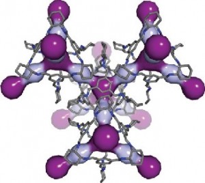
Thallapally’s collaborator Andrew Cooper at the University of Liverpool and others had been researching materials called porous organic cages, whose molecular structures are made up of repeating units that form 3-D cages. Cages built from a molecule called CC3 are the right size to hold about three atoms of xenon, krypton or radon.

To test whether CC3 might be useful here, the team simulated on a computer CC3 interacting with atoms of xenon and other noble gases. The molecular structure of CC3 naturally expands and contracts. The researchers found this breathing created a hole in the cage that grew to 4.5 angstroms wide and shrunk to 3.6 angstroms. One atom of xenon is 4.1 angstroms wide, suggesting it could fit within the window if the cage opens long enough. (Krypton and radon are 3.69 angstroms and 4.17 angstroms wide, respectively, and it takes 10 million angstroms to span a millimeter.)

The computer simulations revealed that CC3 opens its windows big enough for xenon about 7 percent of the time, but that is enough for xenon to hop in. In addition, xenon has a higher likelihood of hopping in than hopping out, essentially trapping the noble gas inside.

The team then tested how well CC3 could pull low concentrations of xenon and krypton out of air, a mix of gases that included oxygen, argon, carbon dioxide and nitrogen. With xenon at 400 parts per million and krypton at 40 parts per million, the researchers sent the mix through a sample of CC3 and measured how long it took for the gases to come out the other side.

Oxygen, nitrogen, argon and carbon dioxide — abundant components of air — traveled through the CC3 and continued to be measured for the experiment’s full 45 minute span. Xenon however stayed within the CC3 for 15 minutes, showing that CC3 could separate xenon from air.

In addition, CC3 trapped twice as much xenon as the leading MOF material. It also caught xenon 20 times more often than it caught krypton, a characteristic known as selectivity. The leading MOF only preferred xenon 7 times as much. These experiments indicated improved performance in two important characteristics of such a material, capacity and selectivity.

“We know that CC3 does this but we’re not sure why. Once we understand why CC3 traps the noble gases so easily, we can improve on it,” said Thallapally.

To explore whether MOFs and porous organic cages offer economic advantages, the researchers estimated the cost compared to cryogenic separations and determined they would likely be less expensive.

“Because these materials function well at ambient or close to ambient temperatures, the processes based on them are less energy intensive to use,” said PNNL’s Denis Strachan.

The material might also find use in pharmaceuticals. Most molecules come in right- and left-handed forms and often only one form works in people. In additional experiments, Cooper and colleagues in the U.K. tested CC3’s ability to distinguish and separate left- and right-handed versions of an alcohol. After separating left- and right-handed forms of CC3, the team showed in biochemical experiments that each form selectively trapped only one form of the alcohol.

The researchers have provided an image illustrating a CC3 cage,

Breathing room: In this computer simulation, light and dark purple highlight the cavities within the 3D pore structure of CC3. Courtesy:  PNNL

Breathing room: In this computer simulation, light and dark purple highlight the cavities within the 3D pore structure of CC3. Courtesy: PNNL

Here’s a link to and a citation for the paper,

Separation of rare gases and chiral molecules by selective binding in porous organic cages by Linjiang Chen, Paul S. Reiss, Samantha Y. Chong, Daniel Holden, Kim E. Jelfs, Tom Hasell, Marc A. Little, Adam Kewley, Michael E. Briggs, Andrew Stephenson, K. Mark Thomas, Jayne A. Armstrong, Jon Bell, Jose Busto, Raymond Noel, Jian Liu, Denis M. Strachan, Praveen K. Thallapally, & Andrew I. Cooper. Nature Material (2014) doi:10.1038/nmat4035 Published online 20 July 2014

This paper is behind a paywall.

Sifting through Twitter with your computer cluster of more than 600 nodes named Olympus—one of the Top 500 fastest supercomputers in the world.

Here are two (seemingly) contradictory pieces of information (1) the US Library of Congress takes over 24 hours to complete a single search of tweets archived from 2006 – 2010, according to my Jan. 16, 2013 posting, and (2) Court (Courtney) Corley, a data scientist at the US Dept. of Energy’s Pacific Northwest National Laboratory (PNNL), has a system (SALSA; SociAL Sensor Analytics) that analyzes billions of tweets in seconds. It’s a little hard to make sense out of these two very different perspectives on accessing data from tweets.

The news from Corley and the PNNL is more recent and, before I speculate further, here’s a bit more about Corley’s work, from the June 6, 2013 PNNL news release (also on EurekAlert)

If you think keeping up with what’s happening via Twitter, Facebook and other social media is like drinking from a fire hose, multiply that by 7 billion – and you’ll have a sense of what Court Corley wakes up to every morning.

Corley, a data scientist at the Department of Energy’s Pacific Northwest National Laboratory, has created a powerful digital system capable of analyzing billions of tweets and other social media messages in just seconds, in an effort to discover patterns and make sense of all the information. His social media analysis tool, dubbed “SALSA” (SociAL Sensor Analytics), combined with extensive know-how – and a fair degree of chutzpah – allows someone like Corley to try to grasp it all.

“The world is equipped with human sensors – more than 7 billion and counting. It’s by far the most extensive sensor network on the planet. What can we learn by paying attention?” Corley said.

Among the payoffs Corley envisions are emergency responders who receive crucial early information about natural disasters such as tornadoes; a tool that public health advocates can use to better protect people’s health; and information about social unrest that could help nations protect their citizens. But finding those jewels amidst the effluent of digital minutia is a challenge.

“The task we all face is separating out the trivia, the useless information we all are blasted with every day, from the really good stuff that helps us live better lives. There’s a lot of noise, but there’s some very valuable information too.”

I was getting a little worried when I saw the bit about separating useless information from the good stuff since that can be a very personal choice. Thankfully, this followed,

One person’s digital trash is another’s digital treasure. For example, people known in social media circles as “Beliebers,” named after entertainer Justin Bieber, covet inconsequential tidbits about Justin Bieber, while “non-Beliebers” send that data straight to the recycle bin.

The amount of data is mind-bending. In social media posted just in the single year ending Aug. 31, 2012, each hour on average witnessed:

  • 30 million comments
  • 25 million search queries
  • 98,000 new tweets
  • 3.8 million blog views
  • 4.5 million event invites
  • 7.1 million photos uploaded
  • 5.5 million status updates
  • The equivalent of 453 years of video watched

Several firms routinely sift posts on LinkedIn, Facebook, Twitter, YouTube and other social media, then analyze the data to see what’s trending. These efforts usually require a great deal of software and a lot of person-hours devoted specifically to using that application. It’s what Corley terms a manual approach.

Corley is out to change that, by creating a systematic, science-based, and automated approach for understanding patterns around events found in social media.

It’s not so simple as scanning tweets. Indeed, if Corley were to sit down and read each of the more than 20 billion entries in his data set from just a two-year period, it would take him more than 3,500 years if he spent just 5 seconds on each entry. If he hired 1 million helpers, it would take more than a day.

But it takes less than 10 seconds when he relies on PNNL’s Institutional Computing resource, drawing on a computer cluster with more than 600 nodes named Olympus, which is among the Top 500 fastest supercomputers in the world.

“We are using the institutional computing horsepower of PNNL to analyze one of the richest data sets ever available to researchers,” Corley said.

At the same time that his team is creating the computing resources to undertake the task, Corley is constructing a theory for how to analyze the data. He and his colleagues are determining baseline activity, culling the data to find routine patterns, and looking for patterns that indicate something out of the ordinary. Data might include how often a topic is the subject of social media, who is putting out the messages, and how often.

Corley notes additional challenges posed by social media. His programs analyze data in more than 60 languages, for instance. And social media users have developed a lexicon of their own and often don’t use traditional language. A post such as “aw my avalanna wristband @Avalanna @justinbieber rip angel pic.twitter.com/yldGVV7GHk” poses a challenge to people and computers alike.

Nevertheless, Corley’s program is accurate much more often than not, catching the spirit of a social media comment accurately more than three out of every four instances, and accurately detecting patterns in social media more than 90 percent of the time.

Corley’s educational background may explain the interest in emergency responders and health crises mentioned in the early part of the news release (from Corley’s PNNL webpage),

B.S. Computer Science from University of North Texas; M.S. Computer Science from University of North Texas; Ph.D. Computer Science and Engineering from University of North Texas; M.P.H (expected 2013) Public Health from University of Washington.

The reference to public health and emergency response is further developed, from the news release,

Much of the work so far has been around public health. According to media reports in China, the current H7N9 flu situation in China was highlighted on Sina Weibo, a China-based social media platform, weeks before it was recognized by government officials. And Corley’s work with the social media working group of the International Society for Disease Surveillance focuses on the use of social media for effective public health interventions.

In collaboration with the Infectious Disease Society of America and Immunizations 4 Public Health, he has focused on the early identification of emerging immunization safety concerns.

“If you want to understand the concerns of parents about vaccines, you’re never going to have the time to go out there and read hundreds of thousands, perhaps millions of tweets about those questions or concerns,” Corley said. “By creating a system that can capture trends in just a few minutes, and observe shifts in opinion minute to minute, you can stay in front of the issue, for instance, by letting physicians in certain areas know how to customize the educational materials they provide to parents of young children.”

Corley has looked closely at reaction to the vaccine that protects against HPV, which causes cervical cancer. The first vaccine was approved in 2006, when he was a graduate student, and his doctoral thesis focused on an analysis of social media messages connected to HPV. He found that creators of messages that named a specific drug company were less likely to be positive about the vaccine than others who did not mention any company by name.

Other potential applications include helping emergency responders react more efficiently to disasters like tornadoes, or identifying patterns that might indicate coming social unrest or even something as specific as a riot after a soccer game. More than a dozen college students or recent graduates are working with Corley to look at questions like these and others.

As to why the US Library of Congress requires 24 hours to search one term in their archived tweets and Corley and the PNNL require seconds to sift through two years of tweets, only two possibilities come to my mind. (1) Corley is doing a stripped down version of an archival search so his searches are not comparable to the Library of Congress searches or (2) Corley and the PNNL have far superior technology.

Bacteria on a battery can be a good thing

In a joint project between the UK’s University of East Anglia (UEA) and the Pacific Northwest National Laboratory (PNNL) in Washington State (US) researchers have published a paper about their work utilizing bacteria to produce electric currents in batteries. From the Mar. 25, 2013 news item on ScienceDaily,

Scientists at the University of East Anglia have made an important breakthrough in the quest to generate clean electricity from bacteria.

Findings published today in the journal Proceedings of the National Academy of Sciences (PNAS) show that proteins on the surface of bacteria can produce an electric current by simply touching a mineral surface.

The research shows that it is possible for bacteria to lie directly on the surface of a metal or mineral and transfer electrical charge through their cell membranes. This means that it is possible to ‘tether’ bacteria directly to electrodes — bringing scientists a step closer to creating efficient microbial fuel cells or ‘bio-batteries’.

The team collaborated with researchers at Pacific Northwest National Laboratory in Washington State in the US.

Shewanella oneidensis (pictured) is part of a family of marine bacteria. The research team created a synthetic version of this bacteria using just the proteins thought to shuttle the electrons from the inside of the microbe to the rock.

Image: Shewanella oneidensis bacteria, Alice Dohnalkova. (downloaded from http://www.uea.ac.uk/mac/comm/media/press/2013/March/bio-batteries)

Image: Shewanella oneidensis bacteria, Alice Dohnalkova. (downloaded from http://www.uea.ac.uk/mac/comm/media/press/2013/March/bio-batteries)

The Mar. 25, 2013 UEA news release,which originated the news item,  describes the work n some detail (Note: A link has been removed),

They inserted these proteins into the lipid layers of vesicles, which are small capsules of lipid membranes such as the ones that make up a bacterial membrane. Then they tested how well electrons travelled between an electron donor on the inside and an iron-bearing mineral on the outside.

Lead researcher Dr Tom Clarke from UEA’s school of Biological Sciences said: “We knew that bacteria can transfer electricity into metals and minerals, and that the interaction depends on special proteins on the surface of the bacteria. But it was not been clear whether these proteins do this directly or indirectly though an unknown mediator in the environment.

“Our research shows that these proteins can directly ‘touch’ the mineral surface and produce an electric current, meaning that is possible for the bacteria to lie on the surface of a metal or mineral and conduct electricity through their cell membranes.

“This is the first time that we have been able to actually look at how the components of a bacterial cell membrane are able to interact with different substances, and understand how differences in metal and mineral interactions can occur on the surface of a cell.

“These bacteria show great potential as microbial fuel cells, where electricity can be generated from the breakdown of domestic or agricultural waste products.

“Another possibility is to use these bacteria as miniature factories on the surface of an electrode, where chemicals reactions take place inside the cell using electrical power supplied by the electrode through these proteins.”

Biochemist Liang Shi of Pacific Northwest National Laboratory said: “We developed a unique system so we could mimic electron transfer like it happens in cells. The electron transfer rate we measured was unbelievably fast – it was fast enough to support bacterial respiration.”

This work reminds me of the biobattery created at Concordia University (my April 20, 2012 posting) and the work on breathable batteries at the Polish Academy of Sciences (my Mar. 8, 2013 posting).

Interested parties can find a full citation for the UEA/PNNL research paper at the bottom of the ScienceDaily news item here.

Graphene material that improves lithium-ion battery performance wins ‘Oscar’ of innovation

Who knew that an ‘Oscar of innovation’ existed? It does and Vorbeck Materials along with its partners,  Pacific Northwest National Laboratory (PNNL) and Princeton University have won it. From the June 22, 2012 news item on Nanowerk,

Vorbeck Materials, in partnership with Pacific Northwest National Labs (PNNL) and Princeton University, was recognized today by R&D Magazine for developing one of the 100 most significant scientific and technological products or advances of the year.

The R&D 100 Award honors Vorbeck’s breakthrough work with PNNL and Princeton to commercialize graphene technology, which will enable greater use of electric vehicles and faster charging consumer electronics.

In collaboration with Professor Ilhan Aksay at Princeton University, PNNL has demonstrated that small quantities of Vor-x™, Vorbeck’s unique graphene material, can dramatically improve the performance and power of lithium-ion batteries. The pioneering work will enable the development of batteries that last longer and recharge quickly, drastically reducing the time it takes to charge an electric vehicle to just a few hours and allowing smartphones to charge in as little as ten minutes. Lithium-ion batteries are also used to power laptops, power tools and other electronic devices.

Vorbeck is working to bring this new technology to market for use in consumer electronic devices, tools, and electric vehicles. Vorbeck is also partnering with Hardwire LLC of Maryland to integrate the new batteries into hybrid military vehicles.

You can find out more about the R&D 100 awards at the R&D (Research and Development) Magazine’s Award page,

The Awards, widely recognized as the “Oscars of Innovation”, identifies and celebrates the top high technology products of the year. Sophisticated testing equipment, innovative new materials, chemistry breakthroughs, biomedical products, consumer items, high-energy physics: the R&D 100 Awards spans industry, academia, and government-sponsored research. …

Since 1963, the R&D 100 Awards have identified revolutionary technologies newly introduced to the market. Many of these have become household names, helping shape everyday life for many Americans. These include the flashcube (1965), the automated teller machine (1973), the halogen lamp (1974), the fax machine (1975), the liquid crystal display (1980), the Kodak Photo CD (1991), the Nicoderm anti-smoking patch (1992), Taxol anticancer drug (1993), lab on a chip (1996), and HDTV (1998).

That’s a very impressive list of innovations.

Canadian scientists get more light in deal with the US Argonne National Laboratory

Canada’s synchrotron, Canadian Light Source (based in Saskatchewan), has signed a new three-year deal with the US Dept. of Energy’s Argonne National Laboratory’s Advanced Photon Source (APS)  that will give Canadian scientists more access to the APS facilities, according to the June 18, 2012 news item at the  Nanowerk website,

Seeking to solve some of today’s greatest global problems, scientists using x-ray light source facilities at national research laboratories in the United States and Canada are sharing more expertise.

The Canadian Light Source (CLS) and the Advanced Photon Source (APS) at the U.S. Department of Energy’s (DOE’s) Argonne National Laboratory agreed in January 2012 to a Partner User Proposal that cements a stronger working relationship between the two facilities for the next three years. These two premier light sources use different but complementary x-ray techniques to probe materials in order to understand chemical and structural behavior.

Tone Kunz’s June 18, 2012 news release for the APS provides details about the deal,

This new agreement will provide Canadian scientists with more research time to use the x-ray light source facilities and more time on a larger number of APS beamlines. Using varied x-ray and imaging capabilities will broaden the range of experiments Canadians may undertake at the APS to augment their research done at the Canadian Light Source. X-ray science offers potential solutions to a broad range of problems in surface, material, environmental and earth sciences, condensed matter physics, chemistry, and geosciences.

Since the Sector 20 beamlines became fully operational, scientists from Canada and other areas who have used these beamlines at the APS have produced an average of 51 scientific publications a year. This research includes the study of more effective mineral exploration strategies, ways to mitigate mine waste and mercury contamination, and novel ways to fabricate nanomaterials for use in fuel cells, batteries, and LEDs.

I had not realized how longstanding the  CLS/APS relationship has been,

Before the Canadian Light Source began operation in 2004, a Canadian group led by Daryl Crozier of Simon Fraser University, working in partnership with colleagues at the University of Washington and the Pacific Northwest National Laboratory, helped found the Sector 20 beamlines at the APS as part of the Pacific Northwest Consortium Collaborative Access Team, or PNC-CAT. Parts of this team were included in the X-ray Science Division of the APS when it was formed.

This long-standing partnership has led to scientifically significant upgrades to the beamline. The new agreement will provide the valuable manpower and expertise to allow the APS to continue to push the innovation envelope. [emphasis mine]

As I was reading Kunz’s news release I kept asking, what’s in it for the APS? Apparently they need more “manpower and expertise.” Unfortunately, their future plans are a little shy of detail,

Scientists from the APS and the Canadian Light Source will work together on R&D projects to improve light-source technology. In particular, scientists will upgrade even further the two beamlines at Sector 20 in four key areas. This will provide a unique capability to prepare and measure in situ films and interfaces, a new technique to create quantitative three-dimensional chemical maps of samples, and improved forms of spectroscopy to expand the range of elements and types of environments that can be examined.

What are the four key areas? For that matter, what is Sector 20? I suspect some of my readers have similar questions about my postings. It’s easy (especially if you write frequently) to forget that your readers may not be as familiar as you are with the subject matter.

(I wrote about the CLS and another deal with a synchrotron in the UK in my May 31, 2011 posting.)