Tag Archives: Parkinson’s Disease

Recording brain activity with flexible tentacle electrodes

A September 4, 2024 news item on ScienceDaily announced some research in Switzerland that improves on electrodes used in brain implants, e.g., like Elon Musk’s company, Neuralink,

Neurostimulators, also known as brain pacemakers, send electrical impulses to specific areas of the brain via special electrodes. It is estimated that some 200,000 people worldwide are now benefiting from this technology, including those who suffer from Parkinson’s disease or from pathological muscle spasms. According to Mehmet Fatih Yanik, Professor of Neurotechnology at ETH Zurich, further research will greatly expand the potential applications: instead of using them exclusively to stimulate the brain, the electrodes can also be used to precisely record brain activity and analyse it for anomalies associated with neurological or psychiatric disorders. In a second step, it would be conceivable in future to treat these anomalies and disorders using electrical impulses.

A September 4, 2024 ETH Zurich press release (also on EurekAlert), which originated the news item, provides more technical detail about the work,

To this end, Yanik and his team have now developed a new type of electrode that enables more detailed and more precise recordings of brain activity over an extended period of time. These electrodes are made of bundles of extremely fine and flexible fibres of electrically conductive gold encapsulated in a polymer. Thanks to a process developed by the ETH Zurich researchers, these bundles can be inserted into the brain very slowly, which is why they do not cause any detectable damage to brain tissue.

This sets the new electrodes apart from rival technologies. Of these, perhaps the best known in the public sphere is the one from Neuralink, an Elon Musk company [emphasis mine]. In all such systems, including Neuralink’s, the electrodes are considerably wider. “The wider the probe, even if it is flexible, the greater the risk of damage to brain tissue,” Yanik explains. “Our electrodes are so fine that they can be threaded past the long processes that extend from the nerve cells in the brain. They are only around as thick as the nerve-cell processes themselves.”

The research team tested the new electrodes on the brains of rats using four bundles, each made up of 64 fibres. In principle, as Yanik explains, up to several hundred electrode fibres could be used to investigate the activity of an even greater number of brain cells. In the study, the electrodes were connected to a small recording device attached to the head of each rat, thereby enabling them to move freely.

No influence on brain activity

In the experiments, the research team was able to confirm that the probes are biocompatible and that they do not influence brain function. Because the electrodes are very close to the nerve cells, the signal quality is very good compared to other methods.

At the same time, the probes are suitable for long-term monitoring activities, with researchers recording signals from the same cells in the brains of animals for the entire duration of a ten-month experiment. Examinations showed that no brain-tissue damage occurred during this time. A further advantage is that the bundles can branch out in different directions, meaning that they can reach multiple brain areas.

Human testing to begin soon

In the study, the researcher used the new electrodes to track and analyse nerve-cell activity in various areas of the brains of rats over a period of several months. They were able to determine that nerve cells in different regions were “co-activated”. Scientists believe that this large-scale, synchronous interaction of brain cells plays a key role in the processing of complex information and memory formation. “The technology is of high interest for basic research that investigates these functions and their impairments in neurological and psychiatric disorders,” Yanik explains.

The group has teamed up with fellow researchers at the University College London in order to test diagnostic use of the new electrodes in the human brain. Specifically, the project involves epilepsy sufferers who do not respond to drug therapy. In such cases, neurosurgeons may remove a small part of the brain where the seizures originate. The idea is to use the group’s method to precisely localise the affected area of the brain prior to tissue removal.

Brain-machine interfaces

There are also plans to use the new electrodes to stimulate brain cells in humans. “This could aid the development of more effective therapies for people with neurological and psychiatric disorders”, says Yanik. In disorders such as depression, schizophrenia or OCD, there is often impairments in specific regions of the brain, which leads to problems in evaluation of information and decision making. Using the new electrodes, it might be possible to detect the pathological signals generated by the neural networks in the brain in advance, and then stimulate the brain in a way that would alleviate such disorders. Yanik also thinks that this technology may give rise to brain-machine interfaces for people with brain injuries. In such cases, the electrodes might be used to read their intentions and thereby, for example, to control prosthetics or a voice-output system.

A bundle of extremely fine electrode fibres in the brain (microscope image). (Image: Yasar TB et al. Nature Communications 2024, modified) Courtesy: ETH Zurich

Here’s a link to and a citation for the paper,

Months-long tracking of neuronal ensembles spanning multiple brain areas with Ultra-Flexible Tentacle Electrodes by Tansel Baran Yasar, Peter Gombkoto, Alexei L. Vyssotski, Angeliki D. Vavladeli, Christopher M. Lewis, Bifeng Wu, Linus Meienberg, Valter Lundegardh, Fritjof Helmchen, Wolfger von der Behrens & Mehmet Fatih Yanik. Nature Communications volume 15, Article number: 4822 (2024) DOI https://doi.org/10.1038/s41467-024-49226-9 Published online: 06 June 2024

This paper is open access.

“Eat up your ceramic nanoparticles” says the European Space Agency

A Sept. 4, 2020 news item on phys.org showcases some intriguing research from the European Space Agency (ESA),

“Eat your vitamins” might be replaced with “ingest your ceramic nano-particles” in the future as space research is giving more weight to the idea that nanoscopic particles could help protect cells from common causes of damage.

A Sept, 4, 2020 ESA press release, which originated the news item, fills in some of the details and raises a question,

Oxidative stress occurs in our bodies when cells lose the natural balance of electrons in the molecules that we are made of. This is a common and constant occurrence that is part of our metabolism but also plays a role in the aging process and several pathological conditions, such as heart failure, muscle atrophy and Parkinson’s disease.

The best advice for keeping your body in balance and avoiding oxidative stress is still to have a healthy diet and eat enough vitamins, but nanoparticles are showing promising results in keeping cells in shape.

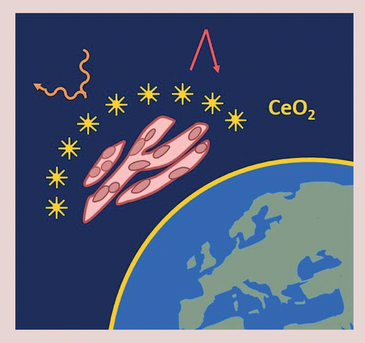
When in space, astronauts have been shown to suffer from more oxidative stress due to the extra radiation they receive and as a by-product of floating in weightlessness, so researchers in Italy were keen to see if nanoparticles would have the same protective effect on cells on the International Space Station as on Earth.

They prepared muscle cells that flew to the International Space Station and were cultured in ESA’s Kubik incubator before being frozen for storage.

A year ago [emphasis mine] our frozen samples splashed down in the Pacific Ocean on the Dragon spacecraft, and after comparing the samples we saw a marked effect in the cells treated with ceramic nanoparticles,” says Gianni Ciofani from the Istituto Italiano di Tecnologia in Italy. “The effect we observed seems to imply that nanoparticles work better and longer than traditional antioxidants such as vitamins.”

“The experiment setup resulted in excellent samples to analyze using state-of-the art RNA sequencing,” continues Gianni. “Conducting space research is nothing like traditional lab work, as we have less samples, we cannot do the work ourselves and we have to work around deadlines such as launch days, landing and storing the samples, it is challenging but thrilling research!” The team even found ways to improve and simplify the process for future studies.

Baby astronauts hypothesis

The research adds weight to the baby-astronaut hypothesis of weightlessness. The changes in muscle tissue observed are similar to how babies’ tissues develop in the womb.

“Some researchers see similarities to how human bodies adapt to living in space with pre-natal conditions: there are similarities with floating in a warm environment with different oxygen intake and we consider it a possibility of return to the state,” says Giada Genchi, also of the Istituto Italiano di Tecnologia’s Smart Bio-Interfaces department.

The team’s high-quality muscle tissue samples are being further analyzed and compared to samples from similar experiments that flew earlier. There is still much more to learn, such as what is the best way to administer nano-ceramics and how long do their protective effects last as well as possible unwanted side effects.

I highlighted a “A year ago” because that should mean 2019 but the research the ESA press release linked to was published in 2018. I cannot find anything more recent. So, for the curious, here’s a link to and a citation for the 2018 research paper,

Modulation of gene expression in rat muscle cells following treatment with nanoceria in different gravity regimes by Giada Graziana Genchi, Andrea Degl’Innocenti, Alice Rita Salgarella, Ilaria Pezzini, Attilio Marino, Arianna Menciassi, Sara Piccirillo, Michele Balsamo & Gianni Ciofani. Nanomedicine Vol. 13, No. 22 Preliminary Communication DOI: https://doi.org/10.2217/nnm-2018-0316 Published Online: 18 Oct 2018 Print Version: 2018 Nov;13 (22): 2821-2833. DOI: 10.2217/nnm-2018-0316.

The paper is behind a paywall.

This image was used to illustrate the work,

Courtesy Nanomedicine (journal)

Regardless of when the research was published, it’s still pretty interesting work and I hope to hear more about it in the future.

Structure of tunneling nanotubes (TNTs) challenges the dogma of the cell

There is a video that accompanies the news but I strongly advise reading the press release first, unless you already know a lot about cells and tunneling nanotubes.

A January 30, 2019 Institut Pasteur press release (also on EurekAlert but published Jan.31, 2019) announces the work,

Cells in our bodies have the ability to speak with one another much like humans do. This communication allows organs in our bodies to work synchronously, which in turn, enables us to perform the remarkable range of tasks we meet on a daily basis. One of this mean of communication is ‘tunneling nanotubes’ or TNTs. In an article published in Nature Communications, researchers from the Institut Pasteur leaded by Chiara Zurzolo discovered, thanks to advanced imaging techniques, that the structure of these nanotubes challenged the very concept of cell.

As their name implies, TNTs are tiny tunnels that link two (or more cells) and allow the transport of a wide variety of cargoes between them, including ions, viruses, and entire organelles. Previous research by the same team (Membrane Traf?c and Pathogenesis Unit) at the Institut Pasteur have shown that TNTs are involved in the intercellular spreading of pathogenic amyloid proteins involved in Alzheimer and Parkinson’s disease. This led researchers to propose that they serve as a major avenue for the spreading of neurodegenerative diseases in the brain and therefore represent a novel therapeutic target to stop the progression of these incurable diseases. TNTs also appear to play a major role in cancer resistance to therapy. But as scientists still know very little about TNTs and how they relate or differ from other cellular protrusions such as filopodia, they decided to pursue their research to deal with these tiny tubular connections in depth.

The dogma of cell unit questioned

A better understanding of these tiny tubular connections is therefore required as TNTs might have tremendous implications in human health and disease. Addressing this issue has been very difficult due to the fragile and transitory nature of these structures, which do not survive classical microscopic techniques. In order to overcome these obstacles, researchers combined various state-of-the-art electron microscopy approaches, and imaged TNTs at below-freezing temperatures.

Using this imaging strategy, researchers were able to decipher the structure of TNTs in high detail. Specifically, they show that most TNTs – previously shown to be single connections – are in fact made up of multiple, smaller, individual tunneling nanotubes (iTNTs). Their images also show the existence of thin wires that connect iTNTs, which could serve to increase their mechanical stability. They demonstrate the functionality of iTNTs by showing the transport of organelles using time-lapse imaging. Finally, researchers employed a type of microscopy known as ‘FIB-SEM’ to produce 3D images with sufficient resolution to clearly identify that TNTs are ‘open’ at both ends, and thus create continuity between two cells. “This discovery challenges the dogma of cells as individual units, showing that cells can open up to neighbors and exchange materials without a membrane barrier” explains Chiara Zurzolo, head of the Membrane Traf?c and Pathogenesis Unit at the Institut Pasteur.

A news step in cell-to-cell communication decoding

By applying an imaging work-flow that improves upon, and avoids, previous limitations of tools used to study the anatomy of TNTs, researchers provide the first structural description of TNTs. Importantly, they provide the absolute demonstration that these are novel cellular organelles with a defined structure, very different from known cell protrusions. “The description of the structure allows the understanding of the mechanisms involved in their formation and provides a better comprehension of their function in transferring material directly between (the cytosol of) two connected cells” says Chiara Zurzolo. Furthermore, their strategy, which preserves these delicate structures, will be useful for studying the role TNTs play in other physiological and pathological conditions

This work is an essential step toward understanding cell-to-cell communication via TNTs and lays the groundwork for investigations into their physiological functions and their role in spreading of particles linked to diseases such as viruses, bacteria, and misfolded proteins.

The researchers have kindly produced a version of the video in English,

Here’s a link to and a citation for the paper,

Correlative cryo-electron microscopy reveals the structure of TNTs in neuronal cells by Anna Sartori-Rupp, Diégo Cordero Cervantes, Anna Pepe, Karine Gousset, Elise Delage, Simon Corroyer-Dulmont, Christine Schmitt, Jacomina Krijnse-Locker & Chiara Zurzolo. Nature Communications volume 10, Article number: 342 (2019) DOI https://doi.org/10.1038/s41467-018-08178-7 Published 21 January 2019

This paper is open access.

Emergence in Toronto and Ottawa and brains in Vancouver (Canada): three April 2018 events

April 2018 is shaping up to be quite the month where art/sci events are concerned. I just published a March 27, 2018 posting titled ‘Curiosity collides with the quantum and with the Science Writers and Communicators of Canada in Vancouver (Canada)‘ and I’ve now received news about more happenings in Toronto and Ottawa.  Plus, there’s a science-themed meeting organized by ARPICO (Society of Italian Researchers &; Professionals in Western Canada) featuring brains and brain imaging in Vancouver.

Toronto’s and Ottawa’s Emergence

There’s an art/sci exhibit opening, from a March 27, 2018 Art/Sci Salon announcement (received via email),
You are invited!

FaceBook event:

The Oakwood Village Library and Arts Centre event:

341 Oakwood Avenue, Toronto, ON  M6E 2W1

I check the library webpage listed in the above and found this artist’s statement,

Artist / Scientist Statement [Stephen Morris]

I am interested in self-organized, emergent patterns and textures. I make images of patterns both from the natural world and of experiments in my laboratory in the Department of Physics at the University of Toronto. Patterns naturally attract casual attention but are also the subject of serious scientific research. Some things just evolve all by themselves into strikingly regular shapes and textures. Why? These shapes emerge spontaneously from a dynamic process of growing, folding, cracking, wrinkling, branching, flowing and other kinds of morphological development. My photos are informed by the scientific aesthetic of nonlinear physics, and celebrate the subtle interplay of order and complexity in emergent patterns. They are a kind of “Scientific Folk Art” of the science of Emergence.

While the official opening is April 5, 2018, the event itself runs from April 1 – 30, 2018.

Next, there’s another March 27, 2018 announcement (received via email) from the Art/Sci Salon but this one concerns a series of talks about ’emergence’, Note: Some of the event information was a little difficult to decipher so I’ve added a note to the relevant section).

What is Emergent Form?

Nature teems with self-organized forms that seem to spring spontaneously from the smooth background of things, by mechanisms that are not always apparent. Think of rippled sand on a beach or regular stripes in the clouds.  Plants, insects and animals exhibit spirals and spots and stripes in an exuberant riot of colours.  Fluid flows in amazingly regular swirls and eddies.  The emergence of form is ubiquitous, and presents a challenge and an inspiration to both artists and scientists. In mathematics, patterns appear as solutions of the nonlinear partial differential equations in the continuum limit of classical physics, chemistry and biology. In the arts and humanities, “emergent form” addresses the entangled ways in which humans, plants animals, microorganisms inevitably co-exist in the universe; the way that human intervention and natural transformation can generate new landscapes and new forms of life.

With Emergent Form, we want to question the idea of a fixed world.

For us, Emergent Form is not just a series of natural and human phenomena too complicated to understand, measure or predict, but also a concept to help us identify ways in which we can come to term with, and embrace their complexity as a source of inspiration.

Join us in Toronto and Ottawa for a series of interdisciplinary discussions, performances and exhibitions on Emergent Form on Apr 10, 11, 12 (Toronto) and Apr. 14 [2018] (Ottawa).

This series is the result of a collaboration among several parties. Each event of the series is different and has its dedicated RSVP 

Tue. Apr 10 The Fields Institute, 222 College Street

Emergent form: an interdisciplinary concept 6:00-8:00 pm Pier Luigi Capucci, Accademia di Belle Arti Urbino. Founder and director, Noemalab*, Charles Sowers, Independent artist and exhibit designer, the Exploratorium, Stephen Morris, Professor of of Physics University of Toronto, Ron Wild, smART Maps

CLICK HERE FOR MORE AND TO RSVP

Wed. Apr 11 The Fields Institute6:00-8:00 pm

Anatomy of an Interconnected SystemA Performative Lecture with Margherita Pevere, Aalto University, Helsinki

CLICK HERE FOR MORE AND TO RSVP

Thu. Apr 12 (Note: I believe that from 5 – 6 pm, you’re invited to see Pevere’s exhibit and then proceed to Luella Massey Studio Theatre for performances)

5:00 pm  Cabinets in the Koffler Student Centre [I believe this is at the University of Toronto] Anatomy of an Interconnected System An exhibition by Margherita Pevere

6:00 pm Luella Massey Studio Theatre, 4 Glen Morris Ave., Toronto biopoetriX – conFiGURing AI

6:00-8:00 pm Performance: 

6:00pm Performance “Corpus Nil. A Ritual of Birth for a Modified Body” conceived and performed by Marco Donnarumma

6.30pm LAB dance: Blitz media posters on labs in the arts, sciences and engineering

7.10pm Panel: Performing AI, hybrid media and humans in/as technologyMarco Donnarumma, Doug van Nort (Dispersion Lab, York U.), Jane Tingley (Stratford User Research & Gameful Experiences Lab –SURGE-, U of Waterloo), Angela Schoellig (Dynamic Systems Lab, U of T)

Panel animators: Antje Budde (Digital Dramaturgy Lab) and Roberta Buiani (ArtSci Salon)

8.15pm Reception at the Italian Cultural Institute, 496 Huron St, Toronto

CLICK HERE FOR MORE AND TO RSVP

Ottawa. Sat. Apr. 14 National Arts Centre, 1 Elgin Street11:00 am-1:00 pm

Emergent Form and complex phenomenaA creative panel discussion and surprise demonstrationsWith Pier Luigi Capucci, Margherita Pevere, Marco Donnarumma, Stephen Morris

CLICK HERE FOR MORE AND TO RSVP

This event would not be possible without the support of The Fields Institute for Research in Mathematical Science, The Italian Embassy, the Centre for Drama, Theatre and Performance Studies at the University of Toronto, the Digital Dramaturgy Lab, and the Istituto Italiano di Cultura. Many thanks to our community partner BYOR (Bring your own Robot)

I wonder if some of the funding from Italy is in support of Italian Research in World Day. This is the inaugural year for the event, which will be held annually on April 15.

Vancouver’s brains

The Society of Italian Researchers and Professionals in Western Canada (ARPICO) is hosting an event in Vancouver (from a March 22, 2018 ARICO announcement received via email),

Our second speaking event of the year, in collaboration with the Consulate General of Italy in Vancouver, has been scheduled for Wednesday, April 11th, 2018 at the Roundhouse Community Centre. Professor Vesna Sossi’s talk will be examining how positron emission tomography (PET) imaging has contributed to better understanding of the brain function and disease with particular focus on Parkinson’s disease. You can read a summary of Prof. Sossi’s lecture as well as her short professional biography at the bottom of this message.

This event is organized in collaboration with the Consulate General of Italy in Vancouver to celebrate the newly instituted Italian Research in the World Day, as part of the Piano Straordinario “Vivere all’Italiana” – Giornata della ricerca Italiana nel mondo. You can read more on our website event page.

We look forward to seeing everyone there.

Please register for the event by visiting the EventBrite link or RSVPing to info@arpico.ca.

The evening agenda is as follows:

  • 6:45 pm – Doors Open
  • 7:00 pm – Lecture by Prof. Vesna Sossi
  • ~8:00 pm – Q & A Period
  • Mingling & Refreshments until about 9:30 pm

If you have not yet RSVP’d, please do so on our EventBrite page.

Further details are also available at arpico.ca, our facebook page, and Eventbrite.


Imaging: A Window into the Brain

Brain illness, comprising neurological disorders, mental illness and addiction, is considered the major health challenge in the 21st century with a socio-economic cost greater than cancer and cardiovascular disease combined. There are at least three unique challenges hampering brain disease management: relative inaccessibility, disease onset often preceding the onset of clinical symptoms by many years and overlap between clinical and pathological symptoms that makes accurate disease identification often difficult. This talk will give examples of how positron emission tomography (PET) imaging has contributed to better understanding of the brain function and disease with particular focus on Parkinson’s disease. Emphasis will be placed on the interplay between scientific discoveries and instrumentation and data analysis development as exemplified by the current understanding of the brain function as comprised by interactions between connectivity networks and neurochemistry and advancement in multi-modal imaging such as simultaneous PET and magnetic resonance imaging (MRI).

Vesna Sossi is a Professor in the University of British Columbia (UBC) Physics and Astronomy Department and at the UBC Djavad Mowafaghian Center for Brain Health. She directs the UBC Positron Emission Tomography (PET) imaging centre, which is known for its use of imaging as applied to neurodegeneration with emphasis on Parkinson’s disease. Her main areas of interest comprise development of imaging methods to enhance the investigation of neurochemical mechanisms that lead to an increased risk of Parkinson’s disease (PD) and mechanisms that contribute to treatment-related complications. She uses PET imaging to explore how alterations of the different neurotransmitter systems contribute to different trajectories of disease progression. Her other areas of interest are PET image analysis, instrumentation and multi-modal, multi-parameter data analysis. She published more than 180 peer review papers, is funded by several granting agencies, including the Michael J Fox Foundation, and sits on several national and international review panels.


WHEN: Wednesday, April 11th, 2018 at 7:00pm (doors open at 6:45pm)
WHERE: Roundhouse Community Centre, Room B – 181 Roundhouse Mews, Vancouver, BC, V6Z 2W3
RSVP: Please RSVP at EventBrite (https://imaging-a-window-into-the-brain.eventbrite.ca) or email info@arpico.ca


Tickets are Needed

  • Tickets are FREE, but all individuals are requested to obtain “free-admission” tickets on EventBrite site due to limited seating at the venue. Organizers need accurate registration numbers to manage wait lists and prepare name tags.
  • All ARPICO events are 100% staffed by volunteer organizers and helpers, however, room rental, stationery, and guest refreshments are costs incurred and underwritten by members of ARPICO. Therefore to be fair, all audience participants are asked to donate to the best of their ability at the door or via EventBrite to “help” defray costs of the event.

You can find directions for the Roundhouse Community Centre here

I have one idle question. What’s going to happen these groups if Canadians change their use of  Facebook or abandon the platform as they are threatening to do in the face of Cambridge Analytica’s use of their data? A March 25, 2018 article on huffingtonpost.ca outlines the latest about Canadians’ reaction to the Cambridge Analytical news according to an Angus Reid poll,

A survey by Angus Reid Institute suggests 73 per cent of Canadian Facebook users say they will make changes, while 27 per cent say it will be “business as usual.”

Nearly a quarter (23 per cent) said they would use Facebook less in the future, and 41 per cent of users said they would check and/or change their privacy settings.

The survey also found that one in 10 say they plan to abandon the platform, at least temporarily.

Facebook has been under fire for its ability to protect user privacy after Cambridge Analytica was accused of lifting the Facebook profiles of more than 50 million users without their permission.

There you have it.

*Well, a bit more information about one of the “Emergent’ speakers was received in an April 4, 2018 ArtSci Salon email announcement,

Do make sure to check out Pier Luigi Capucci’s EU-based (but with international breadth) Noemalab platform. https://noemalab.eu/ since the mid-nineties, this platform has been an important node of information for New Media Art and the relation between the arts and science.

noemalab’s blog regularly hosts reviews of events and conferences occurring around the world, including  the Subtle Technologies Festival between 2007 and 2014. you can search its archives here http://blogs.noemalab.eu/

Capucci has been writing several reflections on emergent forms of Life and theorized what he called the “third life”. See a recent essay https://noemalab.eu/memo/events/evolutionary-creativity-the-inner-life-and-meaning-of-art/ here is a picture which I would love him to explain during Emergent Form. Intrigued? come listen to him!
Gently measuring electrical signals in small animals with nano-SPEARs

This work comes from Rice University (Texas, US) according to an April 17, 2017 news item on Nanowerk,

Microscopic probes developed at Rice University have simplified the process of measuring electrical activity in individual cells of small living animals. The technique allows a single animal like a worm to be tested again and again and could revolutionize data-gathering for disease characterization and drug interactions.

The Rice lab of electrical and computer engineer Jacob Robinson has invented “nanoscale suspended electrode arrays” — aka nano-SPEARs — to give researchers access to electrophysiological signals from the cells of small animals without injuring them. Nano-SPEARs replace glass pipette electrodes that must be aligned by hand each time they are used.”

An April 17, 2017 Rice University news release (also on EurekAltert), which originated the news item, details the work,

“One of the experimental bottlenecks in studying synaptic behavior and degenerative diseases that affect the synapse is performing electrical measurements at those synapses,” Robinson said. “We set out to study large groups of animals under lots of different conditions to screen drugs or test different genetic factors that relate to errors in signaling at those synapses.”

Robinson’s early work at Rice focused on high-quality, high-throughput electrical characterization of individual cells. The new platform adapts the concept to probe the surface cells of nematodes, worms that make up 80 percent of all animals on Earth.

Most of what is known about muscle activity and synaptic transmission in the worms comes from the few studies that successfully used manually aligned glass pipettes to measure electrical activity from individual cells, Robinson said. However, this patch clamp technique requires time-consuming and invasive surgery that could negatively affect the data that is gathered from small research animals.

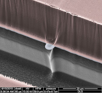
The platform developed by Robinson’s team works something like a toll booth for traveling worms. As each animal passes through a narrow channel, it is temporarily immobilized and pressed against one or several nano-SPEARS that penetrate its body-wall muscle and record electrical activity from nearby cells. That animal is then released, the next is captured and measured, and so on. Robinson said the device proved much faster to use than traditional electrophysiological cell measurement techniques.

The nano-SPEARs are created using standard thin-film deposition procedures and electron-beam or photolithography and can be made from less than 200 nanometers to more than 5 microns thick, depending on the size of animal to be tested. Because the nano-SPEARs can be fabricated on either silicon or glass, the technique easily combines with fluorescence microscopy, Robinson said.

The animals suitable for probing with a nano-SPEAR can be as large as several millimeters, like hydra, cousins of the jellyfish and the subject of an upcoming study. But nematodes known as Caenorhabditis elegans were practical for several reasons: First, Robinson said, they’re small enough to be compatible with microfluidic devices and nanowire electrodes. Second, there were a lot of them down the hall at the lab of Rice colleague Weiwei Zhong, who studies nematodes as transparent, easily manipulated models for signaling pathways that are common to all animals.

“I used to shy away from measuring electrophysiology because the conventional method of patch clamping is so technically challenging,” said Zhong, an assistant professor of biochemistry and cell biology and co-author of the paper. “Only a few graduate students or postdocs can do it. With Jacob’s device, even an undergraduate student can measure electrophysiology.”

“This meshes nicely with the high-throughput phenotyping she does,” Robinson said. “She can now correlate locomotive phenotypes with activity at the muscle cells. We believe that will be useful to study degenerative diseases centered around neuromuscular junctions.”

In fact, the labs have begun doing so. “We are now using this setup to profile worms with neurodegenerative disease models such as Parkinson’s and screen for drugs that reduce the symptoms,” Zhong said. “This would not be possible using the conventional method.”

Initial tests on C. elegans models for amyotrophic lateral sclerosis and Parkinson’s disease revealed for the first time clear differences in electrophysiological responses between the two, the researchers reported.

Testing the efficacy of drugs will be helped by the new ability to study small animals for long periods. “What we can do, for the first time, is look at electrical activity over a long period of time and discover interesting patterns of behavior,” Robinson said.

Some worms were studied for up to an hour, and others were tested on multiple days, said lead author Daniel Gonzales, a Rice graduate student in Robinson’s lab who took charge of herding nematodes through the microfluidic devices.

“It was in some way easier than working with isolated cells because the worms are larger and fairly sturdy,” Gonzales said. “With cells, if there’s too much pressure, they die. If they hit a wall, they die. But worms are really sturdy, so it was just a matter of getting them up against the electrodes and keeping them there.”

The team constructed microfluidic arrays with multiple channels that allowed testing of many nematodes at once. In comparison with patch-clamping techniques that limit labs to studying about one animal per hour, Robinson said his team measured as many as 16 nematodes per hour.

“Because this is a silicon-based technology, making arrays and producing recording chambers in high numbers becomes a real possibility,” he said.

A scanning electron micrograph shows a nano-SPEAR suspended midway between layers of silicon (grey) and photoresist material (pink) that form a recording chamber for immobilized nematodes. The high-throughput technology developed at Rice University can be adapted for other small animals and could enhance data-gathering for disease characterization and drug interactions. Courtesy of the Robinson Lab

Here’s a link to and a citation for the paper,

Scalable electrophysiology in intact small animals with nanoscale suspended electrode arrays by Daniel L. Gonzales, Krishna N. Badhiwala, Daniel G. Vercosa, Benjamin W. Avants, Zheng Liu, Weiwei Zhong, & Jacob T. Robinson. Nature Nanotechnology (2017) doi:10.1038/nnano.2017.55 Published online 17 April 2017

This paper is behind a paywall.

Sniffing out disease (Na-Nose)

The ‘artificial nose’ is not a newcomer to this blog. The most recent post prior to this is a March 15, 2016 piece about Disney using an artificial nose for art conservation. Today’s (Jan. 9, 2016) piece concerns itself with work from Israel and ‘sniffing out’ disease, according to a Dec. 30, 2016 news item in Sputnik News,

A team from the Israel Institute of Technology has developed a device that from a single breath can identify diseases such as multiple forms of cancer, Parkinson’s disease, and multiple sclerosis. While the machine is still in the experimental stages, it has a high degree of promise for use in non-invasive diagnoses of serious illnesses.

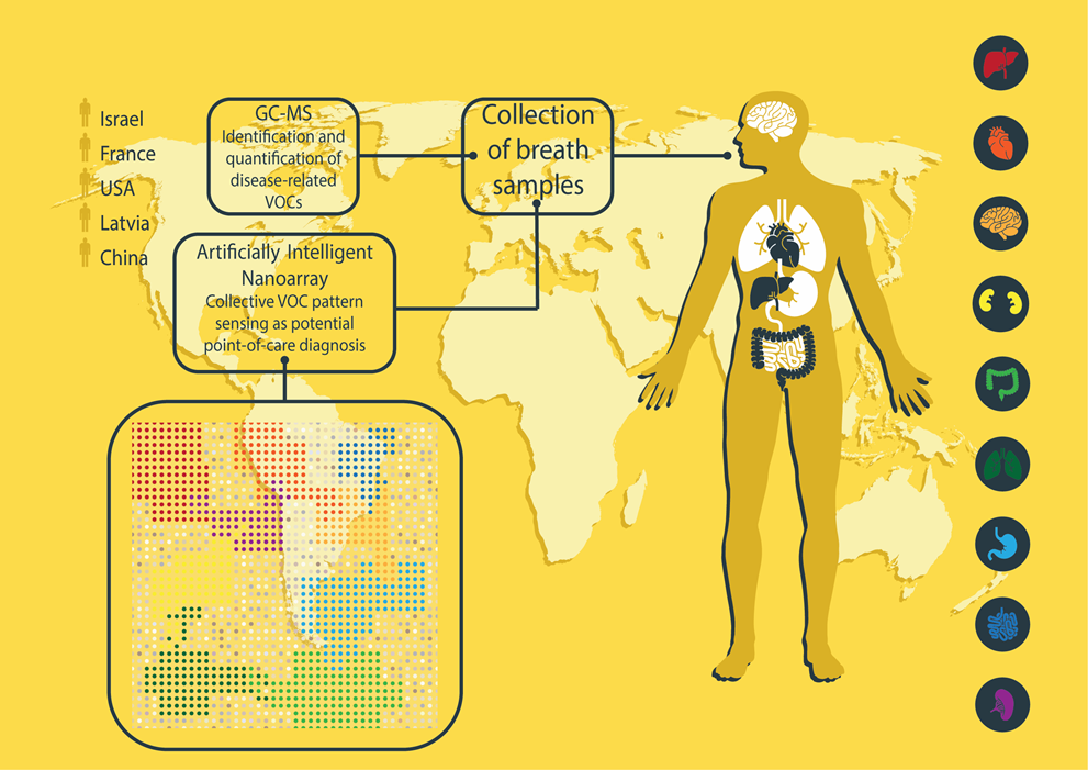
The international team demonstrated that a medical theory first proposed by the Greek physician Hippocrates some 2400 years ago is true, certain diseases leave a “breathprint” on the exhalations of those afflicted. The researchers created a prototype for a machine that can pick up on those diseases using the outgoing breath of a patient. The machine, called the Na-Nose, tests breath samples for the presence of trace amounts of chemicals that are indicative of 17 different illnesses.

A Dec. 22, 2016 Technion Israel Institute of Technology press release offers more detail about the work,

An international team of 56 researchers in five countries has confirmed a hypothesis first proposed by the ancient Greeks – that different diseases are characterized by different “chemical signatures” identifiable in breath samples. …

Diagnostic techniques based on breath samples have been demonstrated in the past, but until now, there has not been scientific proof of the hypothesis that different and unrelated diseases are characterized by distinct chemical breath signatures. And technologies developed to date for this type of diagnosis have been limited to detecting a small number of clinical disorders, without differentiation between unrelated diseases.

The study of more than 1,400 patients included 17 different and unrelated diseases: lung cancer, colorectal cancer, head and neck cancer, ovarian cancer, bladder cancer, prostate cancer, kidney cancer, stomach cancer, Crohn’s disease, ulcerative colitis, irritable bowel syndrome, Parkinson’s disease (two types), multiple sclerosis, pulmonary hypertension, preeclampsia and chronic kidney disease. Samples were collected between January 2011 and June 2014 from in 14 departments at 9 medical centers in 5 countries: Israel, France, the USA, Latvia and China.

The researchers tested the chemical composition of the breath samples using an accepted analytical method (mass spectrometry), which enabled accurate quantitative detection of the chemical compounds they contained. 13 chemical components were identified, in different compositions, in all 17 of the diseases.

According to Prof. Haick, “each of these diseases is characterized by a unique fingerprint, meaning a different composition of these 13 chemical components.  Just as each of us has a unique fingerprint that distinguishes us from others, each disease has a chemical signature that distinguishes it from other diseases and from a normal state of health. These odor signatures are what enables us to identify the diseases using the technology that we developed.”

With a new technology called “artificially intelligent nanoarray,” developed by Prof. Haick, the researchers were able to corroborate the clinical efficacy of the diagnostic technology. The array enables fast and inexpensive diagnosis and classification of diseases, based on “smelling” the patient’s breath, and using artificial intelligence to analyze the data obtained from the sensors. Some of the sensors are based on layers of gold nanoscale particles and others contain a random network of carbon nanotubes coated with an organic layer for sensing and identification purposes.

The study also assessed the efficiency of the artificially intelligent nanoarray in detecting and classifying various diseases using breath signatures. To verify the reliability of the system, the team also examined the effect of various factors (such as gender, age, smoking habits and geographic location) on the sample composition, and found their effect to be negligible, and without impairment on the array’s sensitivity.

“Each of the sensors responds to a wide range of exhalation components,” explain Prof. Haick and his previous Ph.D student, Dr. Morad Nakhleh, “and integration of the information provides detailed data about the unique breath signatures characteristic of the various diseases. Our system has detected and classified various diseases with an average accuracy of 86%.

This is a new and promising direction for diagnosis and classification of diseases, which is characterized not only by considerable accuracy but also by low cost, low electricity consumption, miniaturization, comfort and the possibility of repeating the test easily.”

“Breath is an excellent raw material for diagnosis,” said Prof. Haick. “It is available without the need for invasive and unpleasant procedures, it’s not dangerous, and you can sample it again and again if necessary.”

Here’s a schematic of the study, which the researchers have made available,

Diagram: A schematic view of the study. Two breath samples were taken from each subject, one was sent for chemical mapping using mass spectrometry, and the other was analyzed in the new system, which produced a clinical diagnosis based on the chemical fingerprint of the breath sample. Courtesy: Tech;nion

There is also a video, which covers much of the same ground as the press release but also includes information about the possible use of the Na-Nose technology in the European Union’s SniffPhone project,

Here’s a link to and a citation for the paper,

Diagnosis and Classification of 17 Diseases from 1404 Subjects via Pattern Analysis of Exhaled Molecules by Morad K. Nakhleh, Haitham Amal, Raneen Jeries, Yoav Y. Broza, Manal Aboud, Alaa Gharra, Hodaya Ivgi, Salam Khatib, Shifaa Badarneh, Lior Har-Shai, Lea Glass-Marmor, Izabella Lejbkowicz, Ariel Miller, Samih Badarny, Raz Winer, John Finberg, Sylvia Cohen-Kaminsky, Frédéric Perros, David Montani, Barbara Girerd, Gilles Garcia, Gérald Simonneau, Farid Nakhoul, Shira Baram, Raed Salim, Marwan Hakim, Maayan Gruber, Ohad Ronen, Tal Marshak, Ilana Doweck, Ofer Nativ, Zaher Bahouth, Da-you Shi, Wei Zhang, Qing-ling Hua, Yue-yin Pan, Li Tao, Hu Liu, Amir Karban, Eduard Koifman, Tova Rainis, Roberts Skapars, Armands Sivins, Guntis Ancans, Inta Liepniece-Karele, Ilze Kikuste, Ieva Lasina, Ivars Tolmanis, Douglas Johnson, Stuart Z. Millstone, Jennifer Fulton, John W. Wells, Larry H. Wilf, Marc Humbert, Marcis Leja, Nir Peled, and Hossam Haick. ACS Nano, Article ASAP DOI: 10.1021/acsnano.6b04930 Publication Date (Web): December 21, 2016

Copyright © 2017 American Chemical Society

This paper appears to be open access.

As for SniffPhone, they’re hoping that Na-Nose or something like it will allow them to modify smartphones in a way that will allow diseases to be detected.

I can’t help wondering who will own the data if your smartphone detects a disease. If you think that’s an idle question, here’s an excerpt from Sue Halpern’s Dec. 22, 2016 review of two books (“Weapons of Math Destruction: How Big Data Increases Inequality and Threatens Democracy” by Cathy O’Neil and “Virtual Competition: The Promise and Perils of the Algorithm-Driven Economy” by Ariel Ezrachi and Maurice E. Stucke) for the New York Times Review of Books,

We give our data away. We give it away in drips and drops, not thinking that data brokers will collect it and sell it, let alone that it will be used against us. There are now private, unregulated DNA databases culled, in part, from DNA samples people supply to genealogical websites in pursuit of their ancestry. These samples are available online to be compared with crime scene DNA without a warrant or court order. (Police are also amassing their own DNA databases by swabbing cheeks during routine stops.) In the estimation of the Electronic Frontier Foundation, this will make it more likely that people will be implicated in crimes they did not commit.

Or consider the data from fitness trackers, like Fitbit. As reported in The Intercept:

During a 2013 FTC panel on “Connected Health and Fitness,” University of Colorado law professor Scott Peppet said, “I can paint an incredibly detailed and rich picture of who you are based on your Fitbit data,” adding, “That data is so high quality that I can do things like price insurance premiums or I could probably evaluate your credit score incredibly accurately.”

Halpern’s piece is well worth reading in its entirety.

Electrochemical measurements of biomolecules

This work comes from Finland and features some new nano shapes. From a Nov. 10, 2016 news item on phys.org,

Tomi Laurila’s research topic has many quirky names.

“Nanodiamond, nanohorn, nano-onion…,” lists off the Aalto University Professor, recounting the many nano-shapes of carbon. Laurila is using these shapes to build new materials: tiny sensors, only a few hundred nanometres across, that can achieve great things due to their special characteristics.

For one, the sensors can be used to enhance the treatment of neurological conditions. That is why Laurila, University of Helsinki Professor Tomi Taira and experts from HUS (the Hospital District of Helsinki and Uusimaa) are looking for ways to use the sensors for taking electrochemical measurements of biomolecules. Biomolecules are e.g. neurotransmitters such as glutamate, dopamine and opioids, which are used by nerve cells to communicate with each other.

A Nov. 10, 2016 Aalto University press release, which originated the news item, expands on the theme,

Most of the drugs meant for treating neurological diseases change the communication between nerve cells that is based on neurotransmitters. If we had real time and individual information on the operation of the neurotransmitter system, it would make it much easier to for example plan precise treatments’, explains Taira.

Due to their small size, carbon sensors can be taken directly next to a nerve cell, where the sensors will report what kind of neurotransmitter the cell is emitting and what kind of reaction it is inducing in other cells.

‘In practice, we are measuring the electrons that are moving in oxidation and reduction reactions’, Laurila explains the operating principle of the sensors.

‘The advantage of the sensors developed by Tomi and the others is their speed and small size. The probes used in current measurement methods can be compared to logs on a cellular scale – it’s impossible to use them and get an idea of the brain’s dynamic’, summarizes Taira.

Feedback system and memory traces

For the sensors, the journey from in vitro tests conducted in glass dishes and test tubes to in vivo tests and clinical use is long. However, the researchers are highly motivated.

‘About 165 million people are suffering from various neurological diseases in Europe alone. And because they are so expensive to treat, neurological diseases make up as much as 80 per cent of health care costs’, tells Taira.

Tomi Laurila believes that carbon sensors will have applications in fields such as optogenetics. Optogenetics is a recently developed method where a light-sensitive molecule is brought into a nerve cell so that the cell’s electric operation can then be turned on or off by stimulating it with light. A few years ago, a group of scientists proved in the scientific journal Nature that they had managed to use optogenetics to activate a memory trace that had been created previously due to learning. Using the same technique, researchers were able to demonstrate that with a certain type of Alzheimer’s, the problem is not that there are no memory traces being created, but that the brain cannot read the traces.

‘So the traces exist, and they can be activated by boosting them with light stimuli’, explains Taira but stresses that a clinical application is not yet a reality. However, clinical applications for other conditions may be closer by. One example is Parkinson’s disease. In Parkinson’s disease, the amount of dopamine starts to decrease in the cells of a particular brain section, which causes the typical symptoms such as tremors, rigidity and slowness of movement. With the sensors, the level of dopamine could be monitored in real time.

‘A sort of feedback system could be connected to it, so that it would react by giving an electric or optical stimulus to the cells, which would in turn release more dopamine’, envisions Taira.

‘Another application that would have an immediate clinical use is monitoring unconscious and comatose patients. With these patients, the level of glutamate fluctuates very much, and too much glutamate damages the nerve cell – online monitoring would therefore improve their treatment significantly.

Atom by atom

Manufacturing carbon sensors is definitely not a mass production process; it is slow and meticulous handiwork.

‘At this stage, the sensors are practically being built atom by atom’, summarises Tomi Laurila.

‘Luckily, we have many experts on carbon materials of our own. For example, the nanobuds of Professor Esko Kauppinen and the carbon films of Professor Jari Koskinen help with the manufacturing of the sensors. Carbon-based materials are mainly very compatible with the human body, but there is still little information about them. That’s why a big part of the work is to go through the electrochemical characterisation that has been done on different forms of carbon.’

The sensors are being developed and tested by experts from various fields, such as chemistry, materials science, modelling, medicine and imaging. Twenty or so articles have been published on the basic properties of the materials. Now, the challenge is to build them into geometries that are functional in a physiological environment. And taking measurements is not simple, either.

‘Brain tissue is delicate and doesn’t appreciate having objects being inserted in it. But if this were easy, someone would’ve already done it’, conclude the two.

I wish the researchers good luck.

Bioelectronics: creating components that speak the body’s own language

This is work is still in its early stages but the idea that the body could be stimulated to release more of its own pain relievers is exciting. From a Nov. 2, 2016 news item on ScienceDaily,

With a microfabricated ion pump built from organic electronic components, ions can be sent to nerve or muscle cells at the speed of the nervous system and with a precision of a single cell. “Now we can start to develop components that speak the body’s own language,” says Daniel Simon, head of bioelectronics research at the Laboratory of Organic Electronics, Linköping University, Campus Norrköping.

A Nov. 2, 2016 Linköping University press release (also on EurekAlert), which originated the news item, discusses the research in more detail,

Our nerve and muscle cells send signals to each other using ions and molecules. Certain substances, such as the neurotransmitter GABA (gamma aminobutyric acid), are important signal substances throughout the central nervous system. Eighteen months ago, researchers at the Laboratory of Organic Electronics demonstrated an ion pump which researchers at the Karolinska Institutet could use to reduce the sensation of pain in awake, freely-moving rats. The ion pump delivered GABA directly to the rat´s spinal cord. The news that researchers could deliver the body’s own neurotransmitters was published in Science Advances and garnered intense interest all over the world.

The research group at the Laboratory of Organic Electronics has now achieved another major advance and developed a significantly smaller and more rapid ion pump that transmits signals nearly as rapidly as the cells themselves, and with a precision on the scale of an individual cell. …

“Our skilled doctoral students, Amanda Jonsson and Theresia Arbring Sjöström, have succeeded with the last important part of the puzzle in the development of the ion pump. When a signal passes between two synapses it takes 1-10 milliseconds, and we are now very close to the nervous system’s own speed,” says Magnus Berggren, professor of organic electronics and director of the Laboratory of Organic Electronics.

“We conclude that we have produced artificial nerves that can communicate seamlessly with the nervous system. After more than 10 years’ research we have finally got all the parts of the puzzle in place,” he says.

Amanda Jonsson, who together with Theresia Arbring Sjöström is principal author of the article in Science Advances, has developed the pain-alleviating ion pump as part of her doctoral studies. She proudly presents a glass disk with many of the new miniaturized ion pumps. Some pumps have only a single outlet, but others have six tiny point outlets.

“We can make them with several outlets, it’s just as easy as making one. And all of the outlets can be individually controlled. Previously we could only transport ions horizontally and from all outputs at the same time. Now, however, we can deliver the ions vertically, which makes the distance they have to be transported as short as a micrometre,” she explains.

All of the outputs of the ion pump can also be rapidly switched on or off with the aid of micrometre-sized ion diodes.

“The ions are released rapidly by an electrical signal, in the same way that the neurotransmitter is released in a synapse,” says Theresia Arbring Sjöström.

Organic electronic components have a major advantage here: they can conduct both ions and electricity. In this case, the material PEDOT:PSS enables the electrical signals to be converted to chemical signals that the body understands.

The ion diode has recently been developed, as has the material that forms the basis of the new rapid ion pump.

“The new material makes it possible to build with a precision and reliability not possible in previous versions of the ion pump,” says Daniel Simon.

The new ion pump has so far only been tested in the laboratory. The next step will be to test it with live cells and the researchers hope eventually to, for example alleviate pain, stop epileptic seizures, and reduce the symptoms of Parkinsons disease, using exactly the required dose at exactly the affected cells. Communication using the cell´s own language, and the cell´s own speed.

Here’s a link to and a citation for the paper,

Chemical delivery array with millisecond neurotransmitter release by Amanda Jonsson, Theresia Arbring Sjöström, Klas Tybrandt, Magnus Berggren, and Daniel T. Simon. Science Advances  02 Nov 2016: Vol. 2, no. 11, e1601340 DOI: 10.1126/sciadv.1601340

This paper is open access.

Nanotubes tunnel between neurons in Parkinson’s disease

An Aug. 22, 2016 news item on ScienceDaily describes how scientists from the Institut Pasteur (France) have developed insight into one of the processes in Parkinson’s disease,

Scientists have demonstrated the role of lysosomal vesicles in transporting alpha-synuclein aggregates, responsible for Parkinson’s and other neurodegenerative diseases, between neurons. These proteins move from one neuron to the next in lysosomal vesicles which travel along the ‘tunneling nanotubes’ between cells.

An Aug. 22, 2016 Institut Pasteur press release (also on EurekAlert), expands on the theme,

Synucleinopathies, a group of neurodegenerative diseases including Parkinson’s disease, are characterized by the pathological deposition of aggregates of the misfolded α-synuclein protein into inclusions throughout the central and peripheral nervous system. Intercellular propagation (from one neuron to the next) of α-synuclein aggregates contributes to the progression of the neuropathology, but little was known about the mechanism by which spread occurs.

In this study, scientists from the Membrane Traffic and Pathogenesis Unit, directed by Chiara Zurzolo at the Institut Pasteur, used fluorescence microscopy to demonstrate that pathogenic α-synuclein fibrils travel between neurons in culture, inside lysosomal vesicles through tunneling nanotubes (TNTs), a new mechanism of intercellular communication.

After being transferred via TNTs, α-synuclein fibrils are able to recruit and induce aggregation of the soluble α-synuclein protein in the cytosol of cells receiving the fibrils, thus explaining the propagation of the disease. The scientists propose that cells overloaded with α-synuclein aggregates in lysosomes dispose of this material by hijacking TNT-mediated intercellular trafficking. However, this results in the disease being spread to naive neurons.

This study demonstrates that TNTs play a significant part in the intercellular transfer of α-synuclein fibrils and reveals the specific role of lysosomes in this process. This represents a major breakthrough in understanding the mechanisms underlying the progression of synucleinopathies.

These compelling findings, together with previous reports from the same team, point to the general role of TNTs in the propagation of prion-like proteins in neurodegenerative diseases and identify TNTs as a new therapeutic target to combat the progression of these incurable diseases.

Here’s a link to and a citation for the paper,

Tunneling nanotubes spread fibrillar α‐synuclein by intercellular trafficking of lysosomes by Saïda Abounit, Luc Bousset, Frida Loria, Seng Zhu, Fabrice de Chaumont, Laura Pieri, Jean-Christophe Olivo-Marin, Ronald Melki, Chiara Zurzolo. The EMBO Journal (2016) e201593411 DOI 10.15252/embj.201593411 Published online 22.08.2016

This paper is behind a paywall.

With over 150 partners from over 20 countries, the European Union’s Graphene Flagship research initiative unveils its work package devoted to biomedical technologies

An April 11, 2016 news item on Nanowerk announces the Graphene Flagship’s latest work package,

With a budget of €1 billion, the Graphene Flagship represents a new form of joint, coordinated research on an unprecedented scale, forming Europe’s biggest ever research initiative. It was launched in 2013 to bring together academic and industrial researchers to take graphene from the realm of academic laboratories into European society in the timeframe of 10 years. The initiative currently involves over 150 partners from more than 20 European countries. The Graphene Flagship, coordinated by Chalmers University of Technology (Sweden), is implemented around 15 scientific Work Packages on specific science and technology topics, such as fundamental science, materials, health and environment, energy, sensors, flexible electronics and spintronics.

Today [April 11, 2016], the Graphene Flagship announced in Barcelona the creation of a new Work Package devoted to Biomedical Technologies, one emerging application area for graphene and other 2D materials. This initiative is led by Professor Kostas Kostarelos, from the University of Manchester (United Kingdom), and ICREA Professor Jose Antonio Garrido, from the Catalan Institute of Nanoscience and Nanotechnology (ICN2, Spain). The Kick-off event, held in the Casa Convalescència of the Universitat Autònoma de Barcelona (UAB), is co-organised by ICN2 (ICREA Prof Jose Antonio Garrido), Centro Nacional de Microelectrónica (CNM-IMB-CSIC, CIBER-BBN; CSIC Tenured Scientist Dr Rosa Villa), and Institut d’Investigacions Biomèdiques August Pi i Sunyer (IDIBAPS; ICREA Prof Mavi Sánchez-Vives).

An April 11, 2016 ICN2 press release, which originated the news item, provides more detail about the Biomedical Technologies work package and other work packages,

The new Work Package will focus on the development of implants based on graphene and 2D-materials that have therapeutic functionalities for specific clinical outcomes, in disciplines such as neurology, ophthalmology and surgery. It will include research in three main areas: Materials Engineering; Implant Technology & Engineering; and Functionality and Therapeutic Efficacy. The objective is to explore novel implants with therapeutic capacity that will be further developed in the next phases of the Graphene Flagship.

The Materials Engineering area will be devoted to the production, characterisation, chemical modification and optimisation of graphene materials that will be adopted for the design of implants and therapeutic element technologies. Its results will be applied by the Implant Technology and Engineering area on the design of implant technologies. Several teams will work in parallel on retinal, cortical, and deep brain implants, as well as devices to be applied in the periphery nerve system. Finally, The Functionality and Therapeutic Efficacy area activities will centre on development of devices that, in addition to interfacing the nerve system for recording and stimulation of electrical activity, also have therapeutic functionality.

Stimulation therapies will focus on the adoption of graphene materials in implants with stimulation capabilities in Parkinson’s, blindness and epilepsy disease models. On the other hand, biological therapies will focus on the development of graphene materials as transport devices of biological molecules (nucleic acids, protein fragments, peptides) for modulation of neurophysiological processes. Both approaches involve a transversal innovation environment that brings together the efforts of different Work Packages within the Graphene Flagship.

A leading role for Barcelona in Graphene and 2D-Materials

The kick-off meeting of the new Graphene Flagship Work Package takes place in Barcelona because of the strong involvement of local institutions and the high international profile of Catalonia in 2D-materials and biomedical research. Institutions such as the Catalan Institute of Nanoscience and Nanotechnology (ICN2) develop frontier research in a supportive environment which attracts talented researchers from abroad, such as ICREA Research Prof Jose Antonio Garrido, Group Leader of the ICN2 Advanced Electronic Materials and Devices Group and now also Deputy Leader of the Biomedical Technologies Work Package. Until summer 2015 he was leading a research group at the Technische Universität München (Germany).

Further Graphene Flagship events in Barcelona are planned; in May 2016 ICN2 will also host a meeting of the Spintronics Work Package. ICREA Prof Stephan Roche, Group Leader of the ICN2 Theoretical and Computational Nanoscience Group, is the deputy leader of this Work Package led by Prof Bart van Wees, from the University of Groningen (The Netherlands). Another Work Package, on optoelectronics, is led by Prof Frank Koppens from the Institute of Photonic Sciences (ICFO, Spain), with Prof Andrea Ferrari from the University of Cambridge (United Kingdom) as deputy. Thus a number of prominent research institutes in Barcelona are deeply involved in the coordination of this European research initiative.

Kostas Kostarelos, the leader of the Biomedical Technologies Graphene Flagship work package, has been mentioned here before in the context of his blog posts for The Guardian science blog network (see my Aug. 7, 2014 post for a link to his post on metaphors used in medicine).