Tag Archives: Paul Zaslansky

Mother-of-pearl self-assembles from disorder into perfection

Courtesy: Mother-of-pearl Courtesy: Technische Universitaet (TU) Dresden

Mother-of-pearl (also known as nacre) research has been featured here a few times (links at the end of this post). This time it touches on self-assembly, which is the source of much interest and, on occasion, much concern in the field of nanotechnology.

In any case, the latest mother-of-pearl work comes from the Technische Universität (TU) Dresden (Technical University of Dresden), located in Germany. From a January 4, 2021 news item on phys.org,

In a new study published in Nature Physics, researchers from the B CUBE—Center for Molecular Bioengineering at TU Dresden and European Synchrotron Radiation Facility (ESRF) in Grenoble [Grance] describe, for the first time, that structural defects in self-assembling nacre attract and cancel each other out, eventually leading to a perfect periodic structure.

A January 4, 2021 Technische Universität (TU) Dresden press release (also on EurekAlert), which originated the news item, explains the reason for the ongoing interest in mother-of-pearl and reveals an unexpected turn in the research,

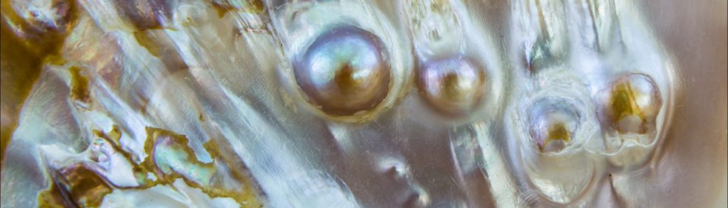
Mollusks build shells to protect their soft tissues from predators. Nacre, also known as the mother of pearl, has an intricate, highly regular structure that makes it an incredibly strong material. Depending on the species, nacres can reach tens of centimeters in length. No matter the size, each nacre is built from materials deposited by a multitude of single cells at multiple different locations at the same time. How exactly this highly periodic and uniform structure emerges from the initial disorder was unknown until now.

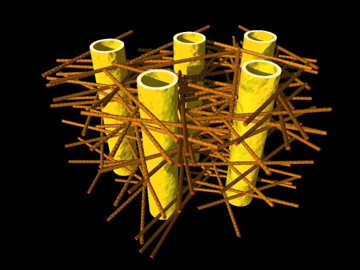
Nacre formation starts uncoordinated with the cells depositing the material simultaneously at different locations. Not surprisingly, the early nacre structure is not very regular. At this point, it is full of defects. “In the very beginning, the layered mineral-organic tissue is full of structural faults that propagate through a number of layers like a helix. In fact, they look like a spiral staircase, having either right-handed or left-handed orientation,” says Dr. Igor Zlotnikov, research group leader at the B CUBE – Center for Molecular Bioengineering at TU Dresden. “The role of these defects in forming such a periodic tissue has never been established. On the other hand, the mature nacre is defect-free, with a regular, uniform structure. How could perfection emerge from such disorder?”

The researchers from the Zlotnikov group collaborated with the European Synchrotron Radiation Facility (ESRF) in Grenoble to take a very detailed look at the internal structure of the early and mature nacre. Using synchrotron-based holographic X-ray nano-tomography the researchers could capture the growth of nacre over time. “Nacre is an extremely fine structure, having organic features below 50 nm in size. Beamline ID16A at the ESRF provided us with an unprecedented capability to visualize nacre in three-dimensions,” explains Dr. Zlotnikov. “The combination of electron dense and highly periodical inorganic platelets with delicate and slender organic interfaces makes nacre a challenging structure to image. Cryogenic imaging helped us to obtain the resolving power we needed,” explains Dr. ‘Alexandra] Pacureanu from the X-ray Nanoprobe group at the ESRF.

The analysis of data was quite a challenge. The researchers developed a segmentation algorithm using neural networks and trained it to separate different layers of nacre. In this way, they were able to follow what happens to the structural defects as nacre grows.

The behavior of structural defects in a growing nacre was surprising. Defects of opposite screw direction were attracted to each other from vast distances. The right-handed and left-handed defects moved through the structure, until they met, and cancelled each other out. These events led to a tissue-wide synchronization. Over time, it allowed the structure to develop into a perfectly regular and defect-free.

Periodic structures similar to nacre are produced by many different animal species. The researchers think that the newly discovered mechanism could drive not only the formation of nacre but also other biogenic structures.

Here’s a link to and a citation for the paper,

Dynamics of topological defects and structural synchronization in a forming periodic tissue by Maksim Beliaev, Dana Zöllner, Alexandra Pacureanu, Paul Zaslansky & Igor Zlotnikov. Nature Physics (2021) First published online: 17 September 2020 Published: 04 January 2021

This paper is behind a paywall.

As promised here are the links for One tough mother, imitating mother-of-pearl for stronger ceramics (a March 14, 2014 posting) and Clues as to how mother of pearl is made (a December 15, 2015 posting).

Discovering why your teeth aren’t perfectly crack-resistant

This helps make your teeth crack-resistant?

Caption: Illustration shows complex biostructure of dentin: the dental tubuli (yellow hollow cylinders, diameters appr. 1 micrometer) are surrounded by layers of mineralized collagen fibers (brown rods). The tiny mineral nanoparticles are embedded in the mesh of collagen fibers and not visible here. Credit: JB Forien @Charité

Caption: Illustration shows complex biostructure of dentin: the dental tubuli (yellow hollow cylinders, diameters appr. 1 micrometer) are surrounded by layers of mineralized collagen fibers (brown rods). The tiny mineral nanoparticles are embedded in the mesh of collagen fibers and not visible here. Credit: JB Forien @Charité

A June 10, 2015 Helmholtz Zentrum Berlin (HZB) press release (also on EurekAlert) explains how the illustration above relates to the research,

Human teeth have to serve for a lifetime, despite being subjected to huge forces. But the high failure resistance of dentin in teeth is not fully understood. An interdisciplinary team led by scientists of Charite Universitaetsmedizin Berlin has now analyzed the complex structure of dentin. At the synchrotron sources BESSY II at HZB, Berlin, Germany, and the European Synchrotron Radiation Facility ESRF, Grenoble, France, they could reveal that the mineral particles are precompressed.

The internal stress works against crack propagation and increases resistance of the biostructure.

Engineers use internal stresses to strengthen materials for specific technical purposes. Now it seems that evolution has long ‘known’ about this trick, and has put it to use in our natural teeth. Unlike bones, which are made partly of living cells, human teeth are not able to repair damage. Their bulk is made of dentin, a bonelike material consisting of mineral nanoparticles. These mineral nanoparticles are embedded in collagen protein fibres, with which they are tightly connected. In every tooth, such fibers can be found, and they lie in layers, making teeth tough and damage resistant. Still, it was not well understood, how crack propagation in teeth can be stopped.

The press release goes on to describe the new research and the teams which investigated the role of the mineral nanoparticles with regard to compression and cracking,

Now researchers from Charite Julius-Wolff-Institute, Berlin have been working with partners from Materials Engineering Department of Technische Universitaets Berlin, MPI of Colloids and Interfaces, Potsdam and Technion – Israel Institute of Technology, Haifa, to examine these biostructures more closely. They performed Micro-beam in-situ stress experiments in the mySpot BESSY facility of HZB, Berlin, Germany and analyzed the local orientation of the mineral nanoparticles using the nano-imaging facility of the European Synchrotron Radiation Facility (ESRF) in Grenoble, France.

When the tiny collagen fibers shrink, the attached mineral particles become increasingly compressed, the science team found out. “Our group was able to use changes in humidity to demonstrate how stress appears in the mineral in the collagen fibers, Dr. Paul Zaslansky from Julius Wolff-Institute of Charite Berlin explains. “The compressed state helps to prevents cracks from developing and we found that compression takes place in such a way that cracks cannot easily reach the tooth inner parts, which could damage the sensitive pulp. In this manner, compression stress helps to prevent cracks from rushing through the tooth.

The scientists also examined what happens if the tight mineral-protein link is destroyed by heating: In that case, dentin in teeth becomes much weaker. We therefore believe that the balance of stresses between the particles and the protein is important for the extended survival of teeth in the mouth, Charite scientist Jean-Baptiste Forien says. Their results may explain why artificial tooth replacements usually do not work as well as healthy teeth do: they are simply too passive, lacking the mechanisms found in the natural tooth structures, and consequently fillings cannot sustain the stresses in the mouth as well as teeth do. “Our results might inspire the development of tougher ceramic structures for tooth repair or replacement, Zaslansky hopes.

Experiments took place as part of the DFG project “Biomimetic Materials Research: Functionality by Hierarchical Structuring of Materials (SPP1420).

Here’s a link to and a citation for the paper,

Compressive Residual Strains in Mineral Nanoparticles as a Possible Origin of Enhanced Crack Resistance in Human Tooth Dentin by Jean-Baptiste Forien, Claudia Fleck, Peter Cloetens, Georg Duda, Peter Fratzl, Emil Zolotoyabko, and Paul Zaslansky. Nano Lett., 2015, 15 (6), pp 3729–3734 DOI: 10.1021/acs.nanolett.5b00143 Publication Date (Web): May 26, 2015

Copyright © 2015 American Chemical Society

This paper is behind a paywall.