Tag Archives: quantum dots

Quantum dots as pollen labels: tracking pollinators

Caption: This bee was caught after it visited a flower of which the pollen grains were labelled with quantum dots. Under the microscope one can see where the pollen was placed, and actually determine which insects carry the most pollen from which flower. Credit: Corneile Minnaar

Fascinating, yes? Next, the news and, then, the video about the research,

A February 14, 2019 news item on ScienceDaily announces research from South Africa,

A pollination biologist from Stellenbosch University in South Africa is using quantum dots to track the fate of individual pollen grains. This is breaking new ground in a field of research that has been hampered by the lack of a universal method to track pollen for over a century.

A February 13, 2019 Stellenbosh University press release (also on EurekAlert but published February 14, 2019) by Wiida Fourie-Basson, which originated the news item, expands on the theme,

In an article published in the journal Methods in Ecology and Evolution this week, Dr Corneile Minnaar describes this novel method, which will enable pollination biologists to track the whole pollination process from the first visit by a pollinator to its endpoint – either successfully transferred to another flower’s stigma or lost along the way.

Despite over two hundred years of detailed research on pollination, Minnaar says, researchers do not know for sure where most of the microscopically tiny pollen grains actually land up once they leave flowers: “Plants produce massive amounts of pollen, but it looks like more than 90% of it never reaches stigmas. For the tiny fraction of pollen grains that make their way to stigmas, the journey is often unclear–which pollinators transferred the grains and from where?”

Starting in 2015, Minnaar decided to tread where many others have thus far failed, and took up the challenge through his PhD research in the Department of Botany and Zoology at Stellenbosch University (SU).

“Most plant species on earth are reliant on insects for pollination, including more than 30% of the food crops we eat. With insects facing rapid global decline, it is crucial that we understand which insects are important pollinators of different plants–this starts with tracking pollen,” he explains.

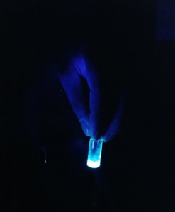
He came upon the idea for a pollen-tracking method after reading an article on the use of quantum dots to track cancer cells in rats (https://doi.org/10.1038/nbt994). Quantum dots are semiconductor nanocrystals that are so small, they behave like artificial atoms. When exposed to UV light, they emit extremely bright light in a range of possible colours. In the case of pollen grains, he figured out that quantum dots with “fat-loving” (lipophilic) ligands would theoretically stick to the fatty outer layer of pollen grains, called pollenkitt, and the glowing colours of the quantum dots can then be used to uniquely “label” pollen grains to see where they end up.

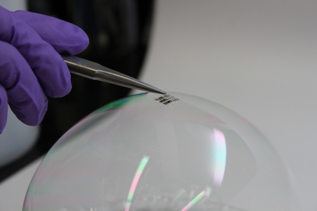
The next step was to find a cost-effective way to view the fluorescing pollen grains under a field dissection microscope. At that stage Minnaar was still using a toy pen from a family restaurant with a little UV LED light that he borrowed from one of his professors.
“I decided to design a fluorescence box that can fit under a dissection microscope. And, because I wanted people to use this method, I designed a box that can easily be 3D-printed at a cost of about R5,000, including the required electronic components.” (view video at https://youtu.be/YHs925F13t0

[or you can scroll down to the bottom of this post]

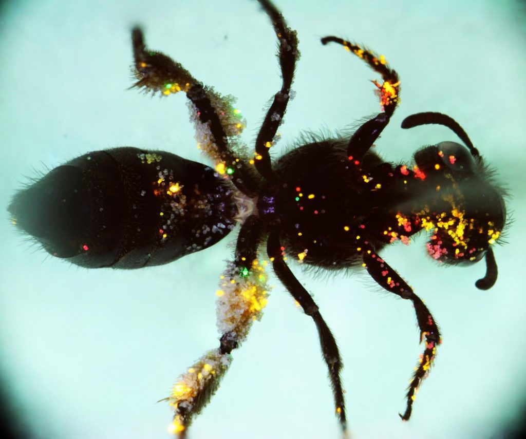
So far, the method and excitation box have proven itself as an easy and relatively inexpensive method to track individual pollen grains: “I’ve done studies where I caught the insects after they have visited the plant with quantum-dot labelled anthers, and you can see where the pollen is placed, and which insects actually carry more or less pollen.”
But the post-labelling part of the work still requires hours and hours of painstaking counting and checking: “I think I’ve probably counted more than a hundred thousand pollen grains these last three years,” he laughs.

As a postdoctoral fellow in the research group of Prof Bruce Anderson in the Department of Botany and Zoology at Stellenbosch University, Minnaar will continue to use the method to investigate the many unanswered questions in this field.

Here’s a link to and a citation for the paper,

Using quantum dots as pollen labels to track the fates of individual pollen grains by Corneile Minnaar and Bruce Anderson. Methods in Ecology and Evolution DOI: https://doi.org/10.1111/2041-210X.13155 First published: 25 January 2019

This paper is behind a paywall.

Here is the video,

Quantum dots derived from tea leaves inhibit growth of lung cancer cells

A May 21, 2018 news item on phys.org announces some intriguing work borne of a UK-India research collaboration,

Nanoparticles derived from tea leaves inhibit the growth of lung cancer cells, destroying up to 80% of them, new research by a joint Swansea University and Indian team has shown.

The team made the discovery while they were testing out a new method of producing a type of nanoparticle called quantum dots. These are tiny particles which measure less than 10 nanometres. A human hair is 40,000 nanometres thick.

A May 21, 2018 Swansea University (UK) press release (also on EurekAlert but dated May 20, 2018), which originated the news item, fills in the details,

Although nanoparticles are already used in healthcare, quantum dots have only recently attracted researchers’ attention.  Already they are showing promise for use in different applications, from computers and solar cells to tumour imaging and treating cancer.

600 x 292

Picture: Size comparison of quantum dots with football and with human hair, in nanometers.

Quantum dots can be made chemically, but this is complicated and expensive and has toxic side effects.  The Swansea-led research team were therefore exploring a non-toxic plant-based alternative method of producing the dots, using tea leaf extract.

Tea leaves contain a wide variety of compounds, including polyphenols, amino acids, vitamins and antioxidants.   The researchers mixed tea leaf extract with cadmium sulphate (CdSO4) and sodium sulphide (Na2S) and allowed the solution to incubate, a process which causes quantum dots to form.   They then applied the dots to lung cancer cells.

The researchers found: 

  • Tea leaves are a simpler, cheaper and less toxic method of producing quantum dots, compared with using chemicals, confirming the results of other research in the field.
  • Quantum dots produced from tea leaves inhibit the growth of lung cancer cellsThey penetrated into the nanopores of the cancer cells and destroyed up to 80% of them.  This was a brand new finding, and came as a surprise to the team.

The research, published in “Applied Nano Materials”, is a collaborative venture between Swansea University experts and colleagues from two Indian universities.

600 x 281

Picture: microscope images of A549 lung cancer cells:  left, untreated; right, treated with quantum dots

Dr Sudhagar Pitchaimuthu of Swansea University, lead researcher on the project, and a Ser Cymru-II Rising Star Fellow, said:

“Our research confirmed previous evidence that tea leaf extract can be a non-toxic alternative to making quantum dots using chemicals.

The real surprise, however, was that the dots actively inhibited the growth of the lung cancer cells.  We hadn’t been expecting this.

The CdS quantum dots derived from tea leaf extract showed exceptional fluorescence emission in cancer cell bioimaging compared to conventional CdS nanoparticles.

Quantum dots are therefore a very promising avenue to explore for developing new cancer treatments.

They also have other possible applications, for example in anti-microbial paint used in operating theatres, or in sun creams.”

Dr Pitchaimuthu outlined the next steps for research:

“Building on this exciting discovery, the next step is to scale up our operation, hopefully with the help of other collaborators.   We want to investigate the role of tea leaf extract in cancer cell imaging, and the interface between quantum dots and the cancer cell.

We would like to set up a “quantum dot factory” which will allow us to explore more fully the ways in which they can be used.”

Here’s a link to and a citation for the paper,

Green-Synthesis-Derived CdS Quantum Dots Using Tea Leaf Extract: Antimicrobial, Bioimaging, and Therapeutic Applications in Lung Cancer Cells by Kavitha Shivaji, Suganya Mani, Ponnusamy Ponmurugan, Catherine Suenne De Castro, Matthew Lloyd Davies, Mythili Gnanamangai Balasubramanian, and Sudhagar Pitchaimuthu. ACS Appl. Nano Mater., 2018, 1 (4), pp 1683–1693 DOI: 10.1021/acsanm.8b00147 Publication Date (Web): March 9, 2018

Copyright © 2018 American Chemical Society

This paper is behind a paywall.

A new graphene-based contrast agent for magnetic resonance imaging (MRI)

After teaching a continuing studies course on bioelectronics for Simon Fraser University (Vancouver, Canada), I’ve developed a mild interest in magnetic resonance imaging and contrast agents which this Nov. 11, 2016 news item on phys.org satisfies,

Graphene, the atomically thin sheets of carbon that materials scientists are hoping to use for everything from nanoelectronics and aircraft de-icers to batteries and bone implants, may also find use as contrast agents for magnetic resonance imaging (MRI), according to new research from Rice University.

“They have a lot of advantages compared with conventionally available contrast agents,” Rice researcher Sruthi Radhakrishnan said of the graphene-based quantum dots she has studied for the past two years. “Virtually all of the widely used contrast agents contain toxic metals, but our material has no metal. It’s just carbon, hydrogen, oxygen and fluorine, and in all of our tests so far it has shown no signs of toxicity.”

The initial findings for Rice’s nanoparticles—disks of graphene that are decorated with fluorine atoms and simply organic molecules that make them magnetic—are described in a new paper in the journal Particle and Particle Systems characterization.

A Nov. 10, 2016 Rice University (Texas, US) news release, which originated the news item, describes the work in more detail,

Pulickel Ajayan, the Rice materials scientist who is directing the work, said the fluorinated graphene oxide quantum dots could be particularly useful as MRI contrast agents because they could be targeted to specific kinds of tissues.

“There are tried-and-true methods for attaching biomarkers to carbon nanoparticles, so one could easily envision using these quantum dots to develop tissue-specific contrast agents,” Ajayan said. “For example, this method could be used to selectively target specific types of cancer or brain lesions caused by Alzheimer’s disease. That kind of specificity isn’t available with today’s contrast agents.”

MRI scanners make images of the body’s internal structures using strong magnetic fields and radio waves. As diagnostic tests, MRIs often provide greater detail than X-rays without the harmful radiation, and as a result, MRI usage has risen sharply over the past decade. More than 30 million MRIs are performed annually in the U.S.

Radhakrishnan said her work began in 2014 after Ajayan’s research team found that adding fluorine to either graphite or graphene caused the materials to show up well on MRI scans.

All materials are influenced by magnetic fields, including animal tissues. In MRI scanners, a powerful magnetic field causes individual atoms throughout the body to become magnetically aligned. A pulse of radio energy is used to disrupt this alignment, and the machine measures how long it takes for the atoms in different parts of the body to become realigned. Based on these measures, the scanner can build up a detailed image of the body’s internal structures.

MRI contrast agents shorten the amount of time it takes for tissues to realign and significantly improve the resolution of MRI scans. Almost all commercially available contrast agents are made from toxic metals like gadolinium, iron or manganese.

“We worked with a team from MD Anderson Cancer Center to assess the cytocompatibility of fluorinated graphene oxide quantum dots,” Radhakrishnan said. “We used a test that measures the metabolic activity of cell cultures and detects toxicity as a drop in metabolic activity. We incubated quantum dots in kidney cell cultures for up to three days and found no significant cell death in the cultures, even at the highest concentrations.”

The fluorinated graphene oxide quantum dots Radhakrishnan studies can be made in less than a day, but she spent two years perfecting the recipe for them. She begins with micron-sized sheets of graphene that have been fluorinated and oxidized. When these are added to a solvent and stirred for several hours, they break into smaller pieces. Making the material smaller is not difficult, but the process for making small particles with the appropriate magnetic properties is exacting. Radhakrishnan said there was no “eureka moment” in which she suddenly achieved the right results by stumbling on the best formula. Rather, the project was marked by incremental improvements through dozens of minor alterations.

“It required a lot of optimization,” she said. “The recipe matters a lot.”

Radhakrishnan said she plans to continue studying the material and hopes to eventually have a hand in proving that it is safe and effective for clinical MRI tests.

“I would like to see it applied commercially in clinical ways because it has a lot of advantages compared with conventionally available agents,” she said.

Here’s a link to and a citation for the paper,

Metal-Free Dual Modal Contrast Agents Based on Fluorographene Quantum Dots by Sruthi Radhakrishnan, Atanu Samanta, Parambath M. Sudeep, Kiersten L. Maldonado, Sendurai A. Mani, Ghanashyam Acharya, Chandra Sekhar Tiwary, Abhishek K. Singh, and Pulickel M. Ajayan. Particle & Particle Systems Characterization DOI: 10.1002/ppsc.201600221 Version of Record online: 21 OCT 2016

This paper is behind a paywall.

Explaining research into matching plasmonic nanoantenna resonances with atoms, molecules, and quantum dots

There’s a very nice explanation of the difficulties associeated with using plasmonic nanoantennas as sensors in a March 21, 2016 news item on phys.org,

Plasmonic nanoantennas are among the hot topics in science at the moment because of their ability to interact strongly with light, which for example makes them useful for different kinds of sensing. But matching their resonances with atoms, molecules or so called quantum dots has been difficult so far because of the very different length scales involved. Thanks to a grant from the Engkvist foundation, Timur Shegai, assistant professor at Chalmers University of Technology, hopes to find a way to do this and by that open doors for applications such as safe long distance communication channels.

A molecule being illuminated by two gold nanoantennas. By: Alexander Ericson Courtesy: Chalmers University of Technology

A molecule being illuminated by two gold nanoantennas. By: Alexander Ericson Courtesy: Chalmers University of Technology

The image, looking like a stylized butterfly or bow tie, above accompanies Karin Weijdegård’s March ??, 2016 Chalmers University of Technology press release, which originated the news item, expands on the research theme,

The diffraction limit makes it very hard for light to interact with the very smallest particles or so called quantum systems such as atoms, molecules or quantum dots. The size of such a particle is simply so much smaller than the wavelength of light that there cannot be a strong interaction between the two. But by using plasmonic nanoantennas, which can be described as metallic nanostructures that are able to focus light very strongly and in wavelengths smaller than those of the visible light, one can build a bridge between the light and the atom, molecule or quantum dot and that is what Timur Shegai is working on.

“Plasmonic nanostructures are themselves smaller than wavelengths of light, but because they have a lot of free electrons they can store the electromagnetic energy in a volume which is actually a lot smaller than the diffraction limit, which helps to bridge the gap between really small objects such as molecules and the larger wavelengths of light,” he says.

Matching the harmonic with the un-harmonic

This might sound easy enough, but the problem with combining the two is that they behave in very different ways. The behaviour of plasmonic nanostructures is very linear, like a harmonic oscillator it will regularly move from side to side no matter how much energy or in other words how many excitations are stored in it. On the other hand, so called quantum systems like atoms, molecules or quantum dots are very much the opposite – their optical properties are highly un-harmonic. Here it makes a big difference if you excite the system with one or two or hundreds of photons.

“Now imagine that you couple together this un-harmonic resonator and a harmonic resonator, and add the possibility to interact with light much stronger than the un-harmonic system alone would have allowed. That opens up very interesting possibilities for quantum technologies and for non-linear optics for example. But as opposed to previous attempts that have been done at very low temperatures and in a vacuum, we will do it at room temperature.”

Communication channels impossible to hack

One possible application where this technology could be useful in the future is to create channels for long distance communications that are impossible to hack. With the current technology this kind of safe communication is only possible if the persons communicating is within a distance of about one hundred kilometres from each other, because that is the maximum distance that an individual photon can run in fibres before it scatters and the signal is lost.

“The kind of ultra small and ultra fast technology we want to develop could be useful in a so called quantum repeater, a device that could be installed across the line from for example New York to London, that would repeat the photon every time it is about to be scattered,” says Timur Shegai.

At the moment though, it is the fundamental aspects of merging plasmons with quantum systems that interest Timur Shegai. To be able to experimentally prove that the there can be interactions between the two systems, he first of all needs to fabricate model systems at the nano level. This is a big challenge, but with the grant of 1,6 million SEK over a period of two years that he just received from the Engkvist foundation, the chances of success have improved.

“Since I am a researcher at the beginning of my career every person is a huge improvement and now I can hire a post doc to work with my group. This means that the project can be divided into sub parts and together we will be able to explore more possibilities about this new technology.”

Thank you Karin Weijdegård for the explanation.

Solar cells and soap bubbles

The MIT team has achieved the thinnest and lightest complete solar cells ever made, they say. To demonstrate just how thin and lightweight the cells are, the researchers draped a working cell on top of a soap bubble, without popping the bubble. Photo: Joel Jean and Anna Osherov

The MIT team has achieved the thinnest and lightest complete solar cells ever made, they say. To demonstrate just how thin and lightweight the cells are, the researchers draped a working cell on top of a soap bubble, without popping the bubble. Photo: Joel Jean and Anna Osherov

That’s quite a compelling image and it comes to us courtesy of researchers at MIT (Massachusetts Institute of Technology). From a Feb. 25, 2016 MIT news release (also on EurekAlert),

Imagine solar cells so thin, flexible, and lightweight that they could be placed on almost any material or surface, including your hat, shirt, or smartphone, or even on a sheet of paper or a helium balloon.

Researchers at MIT have now demonstrated just such a technology: the thinnest, lightest solar cells ever produced. Though it may take years to develop into a commercial product, the laboratory proof-of-concept shows a new approach to making solar cells that could help power the next generation of portable electronic devices.

Bulović [Vladimir Bulović ], MIT’s associate dean for innovation and the Fariborz Maseeh (1990) Professor of Emerging Technology, says the key to the new approach is to make the solar cell, the substrate that supports it, and a protective overcoating to shield it from the environment, all in one process. The substrate is made in place and never needs to be handled, cleaned, or removed from the vacuum during fabrication, thus minimizing exposure to dust or other contaminants that could degrade the cell’s performance.

“The innovative step is the realization that you can grow the substrate at the same time as you grow the device,” Bulović says.

In this initial proof-of-concept experiment, the team used a common flexible polymer called parylene as both the substrate and the overcoating, and an organic material called DBP as the primary light-absorbing layer. Parylene is a commercially available plastic coating used widely to protect implanted biomedical devices and printed circuit boards from environmental damage. The entire process takes place in a vacuum chamber at room temperature and without the use of any solvents, unlike conventional solar-cell manufacturing, which requires high temperatures and harsh chemicals. In this case, both the substrate and the solar cell are “grown” using established vapor deposition techniques.

One process, many materials

The team emphasizes that these particular choices of materials were just examples, and that it is the in-line substrate manufacturing process that is the key innovation. Different materials could be used for the substrate and encapsulation layers, and different types of thin-film solar cell materials, including quantum dots or perovskites, could be substituted for the organic layers used in initial tests.

But already, the team has achieved the thinnest and lightest complete solar cells ever made, they say. To demonstrate just how thin and lightweight the cells are, the researchers draped a working cell on top of a soap bubble, without popping the bubble. The researchers acknowledge that this cell may be too thin to be practical — “If you breathe too hard, you might blow it away,” says Jean [Joel Jean, doctoral student] — but parylene films of thicknesses of up to 80 microns can be deposited easily using commercial equipment, without losing the other benefits of in-line substrate formation.

A flexible parylene film, similar to kitchen cling-wrap but only one-tenth as thick, is first deposited on a sturdier carrier material – in this case, glass. Figuring out how to cleanly separate the thin material from the glass was a key challenge, explains Wang [Annie Wang, research scientist], who has spent many years working with parylene.

The researchers lift the entire parylene/solar cell/parylene stack off the carrier after the fabrication process is complete, using a frame made of flexible film. The final ultra-thin, flexible solar cells, including substrate and overcoating, are just one-fiftieth of the thickness of a human hair and one-thousandth of the thickness of equivalent cells on glass substrates — about two micrometers thick — yet they convert sunlight into electricity just as efficiently as their glass-based counterparts.

No miracles needed

“We put our carrier in a vacuum system, then we deposit everything else on top of it, and then peel the whole thing off,” explains Wang. Bulović says that like most new inventions, it all sounds very simple — once it’s been done. But actually developing the techniques to make the process work required years of effort.

While they used a glass carrier for their solar cells, Jean says “it could be something else. You could use almost any material,” since the processing takes place under such benign conditions. The substrate and solar cell could be deposited directly on fabric or paper, for example.

While the solar cell in this demonstration device is not especially efficient, because of its low weight, its power-to-weight ratio is among the highest ever achieved. That’s important for applications where weight is important, such as on spacecraft or on high-altitude helium balloons used for research. Whereas a typical silicon-based solar module, whose weight is dominated by a glass cover, may produce about 15 watts of power per kilogram of weight, the new cells have already demonstrated an output of 6 watts per gram — about 400 times higher.

“It could be so light that you don’t even know it’s there, on your shirt or on your notebook,” Bulović says. “These cells could simply be an add-on to existing structures.”

Still, this is early, laboratory-scale work, and developing it into a manufacturable product will take time, the team says. Yet while commercial success in the short term may be uncertain, this work could open up new applications for solar power in the long term. “We have a proof-of-concept that works,” Bulović says. The next question is, “How many miracles does it take to make it scalable? We think it’s a lot of hard work ahead, but likely no miracles needed.”

Here’s a link to and a citation for the paper,

In situ vapor-deposited parylene substrates for ultra-thin, lightweight organic solar cells by Joel Jean, Annie Wang, Vladimir Bulović. Organic Electronics Volume 31, April 2016, Pages 120–126 doi:10.1016/j.orgel.2016.01.022

This paper is behind a paywall.

University of New Brunswick (Canada), ‘sun in a can’, and buckyballs

Cutting the cost for making solar cells could be a step in the right direction for more widespread adoption. At any rate, that seems to be the motivation for Dr. Felipe Chibante of the University of New Brunswick  and his team as they’ve worked for the past three years or so on cutting production costs for fullerenes (also known as, buckminsterfullerenes, C60, and buckyballs). From a Dec. 23, 2015 article by Michael Tutton for Canadian Press,

A heating system so powerful it gave its creator a sunburn from three metres away is being developed by a New Brunswick engineering professor as a method to sharply reduce the costs of making the carbon used in some solar cells.

Felipe Chibante says his “sun in a can” method of warming carbon at more than 5,000 degrees Celsius helps create the stable carbon 60 needed in more flexible forms of photovoltaic panels.

Tutton includes some technical explanations in his article,

Chibante and senior students at the University of New Brunswick created the system to heat baseball-sized lumps of plasma — a form of matter composed of positively charged gas particles and free-floating negatively charged electrons — at his home and later in a campus lab.

According to a May 22, 2012 University of New Brunswick news release received funding of almost $1.5M from the Atlantic Canada Opportunities Agency for his work with fullerenes,

Dr. Felipe Chibante, associate professor in UNB’s department of chemical engineering, and his team at the Applied Nanotechnology Lab received nearly $1.5 million to lower the cost of fullerenes, which is the molecular form of pure carbon and is a critical ingredient for the plastic solar cell market.

Dr. Chibante and the collaborators on the project have developed fundamental synthesis methods that will be integrated in a unique plasma reactor to result in a price reduction of 50-75 per cent.

Dr. Chibante and his work were also featured in a June 10, 2013 news item on CBC (Canadian Broadcasting Corporation) news online,

Judges with the New Brunswick Innovation Fund like the idea and recently awarded Chibante $460,000 to continue his research at the university’s Fredericton campus.

Chibante has a long history of working with fullerenes — carbon molecules that can store the sun’s energy. He was part of the research team that discovered fullerenes in 1985 [the three main researchers at Rice University, Texas, received Nobel Prizes for the work].

He says they can be added to liquid, spread over plastic and shingles and marketed as a cheaper way to convert sunlight into electricity.

“What we’re trying to do in New Brunswick with the science research and innovation is we’re really trying to get the maximum bang for the buck,” said Chibante.

As it stands, fullerenes cost about $15,000 per kilogram. Chibante hopes to lower the cost by a factor of 10.

The foundation investment brings Chibante’s research funding to about $6.2 million.

Not everyone is entirely sold on this approach to encouraging solar energy adoption (from the CBC news item),

The owner of Urban Pioneer, a Fredericton [New Brunswick] company that sells alternative energy products, likes the concept, but doubts there’s much of a market in New Brunswick.

“We have conventional solar panels right now and they’re not that popular,” said Tony Craft.

“So I can’t imagine, like, when you throw something completely brand new into it, I don’t know how people are going to respond to that even, so it may be a very tough sell,” he said.

Getting back to Chibante’s breakthrough (from Tutton’s Dec. 23, 2015 article),

The 52-year-old researcher says he first set up the system to operate in his garage.

He installed optical filters to watch the melting process but said the light from the plasma was so intense that he later noticed a sunburn on his neck.

The plasma is placed inside a container that can contain and cool the extremely hot material without exposing it to the air.

The conversion technology has the advantage of not using solvents and doesn’t produce the carbon dioxide that other baking systems use, says Chibante.

He says the next stage is finding commercial partners who can help his team further develop the system, which was originally designed and patented by French researcher Laurent Fulcheri.

Chibante said he doesn’t believe the carbon-based, thin-film solar cells will displace the silicon-based cells because they capture less energy.

But he nonetheless sees a future for the more flexible sheets of solar cells.

“You can make fibres, you can make photovoltaic threads and you get into wearable, portable forms of power that makes it more ubiquitous rather than having to carry a big, rigid structure,” he said.

The researcher says the agreement earlier this month [Nov. 30 – Dec. 12, 2015] in Paris among 200 countries to begin reducing the use of fossil fuels and slow global warming may help his work.

By the way,  Chibante estimates production costs for fullerenes, when using his system, would be less that $50/kilogram for what is now the highest priced component of carbon-based solar cells.

There is another researcher in Canada who works in the field of solar energy, Dr. Ted Sargent at the University of Toronto (Ontario). He largely focuses on harvesting solar energy by using quantum dots. I last featured Sargent’s quantum dot work in a Dec. 9, 2014 posting.

Using quantum dots to detect and identify explosives

This research is courtesy of the University College London (UCL) according to a Dec. 9, 2015 news item on Nanowerk,

A new test for detecting multiple explosives simultaneously has been developed by UCL scientists. The proof-of-concept sensor is designed to quickly identify and quantify five commonly used explosives in solution to help track toxic contamination in waste water and improve the safety of public spaces.

Lead researcher, Dr William Peveler (UCL Chemistry), said: “This is the first time multiple explosives have been detected using a single sensor before, demonstrating proof-of-concept for this approach. Our sensor changes colour within 10 seconds to give information about how much and what explosives are present in a sample. Following further development, we hope it will be used to quickly analyse the nature of threats and inform tailored responses.”

A Dec. 9, 2015 UCL press release (also on EurekAlert), which originated the news item, expands on the theme,

Dr Peveler, added: “We analysed explosives which are commonly used for industrial and military purposes to create a useful tool for environmental and security monitoring. For example, DNT is a breakdown product from landmines, and RDX and PETN have been used in terror plots in recent years as they can be hard to detect using sniffer dogs. Our test can quickly identify these compounds so we see it having a variety of applications from monitoring the waste water of munitions factories and military ranges to finding evidence of illicit activities.”

The sensor is made of quantum dots, which are tiny light-emitting particles or nanomaterials, to which explosive targeting receptors are attached. As each explosive binds to the quantum dot, it quenches the light being emitted to a different degree. The distinct changes in colour are analysed computationally in a variety of conditions to give a unique fingerprint for each compound, allowing multiple explosives to be detected with a single test.

Senior author, Professor Ivan Parkin (UCL Chemistry), said: “Our sensor is a significant step forward for multiple explosives detection. Current methods can be laborious and require expensive equipment but our test is designed to be inexpensive, fast and use a much smaller volume of sample than previously possible. Although all of these factors are important, speed and accuracy are crucial when identifying explosive compounds.”

The team plan to take it from the laboratory into the field by blind testing it with contaminated waste water samples. They also hope to improve the sensitivity of the test by tailoring the surfaces of the quantum dots. Currently, its limit is less than one part per million which the team hope to increase into the part per billion range.

Here’s a link to and a citation for the paper,

Multichannel Detection and Differentiation of Explosives with a Quantum Dot Array by William J. Peveler, Alberto Roldan, Nathan Hollingsworth, Michael J. Porte‡, and Ivan P. Parkin. ACS Nano, Article ASAP DOI: 10.1021/acsnano.5b06433 Publication Date (Web): November 18, 2015

Copyright © 2015 American Chemical Society

This paper is behind a paywall.

Light emitting diodes (LEDs) from food and beverage waste

It’s exciting to think that with emerging technologies we’ll be able to make use of waste products rather than sending them off to fill up garbage dumps. An Oct. 13, 2015 news item on Nanowerk highlights some research where food and beverage waste products could be used to produce light emitting diodes (LEDs),

Most Christmas lights, DVD players, televisions and flashlights have one thing in common: they’re made with light emitting diodes (LEDs). LEDs are widely used for a variety of applications and have been a popular, more efficient alternative to fluorescent and incandescent bulbs for the past few decades. Two University of Utah researchers have now found a way to create LEDs from food and beverage waste. In addition to utilizing food and beverage waste that would otherwise decompose and be of no use, this development can also reduce potentially harmful waste from LEDs generally made from toxic elements.

An Oct. 13, 2015 University of Utah news release, which originated the news item, describes some of the issues with our current LEDs and how the researchers went about synthesizing the waste for reuse,

LEDs can be produced by using quantum dots, or tiny crystals that have luminescent properties, to produce light. Quantum dots (QDs) can be made with numerous materials, some of which are rare and expensive to synthesize, and even potentially harmful to dispose of. Some research over the past 10 years has focused on using carbon dots (CDs), or simply QDs made of carbon, to create LEDs instead.

Compared to other types of quantum dots, CDs have lower toxicity and better biocompatibility, meaning they can be used in a broader variety of applications.

U Metallurgical Engineering Research Assistant Professor Prashant Sarswat and Professor Michael Free, over the past year and a half, have successfully turned food waste such as discarded pieces of tortilla into CDs, and subsequently, LEDs.

From bread to bulb

To synthesize waste into CDs, Sarswat and Free employed a solvothermal synthesis, or one in which the waste was placed into a solvent under pressure and high temperature until CDs were formed. In this experiment, the researchers used soft drinks and pieces of bread and tortilla.

The food and beverage waste were each placed in a solvent and heated both directly and indirectly for anywhere from 30 to 90 minutes.

After successfully finding traces of CDs from the synthesis, Sarswat and Free proceeded to illuminate the CDs to monitor their formation and color.

The pair also employed four other tests, Fourier transform infrared spectroscopy, x-ray photoelectron spectroscopy, Raman and AFM [atomic force microscopy] imaging to determine the CDs’ various optical and material properties.

“Synthesizing and characterizing CDs derived from waste is a very challenging task. We essentially have to determine the size of dots which are only 20 nanometers or smaller in diameter, so we have to run multiple tests to be sure CDs are present and to determine what optical properties they possess,” said Sarswat.

For comparison, a human hair is around 75,000 nanometers in diameter.

The various tests Sarswat and Free ran first measured the size of the CDs, which correlates with the intensity of the dots’ color and brightness. The tests then determined which carbon source produced the best CDs. For example, sucrose and D-fructose dissolved in soft drinks were found to be the most effective sources for production of CDs.

An environmentally sustainable alternative

Currently, one of the most common sources of QDs is cadmium selenide, a compound comprised of a two toxic elements. The ability to create QDs in the form of CDs from food and beverage waste would eliminate the need for concern over toxic waste, as the food and beverages themselves are not toxic.

“QDs derived from food and beverage waste are not based on common toxic elements such as cadmium and selenium, which makes their processing and disposal more environmentally friendly than it is for most other QDs.  In addition, the use of food and beverage waste as the starting material for QDs allows for reduced waste and cost to produce a useful material,” said Free.

In addition to being toxic when broken down, cadmium selenide is also expensive—one website listed a price of $529 for 25 ml of the compound.

“With food and beverage waste that are already there, our starting material is much less expensive. In fact, it’s essentially free,” said Sarswat.

According to a report from the US Department of Agriculture, roughly 31% of food produced in 2014 was not available for human consumption. To be able to use this waste for creating LEDs which are widely used in a number of technologies would be an environmentally sustainable approach.

Looking forward, Sarswat and Free hope to continue studying the LEDs produced from food and beverage waste for stability and long term performance.

“The ultimate goal is to do this on a mass scale and to use these LEDs in everyday devices. To successfully make use of waste that already exists, that’s the end goal,” said Sarswat.

Finally, the CDs were suspended in epoxy resins, heated and hardened to solidify the CDs for practical use in LEDs.

The researchers have made an image of the luminescent carbon dots available,

PHOTO CREDIT: Prashant Sarswat The luminescence of carbon dots can be seen when irradiated with UV light.

PHOTO CREDIT: Prashant Sarswat The luminescence of carbon dots can be seen when irradiated with UV light.

Here’s a link to and a citation for the paper,

Light emitting diodes based on carbon dots derived from food, beverage, and combustion wastes by Prashant K. Sarswat and Michael L. Free. Phys. Chem. Chem. Phys., 2015,17, 27642-27652 DOI: 10.1039/C5CP04782J First published online 01 Oct 2015

This paper appears to be behind a paywall. One final note, despite the paper’s title there doesn’t seem to be any mention of combustion waste in the news release which is a bit puzzling.

Windows as solar panels

Thanks to Dexter Johnson’s Aug. 27, 2015 posting, I’ve found another type of ‘smart’ window (I have written many postings about nanotechnology-enabled windows, especially self-cleaning ones); this window is a solar panel (Note: Links have been removed),

In joint research between the Department of Energy’s Los Alamos National Laboratory (LANL) and the University of Milan-Bicocca (UNIMIB) in Italy, researchers have spent the last 16 months perfecting a technique that makes it possible to embed quantum dots into windows so that the window itself becomes a solar panel.

Of course, this is not the first time someone thought that it would be a good idea to make windows into solar collectors. But this latest iteration marks a significant development in the evolution of the technology. Previous technologies used organic emitters that limited the size of the concentrators to just a few centimeters.

The energy conversion efficiency the researchers were able to acheive with the solar windows was around 3.2 percent, which stands up pretty well when compared with state-of-the-art quantum dot-based solar cells that have reached 9 percent conversion efficiency.

An August 24, 2015 US Los Alamos National Laboratory news release, which inspired Dexter’s posting, describes the research and the US-Italian collaboration in more detail,

A luminescent solar concentrator [LSC] is an emerging sunlight harvesting technology that has the potential to disrupt the way we think about energy; It could turn any window into a daytime power source.

“In these devices, a fraction of light transmitted through the window is absorbed by nanosized particles (semiconductor quantum dots) dispersed in a glass window, re-emitted at the infrared wavelength invisible to the human eye, and wave-guided to a solar cell at the edge of the window,” said Victor Klimov, lead researcher on the project at the Department of Energy’s Los Alamos National Laboratory. “Using this design, a nearly transparent window becomes an electrical generator, one that can power your room’s air conditioner on a hot day or a heater on a cold one.”

… The work was performed by researchers at the Center for Advanced Solar Photophysics (CASP) of Los Alamos, led by Klimov and the research team coordinated by Sergio Brovelli and Francesco Meinardi of the Department of Materials Science of the University of Milan-Bicocca (UNIMIB) in Italy.

The news release goes on to describe the precursor work which made this latest step forward possible,

In April 2014, using special composite quantum dots, the American-Italian collaboration demonstrated the first example of large-area luminescent solar concentrators free from reabsorption losses of the guided light by the nanoparticles. This represented a fundamental advancement with respect to the earlier technology, which was based on organic emitters that allowed for the realization of concentrators of only a few centimeters in size.

However, the quantum dots used in previous proof-of-principle devices were still unsuitable for real-world applications, as they were based on the toxic heavy metal cadmium and were capable of absorbing only a small portion of the solar light. This resulted in limited light-harvesting efficiency and strong yellow/red coloring of the concentrators, which complicated their application in residential environments.

Here’s how they solved the problem (from the news release),

Klimov, CASP’s director, explained how the updated approach solves the coloring problem: “Our new devices use quantum dots of a complex composition which includes copper (Cu), indium (In), selenium (Se) and sulfur (S). This composition is often abbreviated as CISeS. Importantly, these particles do not contain any toxic metals that are typically present in previously demonstrated LSCs.”

“Furthermore,” Klimov noted, “the CISeS quantum dots provide a uniform coverage of the solar spectrum, thus adding only a neutral tint to a window without introducing any distortion to perceived colors. In addition, their near-infrared emission is invisible to a human eye, but at the same time is ideally suited for most common solar cells based on silicon.”

Francesco Meinardi, professor of Physics at UNIMIB, described the emerging work, noting, “In order for this technology to leave the research laboratories and reach its full potential in sustainable architecture, it is necessary to realize non-toxic concentrators capable of harvesting the whole solar spectrum.”

“We must still preserve the key ability to transmit the guided luminescence without reabsorption losses, though, so as to complement high photovoltaic efficiency with dimensions compatible with real windows. The aesthetic factor is also of critical importance for the desirability of an emerging technology,” Meinardi said. [emphasis mine]

I couldn’t agree more with Professor Meinardi. You’re much more likely to adopt something that’s good for you and the planet if you like the look. Following on that thought, you’re much more likely to adopt solar panel windows if they’re aesthetically pleasing.

However, there is still a problem to be solved,

Hunter McDaniel, formerly a Los Alamos CASP postdoctoral fellow and presently a quantum dot entrepreneur (UbiQD founder and president), added, “with a new class of low-cost, low-hazard quantum dots composed of CISeS, we have overcome some of the biggest roadblocks to commercial deployment of this technology.”

“One of the remaining problems to tackle is reducing cost, but already this material is significantly less expensive to manufacture than alternative quantum dots used in previous LSC demonstrations,” McDaniel said.

Nonetheless, they have high hopes the technology can be commercialized (although as Dexter notes, it’s probably not going to be in the near future), from the news release,

A key element of this work is a procedure comparable to the cell casting industrial method used for fabricating high optical quality polymer windows. It involves a new UNIMIB protocol for encapsulating quantum dots into a high-optical quality transparent polymer matrix. The polymer used in this study is a cross-linked polylaurylmethacrylate, which belongs to the family of acrylate polymers. Its long side-chains prevent agglomeration of the quantum dots and provide them with the “friendly” local environment, which is similar to that of the original colloidal suspension. This allows one to preserve light emission properties of the quantum dots upon encapsulation into the polymer.

Sergio Brovelli, the lead researcher on the Italian team, concluded: “Quantum dot solar window technology, of which we had demonstrated the feasibility just one year ago, now becomes a reality that can be transferred to the industry in the short to medium term, allowing us to convert not only rooftops, as we do now, but the whole body of urban buildings, including windows, into solar energy generators.”

“This is especially important in densely populated urban area where the rooftop surfaces are too small for collecting all the energy required for the building operations,” he said. He proposes that the team’s estimations indicate that by replacing the passive glazing of a skyscraper such as the One World Trade Center in NYC (72,000 square meters divided into 12,000 windows) with our technology, it would be possible to generate the equivalent of the energy need of over 350 apartments.

“Add to these remarkable figures, the energy that would be saved by the reduced need for air conditioning thanks to the filtering effect by the LSC, which lowers the heating of indoor spaces by sunlight, and you have a potentially game-changing technology towards “net-zero” energy cities,” Brovelli said.

For anyone interested in this latest work on energy harvesting and windows, here’s a link to and a citation for the paper,

Highly efficient large-area colourless luminescent solar concentrators using heavy-metal-free colloidal quantum dots by Francesco Meinardi, Hunter McDaniel, Francesco Carulli, Annalisa Colombo, Kirill A. Velizhanin, Nikolay S. Makarov, Roberto Simonutti, Victor I. Klimov, & Sergio Brovelli. Nature Nanotechnology (2015) doi:10.1038/nnano.2015.178 Published online 24 August 2015

This paper is behind a paywall.

Stress makes quantum dots ‘breathe’

A March 19, 2015 news item on ScienceDaily describes some new research on quantum dots,

Researchers at the Department of Energy’s SLAC National Accelerator Laboratory watched nanoscale semiconductor crystals expand and shrink in response to powerful pulses of laser light. This ultrafast “breathing” provides new insight about how such tiny structures change shape as they start to melt — information that can help guide researchers in tailoring their use for a range of applications.

In the experiment using SLAC’s Linac Coherent Light Source (LCLS) X-ray laser, a DOE Office of Science User Facility, researchers first exposed the nanocrystals to a burst of laser light, followed closely by an ultrabright X-ray pulse that recorded the resulting structural changes in atomic-scale detail at the onset of melting.

“This is the first time we could measure the details of how these ultrasmall materials react when strained to their limits,” said Aaron Lindenberg, an assistant professor at SLAC and Stanford who led the experiment. The results were published March 12 [2015] in Nature Communications.

A March 18, 2015 SLAC news release, which originated the news item, provides a general description of quantum dots,

The crystals studied at SLAC are known as “quantum dots” because they display unique traits at the nanoscale that defy the classical physics governing their properties at larger scales. The crystals can be tuned by changing their size and shape to emit specific colors of light, for example.

So scientists have worked to incorporate them in solar panels to make them more efficient and in computer displays to improve resolution while consuming less battery power. These materials have also been studied for potential use in batteries and fuel cells and for targeted drug delivery.

Scientists have also discovered that these and other nanomaterials, which may contain just tens or hundreds of atoms, can be far more damage-resistant than larger bits of the same materials because they exhibit a more perfect crystal structure at the tiniest scales. This property could prove useful in battery components, for example, as smaller particles may be able to withstand more charging cycles than larger ones before degrading.

The news release then goes on to describe the latest research showing the dots ‘breathe’ (Note: A link has been removed),

In the LCLS experiment, researchers studied spheres and nanowires made of cadmium sulfide and cadmium selenide that were just 3 to 5 nanometers, or billionths of a meter, across. The nanowires were up to 25 nanometers long. By comparison, amino acids – the building blocks of proteins – are about 1 nanometer in length, and individual atoms are measured in tenths of nanometers.

By examining the nanocrystals from many different angles with X-ray pulses, researchers reconstructed how they change shape when hit with an optical laser pulse. They were surprised to see the spheres and nanowires expand in width by about 1 percent and then quickly contract within femtoseconds, or quadrillionths of a second. They also found that the nanowires don’t expand in length, and showed that the way the crystals respond to strain was coupled to how their structure melts.

In an earlier, separate study, another team of researchers had used LCLS to explore the response of larger gold particles on longer timescales.

“In the future, we want to extend these experiments to more complex and technologically relevant nanostructures, and also to enable X-ray exploration of nanoscale devices while they are operating,” Lindenberg said. “Knowing how materials change under strain can be used together with simulations to design new materials with novel properties.”

Participating researchers were from SLAC, Stanford and two of their joint institutes, the Stanford Institute for Materials and Energy Sciences (SIMES) and Stanford PULSE Institute; University of California, Berkeley; University of Duisburg-Essen in Germany; and Argonne National Laboratory. The work was supported by the DOE Office of Science and the German Research Council.

Here’s a link to and a citation for the paper,

Visualization of nanocrystal breathing modes at extreme strains by Erzsi Szilagyi, Joshua S. Wittenberg, Timothy A. Miller, Katie Lutker, Florian Quirin, Henrik Lemke, Diling Zhu, Matthieu Chollet, Joseph Robinson, Haidan Wen, Klaus Sokolowski-Tinten, & Aaron M. Lindenberg. Nature Communications 6, Article number: 6577 doi:10.1038/ncomms7577 Published 12 March 2015

This paper is behind a paywall but there is a free preview available through ReadCube Access.