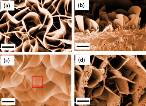
a) Colorized SEM images of iron oxide nanoblades used in the experiment. b) Colorized cross-section of SEM image of the nanoblades. c) Colorized SEM image of nanoblades after 1 hour of reduction reaction at 500 °C in molecular hydrogen, showing the sawtooth shape along the edges (square). d) Colorized SEM image showing the formation of holes after 2 hours of reduction. The scale bar is 1 micrometer. Credit: W. Zhu et al./ACS Nano and K. Irvine/NIST
Here’s more about being able to watch iron transition from one state to the next according to an April 5, 2017 news item on phys.org
Using a state-of-the-art microscopy technique, experimenters at the National Institute of Standards and Technology (NIST) and their colleagues have witnessed a slow-motion, atomic-scale transformation of rust—iron oxide—back to pure iron metal, in all of its chemical steps.
An April 4, 2017 NIST news release describes the role iron plays in modern lifestyles and the purpose of this research,
Among the most abundant minerals on Earth, iron oxides play a leading role in magnetic data storage, cosmetics, the pigmentation of paints and drug delivery. These materials also serve as catalysts for several types of chemical reactions, including the production of ammonia for fertilizer.
To fine-tune the properties of these minerals for each application, scientists work with nanometer-scale particles of the oxides. But to do so, researchers need a detailed, atomic-level understanding of reduction, a key chemical reaction that iron oxides undergo. That knowledge, however, is often lacking because reduction—a process that is effectively the opposite of rusting—proceeds too rapidly for many types of probes to explore at such a fine level.
In a new effort to study the microscopic details of metal oxide reduction, researchers used a specially adapted transmission electron microscope (TEM) at NIST’s NanoLab facility to document the step-by-step transformation of nanocrystals of the iron oxide hematite (Fe2O3) to the iron oxide magnetite (Fe3O4), and finally to iron metal.
“Even though people have studied iron oxide for many years, there have been no dynamic studies at the atomic scale,” said Wenhui Zhu of the State University of New York at Binghamton, who worked on her doctorate in the NanoLab in 2015 and 2016. “We are seeing what’s actually happening during the entire reduction process instead of studying just the initial steps.”
That’s critical, added NIST’s Renu Sharma, “if you want to control the composition or properties of iron oxides and understand the relationships between them.”
By lowering the temperature of the reaction and decreasing the pressure of the hydrogen gas that acted as the reducing agent, the scientists slowed down the reduction process so that it could be captured with an environmental TEM—a specially configured TEM that can study both solids and gas. The instrument enables researchers to perform atomic-resolution imaging of a sample under real-life conditions—in this case the gaseous environment necessary for iron oxides to undergo reduction–rather than under the vacuum needed in ordinary TEMs.
“This is the most powerful tool I’ve used in my research and one of the very few in the United States,” said Zhu. She, Sharma and their colleagues describe their findings in a recent issue of ACS Nano.
The team examined the reduction process in a bicrystal of iron oxide, consisting of two identical iron oxide crystals rotated at 21.8 degrees with respect to each other. The bicrystal structure also served to slow down the reduction process, making it easier to follow with the environmental TEM.
In studying the reduction reaction, the researchers identified a previously unknown intermediate state in the transformation from magnetite to hematite. In the middle stage, the iron oxide retained its original chemical structure, Fe2O3, but changed the crystallographic arrangement of its atoms from rhombohedral (a diagonally stretched cube) to cubic.
This intermediate state featured a defect in which oxygen atoms fail to populate some of the sites in the crystal that they normally would. This so-called oxygen vacancy defect is not uncommon and is known to strongly influence the electrical and catalytic properties of oxides. But the researchers were surprised to find that the defects occurred in an ordered pattern, which had never been found before in the reduction of Fe2O3 to Fe3O4, Sharma said.
The significance of the intermediate state remains under study, but it may be important for controlling the reduction rate and other properties of the reduction process, she adds. “The more we understand, the better we can manipulate the microstructure of these oxides,” said Zhu. By manipulating the microstructure, researchers may be able to enhance the catalytic activity of iron oxides.
Even though a link has already been provided for the paper, I will give it again along with a citation,
In Situ Atomic-Scale Probing of the Reduction Dynamics of Two-Dimensional Fe2O3 Nanostructures by Wenhui Zhu, Jonathan P. Winterstein, Wei-Chang David Yang, Lu Yuan, Renu Sharma, and Guangwen Zhou. ACS Nano, 2017, 11 (1), pp 656–664 DOI: 10.1021/acsnano.6b06950 Publication Date (Web): December 13, 2016
Copyright © 2016 American Chemical Society
This paper is behind a paywall.