Tag Archives: Ruiguo Yang

The inside scoop on beetle exoskeletons

In the past I’ve covered work on the Namib beetle and its bumps which allow it to access condensation from the air in one of the hottest places on earth and work on jewel beetles and how their structural colo(u)r is derived. Now, there’s research into a beetle’s body armor from the University of Nebraska-Lincoln according to a Feb. 22, 2017 news item on ScienceDaily,

Beetles wear a body armor that should weigh them down — think medieval knights and turtles. In fact, those hard shells protecting delicate wings are surprisingly light, allowing even flight.

Better understanding the structure and properties of beetle exoskeletons could help scientists engineer lighter, stronger materials. Such materials could, for example, reduce gas-guzzling drag in vehicles and airplanes and reduce the weight of armor, lightening the load for the 21st-century knight.

But revealing exoskeleton architecture at the nanoscale has proven difficult. Nebraska’s Ruiguo Yang, assistant professor of mechanical and materials engineering, and his colleagues found a way to analyze the fibrous nanostructure. …

A Feb. 22, 2017 University of Nebraska-Lincoln news release by Gillian Klucas (also on EurekAlert), which originated the news item, describes skeletons and the work in more detail,

The lightweight exoskeleton is composed of chitin fibers just around 20 nanometers in diameter (a human hair measures approximately 75,000 nanometers in diameter) and packed and piled into layers that twist in a spiral, like a spiral staircase. The small diameter and helical twisting, known as Bouligand, make the structure difficult to analyze.

Yang and his team developed a method of slicing down the spiral to reveal a surface of cross-sections of fibers at different orientations. From that viewpoint, the researchers were able to analyze the fibers’ mechanical properties with the aid of an atomic force microscope. This type of microscope applies a tiny force to a test sample, deforms the sample and monitors the sample’s response. Combining the experimental procedure and theoretical analysis, the researchers were able to reveal the nanoscale architecture of the exoskeleton and the material properties of the nanofibers.

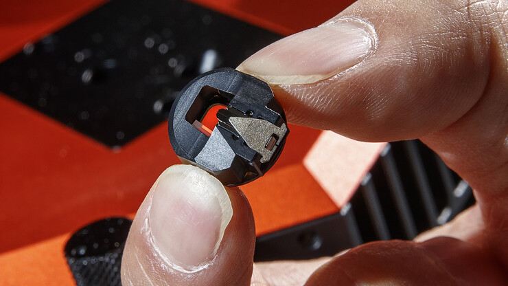
Yang holds a piece of the atomic force microscope used to measure the beetle's surface. A small wire can barely be seen in the middle of the piece. Unseen is a two-nano-size probe attached to the wire, which does the actual measuring.

Craig Chandler | University Communication

Yang holds a piece of the atomic force microscope used to measure the beetle’s surface. A small wire can barely be seen in the middle of the piece. Unseen is a two-nano-size probe attached to the wire, which does the actual measuring.

They made their discoveries in the common figeater beetle, Cotinis mutabilis, a metallic green native of the western United States. But the technique can be used on other beetles and hard-shelled creatures and might also extend to artificial materials with fibrous structures, Yang said.

Comparing beetles with differing demands on their exoskeletons, such as defending against predators or environmental damage, could lead to evolutionary insights as well as a better understanding of the relationship between structural features and their properties.

Yang’s co-authors are Alireza Zaheri and Horacio Espinosa of Northwestern University; Wei Gao of the University of Texas at San Antonio; and Cheryl Hayashi of the University of California, Riverside.

Here’s a link to and a citation for the paper,

Exoskeletons: AFM Identification of Beetle Exocuticle: Bouligand Structure and Nanofiber Anisotropic Elastic Properties by Ruiguo Yang, Alireza Zaheri,Wei Gao, Charely Hayashi, Horacio D. Espinosa. Adv. Funct. Mater. vol. 27 (6) 2017 DOI: 10.1002/adfm.201770031 First published: 8 February 2017

This paper is behind a paywall.
Nanomechanics for deciphering beetle exoskeletons

Beetles carry remarkably light yet strong armor in the form of their exoskeletons and a research team at Northwestern University (US) is looking to those beetle exoskeletons for inspiration according to a Jan. 11, 2017 news item on ScienceDaily,

What can a beetle tell us about good design principles? Quite a lot, actually.

Many insects and crustaceans possess hard, armor-like exoskeletons that, in theory, should weigh the creatures down. But, instead, the exoskeletons are surprisingly light — even allowing the armor-wearing insects, like the beetle, to fly.

Northwestern Engineering’s Horacio D. Espinosa and his group are working to understand the underlying design principles and mechanical properties that result in structures with these unique, ideal properties. This work could ultimately uncover information that could guide the design and manufacturing of new and improved artificial materials by emulating these time-tested natural patterns, a process known as bio-mimicry.

Supported by the Air Force Office of Scientific Research’s Multidisciplinary University Research Initiative (MURI), the research was featured on the cover of Advanced Functional Materials. Postdoctoral fellows Ruiguo Yang and Wei Gao and graduate student Alireza Zaheri, all members of Espinosa’s laboratory, were co-first authors of the paper. Cheryl Hayashi, professor of biology at the University of California, Riverside, was also a co-author.

A Jan. 11, 2017 Northwestern University news release, which originated the news item, expands on the theme,

Though there are more than a million species of beetles, the team is first studying the exoskeleton of the Cotinis mutabilis, a field crop pest beetle native to the western United States. Like all insects and crustaceans, its exoskeleton is composed of twisted plywood structures, known as Bouligand structures, which help protect against predators. Fibers in this Bouligand structure are bundles of chitin polymer chains wrapped with proteins. In this chain structure, each fiber has a higher density along the length than along the transverse.

“It is very challenging to characterize the properties of such fibers given that they are directionally dependent and have a small diameter of just 20 nanometers,” said Espinosa, the James N. and Nancy J. Farley Professor in Manufacturing and Entrepreneurship at Northwestern’s McCormick School of Engineering. “We had to develop a novel characterization method by taking advantage of the spatial distribution of fibers in the Bouligand structure.”

To meet this challenge, Espinosa and his team employed a creative way to identify the geometry and material properties of the fibers that comprise the exoskeleton. They cut the Bouligand structure along a plane, resulting in a surface composed of closely packed cross-sections of fibers with different orientations. They were then able to analyze the mechanics of the fibers.

“With more than a million species, which greatly vary from each other in taxomic relatedness, size, and ecology, the beetle is the largest group of insects,” Hayashi said. “What makes this research exciting is that the methods applied to the Cotinis mutabilis beetle exoskeleton can be extended to other beetle species.”

By correlating the mechanical properties with the exoskeleton geometries from diverse beetle species, Espinosa and his team plan to gain insight into natural selection and better understand structure-function-properties relationships.

Here’s a link to and a citation for the paper,

AFM Identification of Beetle Exocuticle: Bouligand Structure and Nanofiber Anisotropic Elastic Properties by Ruiguo Yang, Alireza Zaheri, Wei Gao, Cheryl Hayashi, and Horacio D. Espinosa. Advanced Functional Materials DOI: 10.1002/adfm.201603993 Version of Record online: 27 DEC 2016

© 2016 WILEY-VCH Verlag GmbH & Co. KGaA, Weinheim

This paper is behind a paywall.

Nanorobotic approach to studying how skin falls apart

Scientists have combined robotic techniques with atomic force microscopy to achieve understanding of how skin falls apart at the nanoscale. From a Sept. 11, 2014 news item on Azonano,

University at Buffalo researchers and colleagues studying a rare, blistering disease have discovered new details of how autoantibodies destroy healthy cells in skin. This information provides new insights into autoimmune mechanisms in general and could help develop and screen treatments for patients suffering from all autoimmune diseases, estimated to affect 5-10 percent of the U.S. population.

“Our work represents a unique intersection between the fields of biology and engineering that allowed for entirely new investigational strategies applied to the study of clinical disease,” says Animesh A. Sinha, MD, PhD, Rita M. and Ralph T. Behling Professor and chair of the Department of Dermatology in the UB School of Medicine and Biomedical Sciences and senior author on the study.

A Sept. 9, 2014 University of Buffalo news release by Ellen Goldbaum (also on EurekAlert dated Sept. 10, 2014), which originated the news item, describes the condition and the research in more detail,

PV [Pemphigus Vulgaris] results in the often painful blistering of the skin and mucous membranes. Generally treated with corticosteroids and other immunosuppressive agents, the condition is life-threatening if untreated.

Sinha’s research team, in collaboration with scientists at Michigan State University, describe the use of atomic force microscopy (AFM), a technique originally developed to study nonbiological materials, to look at cell junctions and how they rupture, a process called acantholysis.

“It has been very difficult to study cell junctions, which maintain the skin’s barrier function by keeping cells attached to each other,” says Sinha. “These junctions, micron-sized spots on cell membranes, are very complex molecular structures. Their small size has made them resistant to detailed investigation.”

Sinha’s interest lies in determining what destroys those junctions in Pemphigus Vulgaris.

“We haven’t understood why some antibodies generated by the condition cause blisters and why other antibodies it generates do not,” says Sinha.

By studying the connections between skin cells using AFM and other techniques that probe cells at the nanoscale, Sinha and his colleagues report that pathogenic antibodies change structural and functional properties of skin cells in distinct ways.

“Our data suggest a new model for the action of autoantibodies in which there are two steps or ‘hits’ in the development of lesions,” says Sinha. “The first hit results in the initial separation of cells but only the pathogenic antibodies drive further intracellular changes that lead to the breaking of the cell junction and blistering.”

The researchers examined the cells using AFM, which requires minimal sample preparation and provides three-dimensional images of cell surfaces.

The AFM tip acts like a little probe, explains Sinha. When tapped against a cell, it sends back information regarding the cell’s mechanical properties, such as thickness, elasticity, viscosity and electrical potential.

“We combined existing and novel nanorobotic techniques with AFM, including a kind of nanodissection, where we physically detached cells from each other at certain points so that we could test what that did to their mechanical and biological functions,” Sinha adds.

Those data were then combined with information about functional changes in cell behavior to develop a nanomechanical profile, or phenotype, for specific cellular states.

He also envisions that this kind of nanomechanical phenotyping should allow for the development of predictive models for cellular behavior for any kind of cell.

“Ultimately, in the case of autoimmunity, we should be able to use these techniques as a high-throughput assay to screen hundreds or thousands of compounds that might block the effects of autoantibodies and identify novel agents with therapeutic potential in given individuals,” says Sinha.  “Such strategies aim to advance us toward a new era of personalized medicine”.

I found some more information about the nanorobotics technique, mentioned in the news release, in the researchers’ paper (Note: A link has been removed),

Nanorobotic surgery

AFM-based nanorobotics enables accurate and convenient sample manipulation and drug delivery. This capability was used in the current study to control the AFM tip position over the intercellular junction area, and apply vertical indentation forces, so that bundles of intercellular adhesion structures can be dissected precisely with an accuracy of less than 100 nm in height. We used a tip sharp enough (2 nm in tip apex diameter) to penetrate the cell membrane and the intermediate filaments. It has been shown that intermediate filaments have extremely high tensile strength by in vitro AFM stretching [19]. Thus, the vertical force and moving speed of the AFM cantilever (0.06 N/m in vertical spring constant) was controlled at a vertical force of 5 nN at an indentation speed of 0.1 µm/s to guarantee the rupture of the filament and to partially dissect cell adhesion structures between two neighboring cells.

For those who want to know more, here’s a link to and a citation for the paper,

Nanorobotic Investigation Identifies Novel Visual, Structural and Functional Correlates of Autoimmune Pathology in a Blistering Skin Disease Model by Kristina Seiffert-Sinha, Ruiguo Yang, Carmen K. Fung, King W. Lai, Kevin C. Patterson, Aimee S. Payne, Ning Xi, Animesh A. Sinha. PLOSONE Published: September 08, 2014 DOI: 10.1371/journal.pone.0106895

This is an open access paper.