Tag Archives: Sang Jin Lee

A pumpkin-shaped molecule for the first real-time methamphetamine and amphetamine sensor

A Sept. 28,2017 news item on Nanowerk announces a portable, inexpensive sensor for drugs (Note: A link has been renewed),

Speed, uppers, chalk, glass, crystal, or whatever you prefer to call them, can be instantly detected from biological fluids with a new portable kit that costs as little as $50. Scientists at the Center for Self-Assembly and Complexity, within the Institute for Basic Science (IBS, South Korea), in collaboration with Pohang University of Science and Technology (POSTECH), have devised the first methamphetamine and amphetamine sensor that can detect minute concentrations of these drugs from a single drop of urine in real-time.

Published in the journal Chem (“Point-of-Use Detection of Amphetamine-Type Stimulants with Host-Molecule-Functionalized Organic Transistors”), this simple and flexible sensor, which can be attached to a wristband and connected to an Android app via Bluetooth, could move drug screening from the labs to the streets.

A Sept. 28 (?), 2017 IBS press release by Letizia Diamante (also on EurekAlert), which originated the news item, expands on the theme,

Easy to synthesize and cheaper than heroin or cocaine, amphetamine-based drugs are the most abused drugs in the world, after cannabis. Conventional drug detection methods require a long time, as the sample must be taken into a lab for the analysis. It also needs experts to run the expensive equipment. The technology reported in this study is instead small, portable, cheap, fast and easy to use.

The idea for this technology came from the IBS chemist HWANG Ilha: “I was watching a TV news report on the usage of illegal drugs, and I thought to check what the chemical structure of methamphetamine looks like.” Soon after, the scientist anticipated that the drug would form a tight complex with a family of hollow pumpkin-shaped molecules, called cucurbituril (CB) members. The team then discovered that cucurbit[7]uril (CB[7])’s empty cavity binds well with amphetamine-based drugs and can be used as the drug recognition unit of a sensor. Cucurbiturils’ hollow chamber has already been studied for various technological uses, but this is the first device application in amphetamine-based drug detection.


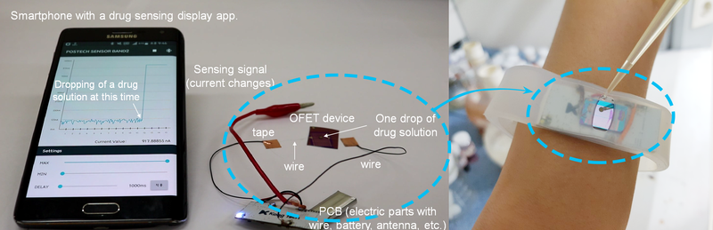
▲ Figure 1: Wireless sensor for amphetamine-based drug detection.The kit is made of an organic field-effect transistor (OFET) device, an electric circuit board with a rechargeable battery and an antenna. The OFET device surface is coated with CB[7], whose function is to bind amphetamine and methamphetamine drugs in solution. The binding event is instantly converted to current, whose magnitude is proportional to the concentration of the drug. The app on the smartphone shows a peak as soon as a drop of urine with the drug is applied to the device. Moreover the entire kit can fit in a handy wristband.


▲ Video 1: The detector in action.
[Click text not image]
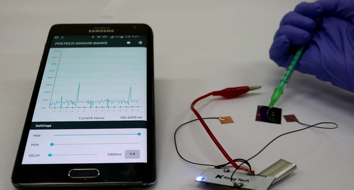
As soon as a drop of water with 0.0001 ng/mL (1 pM) of amphetamine is applied to the kit, the app shows a peak in current proportional to the concentration of drug. When the liquid is removed, the current level goes back to baseline, and the sensor can be reused. (Modified from Jang et al, Chem 2017)

Combining a transistor coated with CB[7], flexible materials, rechargeable batteries and a Bluetooth antenna, the research team developed a detector wristband connected to an app. In the presence of the drug, the molecular recognition between CB[7] and the drug molecule triggers an electrical signal which appears as a peak on the smartphone screen.

Current drug detection based on immunoassay or liquid chromatography/mass spectrometry techniques has a detection limit of about 10 ng/mL. On the contrary, the sensitivity of this new sensor is about 0.0001 ng/mL in water and 0.1 ng/mL in urine. Therefore, it is expected that this method will allow the detection of drug molecules in biological fluids, like urine and sweat, for a longer time after drug consumption.


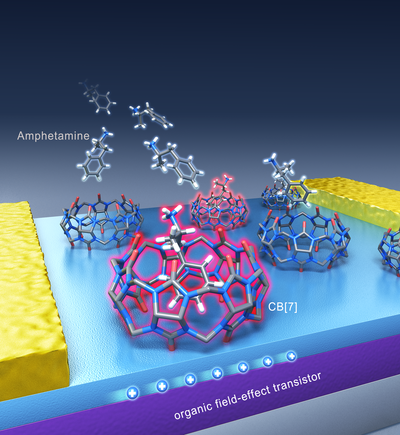
▲ Figure 2: Graphic representation of the drug detection platform.Binding of drug molecules to the hollow cucurbit[7]uril (CB[7])’s cavity changes the current signal flowing in the transistor and therefore can be used as a detection system. The molecular structure of amphetamine and methamphetamine bound to cucurbit[7]uril (CB[7]) was confirmed with X-ray crystallography. Each color indicates a different atom (blue: nitrogen, red: oxygen, gray: carbon, and white: hydrogen). CB[7]’s hydrogen atoms have been omitted for clarity.


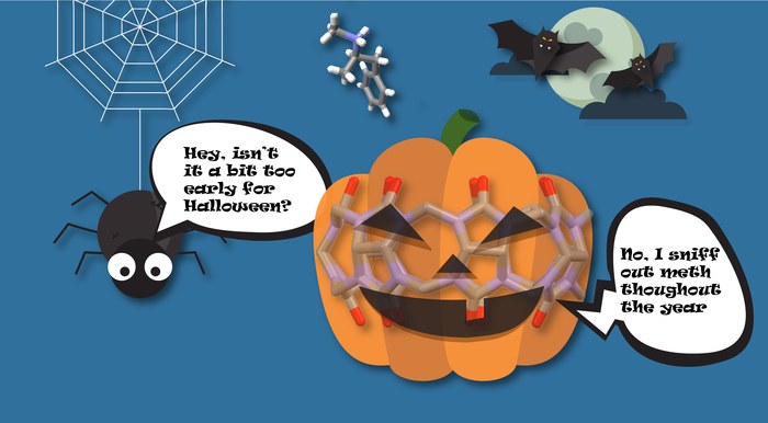
▲ Figure 3: Humorous view of the pumpkin-shaped molecule, cucurbit[7]uril (CB[7]), able to bind and detect amphetamine and methamphetamine molecules.(Credits: Modified from Titusurya – Freepik.com)

“Real time detection of amphetamine drugs on location would bring a big change to society,” explains another corresponding author KIM Kimoon. “In the same way as police can use a breathalyzer to detect alcohol on the spot, we aim to achieve the same with this device.”

False positives cannot be excluded yet, as urine contains a rich mixture of proteins and other metabolites that could affect the reading. Therefore, before commercializing it, clinical trials with drug users’ biological fluids are necessary. The researchers have patented the technology and they will continue to do further research in the near future.s

“Combining basic science with the latest technology, we can expect that this research will also lead to other new sensors, useful for our daily life,” concludes the third corresponding author OH Joon Hak. Indeed, the team is also keen on developing sensors for other kinds of drugs, as well as kits for the detection of dangerous substances, environmental monitoring, healthcare and safety.

Here’s a link to and a citation for the paper,

Point-of-Use Detection of Amphetamine-Type Stimulants with Host-Molecule-Functionalized Organic Transistors by Yoonjung Jang, Moonjeong Jang, Hyoeun Kim, Sang Jin Lee, Eunyeong Jin, Jin Young Koo, In-Chul Hwang, Yonghwi Kim, Young Ho Ko, Ilha Hwang., Joon Hak Oh, Kimoon Kim. Chem (2017). DOI: 10.1016/j.chempr.2017.08.015 Publication stage: In Press Corrected Proof

This paper appears to be behind a paywall.

Feasibility of printing ear, bone, and muscle structures

Over ten years ago I attended a show at the Vancouver (Canada) Art Gallery titled ‘Massive Change’ where I saw part of a nose or ear being grown in a petri dish (the work was from an Israeli laboratory) and that was my introduction to tissue engineering. For anyone who’s been following the tissue engineering story, 3D printers have sped up the growth process considerably. More recently, researchers at Wake Forest Baptist Medical Center (North Carolina, US) have announced another step forward for growing organs and body parts, from a Feb. 15, 2016 Wake Forest Baptist Medical Center news release on EurekAlert,

Using a sophisticated, custom-designed 3D printer, regenerative medicine scientists at Wake Forest Baptist Medical Center have proved that it is feasible to print living tissue structures to replace injured or diseased tissue in patients.

Reporting in Nature Biotechnology, the scientists said they printed ear, bone and muscle structures. When implanted in animals, the structures matured into functional tissue and developed a system of blood vessels. Most importantly, these early results indicate that the structures have the right size, strength and function for use in humans.

“This novel tissue and organ printer is an important advance in our quest to make replacement tissue for patients,” said Anthony Atala, M.D., director of the Wake Forest Institute for Regenerative Medicine (WFIRM) and senior author on the study. “It can fabricate stable, human-scale tissue of any shape. With further development, this technology could potentially be used to print living tissue and organ structures for surgical implantation.”

With funding from the Armed Forces Institute of Regenerative Medicine, a federally funded effort to apply regenerative medicine to battlefield injuries, Atala’s team aims to implant bioprinted muscle, cartilage and bone in patients in the future.

Tissue engineering is a science that aims to grow replacement tissues and organs in the laboratory to help solve the shortage of donated tissue available for transplants. The precision of 3D printing makes it a promising method for replicating the body’s complex tissues and organs. However, current printers based on jetting, extrusion and laser-induced forward transfer cannot produce structures with sufficient size or strength to implant in the body.

The Integrated Tissue and Organ Printing System (ITOP), developed over a 10-year period by scientists at the Institute for Regenerative Medicine, overcomes these challenges. The system deposits both bio-degradable, plastic-like materials to form the tissue “shape” and water-based gels that contain the cells. In addition, a strong, temporary outer structure is formed. The printing process does not harm the cells.

A major challenge of tissue engineering is ensuring that implanted structures live long enough to integrate with the body. The Wake Forest Baptist scientists addressed this in two ways. They optimized the water-based “ink” that holds the cells so that it promotes cell health and growth and they printed a lattice of micro-channels throughout the structures. These channels allow nutrients and oxygen from the body to diffuse into the structures and keep them live while they develop a system of blood vessels.

It has been previously shown that tissue structures without ready-made blood vessels must be smaller than 200 microns (0.007 inches) for cells to survive. In these studies, a baby-sized ear structure (1.5 inches) survived and showed signs of vascularization at one and two months after implantation.

“Our results indicate that the bio-ink combination we used, combined with the micro-channels, provides the right environment to keep the cells alive and to support cell and tissue growth,” said Atala.

Another advantage of the ITOP system is its ability to use data from CT and MRI scans to “tailor-make” tissue for patients. For a patient missing an ear, for example, the system could print a matching structure.

Several proof-of-concept experiments demonstrated the capabilities of ITOP. To show that ITOP can generate complex 3D structures, printed, human-sized external ears were implanted under the skin of mice. Two months later, the shape of the implanted ear was well-maintained and cartilage tissue and blood vessels had formed.

To demonstrate the ITOP can generate organized soft tissue structures, printed muscle tissue was implanted in rats. After two weeks, tests confirmed that the muscle was robust enough to maintain its structural characteristics, become vascularized and induce nerve formation.

And, to show that construction of a human-sized bone structure, jaw bone fragments were printed using human stem cells. The fragments were the size and shape needed for facial reconstruction in humans. To study the maturation of bioprinted bone in the body, printed segments of skull bone were implanted in rats. After five months, the bioprinted structures had formed vascularized bone tissue.

Ongoing studies will measure longer-term outcomes.

###

The research was supported, in part, by grants from the Armed Forces Institute of Regenerative Medicine (W81XWH-08-2-0032), the Telemedicine and Advanced Technology Research Center at the U.S. Army Medical Research and Material Command (W81XWH-07-1-0718) and the Defense Threat Reduction Agency (N66001-13-C-2027).

(Sometimes the information about the funding agencies is almost as interesting as the research.) Here’s a link to and a citation for the paper,

A 3D bioprinting system to produce human-scale tissue constructs with structural integrity by Hyun-Wook Kang, Sang Jin Lee, In Kap Ko, Carlos Kengla, James J Yoo, & Anthony Atala. Nature Biotechnology (2016)  doi:10.1038/nbt.3413 Published online 15 February 2016

This paper is behind a paywall.

As you can see, despite being printed, this latest ear is also spending time in a dish,

WakeBaptistEar

Courtesy: Wake Forest Baptist Medical Center