Tag Archives: self-assembly

Directing self-assembly of multiple molecular patterns within a single material

Self-assembly in this context references the notion of ‘bottom-up engineering’, that is, following nature’s engineering process where elements assemble themselves into a plant, animal, or something else. Humans have for centuries used an approach known as ‘top-down engineering’ where we take materials and reform them, e.g., trees into paper or houses.

Theoretically, bottom-up engineering (self-assembly) is more efficient than top-down engineering but we have yet to become as skilled as Nature at the process.

Scientists at the US Brookhaven National Laboratory believe they have taken a step in the right direction with regard to self-assembly. From an Aug. 8, 2016 Brookhaven National Laboratory news release (also on EurekAlert) by Justin Eure describes the research (Note: A link has been removed),

To continue advancing, next-generation electronic devices must fully exploit the nanoscale, where materials span just billionths of a meter. But balancing complexity, precision, and manufacturing scalability on such fantastically small scales is inevitably difficult. Fortunately, some nanomaterials can be coaxed into snapping themselves into desired formations-a process called self-assembly.

Scientists at the U.S. Department of Energy’s (DOE) Brookhaven National Laboratory have just developed a way to direct the self-assembly of multiple molecular patterns within a single material, producing new nanoscale architectures. The results were published in the journal Nature Communications.

“This is a significant conceptual leap in self-assembly,” said Brookhaven Lab physicist Aaron Stein, lead author on the study. “In the past, we were limited to a single emergent pattern, but this technique breaks that barrier with relative ease. This is significant for basic research, certainly, but it could also change the way we design and manufacture electronics.”

Microchips, for example, use meticulously patterned templates to produce the nanoscale structures that process and store information. Through self-assembly, however, these structures can spontaneously form without that exhaustive preliminary patterning. And now, self-assembly can generate multiple distinct patterns-greatly increasing the complexity of nanostructures that can be formed in a single step.

“This technique fits quite easily into existing microchip fabrication workflows,” said study coauthor Kevin Yager, also a Brookhaven physicist. “It’s exciting to make a fundamental discovery that could one day find its way into our computers.”

The experimental work was conducted entirely at Brookhaven Lab’s Center for Functional Nanomaterials (CFN), a DOE Office of Science User Facility, leveraging in-house expertise and instrumentation.

Cooking up organized complexity

The collaboration used block copolymers-chains of two distinct molecules linked together-because of their intrinsic ability to self-assemble.

“As powerful as self-assembly is, we suspected that guiding the process would enhance it to create truly ‘responsive’ self-assembly,” said study coauthor Greg Doerk of Brookhaven. “That’s exactly where we pushed it.”

To guide self-assembly, scientists create precise but simple substrate templates. Using a method called electron beam lithography-Stein’s specialty-they etch patterns thousands of times thinner than a human hair on the template surface. They then add a solution containing a set of block copolymers onto the template, spin the substrate to create a thin coating, and “bake” it all in an oven to kick the molecules into formation. Thermal energy drives interaction between the block copolymers and the template, setting the final configuration-in this instance, parallel lines or dots in a grid.

“In conventional self-assembly, the final nanostructures follow the template’s guiding lines, but are of a single pattern type,” Stein said. “But that all just changed.”

Lines and dots, living together

The collaboration had previously discovered that mixing together different block copolymers allowed multiple, co-existing line and dot nanostructures to form.

“We had discovered an exciting phenomenon, but couldn’t select which morphology would emerge,” Yager said. But then the team found that tweaking the substrate changed the structures that emerged. By simply adjusting the spacing and thickness of the lithographic line patterns-easy to fabricate using modern tools-the self-assembling blocks can be locally converted into ultra-thin lines, or high-density arrays of nano-dots.

“We realized that combining our self-assembling materials with nanofabricated guides gave us that elusive control. And, of course, these new geometries are achieved on an incredibly small scale,” said Yager.

“In essence,” said Stein, “we’ve created ‘smart’ templates for nanomaterial self-assembly. How far we can push the technique remains to be seen, but it opens some very promising pathways.”

Gwen Wright, another CFN coauthor, added, “Many nano-fabrication labs should be able to do this tomorrow with their in-house tools-the trick was discovering it was even possible.”

The scientists plan to increase the sophistication of the process, using more complex materials in order to move toward more device-like architectures.

“The ongoing and open collaboration within the CFN made this possible,” said Charles Black, director of the CFN. “We had experts in self-assembly, electron beam lithography, and even electron microscopy to characterize the materials, all under one roof, all pushing the limits of nanoscience.”

Here’s a link to and a citation for the paper,

Selective directed self-assembly of coexisting morphologies using block copolymer blends by A. Stein, G. Wright, K. G. Yager, G. S. Doerk, & C. T. Black. Nature Communications 7, Article number: 12366  doi:10.1038/ncomms12366 Published 02 August 2016

This paper is open access.
Watching artificial nanofibres self-sort in real-time

A May 31, 2016 news item on phys.org describes research on self-assembling fibres at Kyoto University (Japan) by referencing the ancient Greek mythological figure, Psyche,

The Greek goddess Psyche borrowed help from ants to sort a room full of different grains. Cells, on the other hand, do something similar without Olympian assistance, as they organize molecules into robust, functional fibers. Now scientists are able to see self-sorting phenomena happen in real time with artificial molecules.

The achievement, reported in Nature Chemistry, elucidates how two different types of nanofibers sort themselves into organized structures under artificial conditions.

“Basic cellular structures, such as actin filaments, come into being through the autonomous self-sorting of individual molecules, even though a tremendous variety of proteins and small molecules are present inside the cell,” says lead author Hajime Shigemitsu, a researcher in Itaru Hamachi’s lab at Kyoto University.

A May 30, 2016 Kyoto University news release (also on EurekAlert), which originated the news item, expands on the theme,

“Imagine a box filled with an assortment of building blocks — it’s as if the same type of blocks started sorting themselves into neat bundles all on their own. In living cells, such phenomena always happen, enabling accurate self-assembling of proteins, which is essential for cell functions.”

“If we are able to control self-sorting with artificial molecules, we can work toward developing intelligent, next-generation biomimics that possess the flexibility and diversity of functions that exist in a living cell.”

Study co-author Ryou Kubota explains that previous studies have already made artificial molecules build themselves into fibers — but only when there was one type of molecule around. Having a jumble of types, on the other hand, made the molecules confused.

“The difficulty in inducing self-assembly with artificial molecules is that they don’t recognize the same type of molecule, unlike molecules in the natural world. Different types of artificial molecules interact with each other and make an unsorted cluster.”

From a database of structural analyses, Hamachi and colleagues discovered a combination of nanofibers — namely a peptide-based and lipid-based hydrogelator — that would make sorted fibers without mixing with the other. They then tethered the fibers with fluorescent probes; with a type of microscope typically used in cell imaging, the team was able to observe directly and in real-time how the artificial molecules sorted themselves.

“Ultimately, this finding could help develop new materials that respond dynamically to different environments and stimuli,” elaborates Hamachi. “This insight is not only useful for materials science, but may also provide useful clues for understanding self-organization in cells.”

Here’s a link to and a citation for the paper,

In situ real-time imaging of self-sorted supramolecular nanofibres by Shoji Onogi, Hajime Shigemitsu, Tatsuyuki Yoshii, Tatsuya Tanida, Masato Ikeda, Ryou Kubota, & Itaru Hamachi. Nature Chemistry (2016) doi:10.1038/nchem.2526 Published online 30 May 2016

This paper is behind a paywall bu the researchers have made a video of the self-sorting proteins freely available,

Self-assembling molecular rings from McMaster University (Canada)

An April 21, 2016 news item on Nanotechnology Now highlights some research from Canada’s McMaster University,

Imagine throwing Lego pieces into the air and seeing them fall to the ground assembled into the shape of a house or plane.

Nature effortlessly does the equivalent all the time, using molecules as building blocks.

The right combination of ingredients and conditions spontaneously assembles structures as complex as viruses or cellular membranes. Chemists marvel at this very efficient approach to creating large molecular structures and keep searching for new ways to emulate the process using their own components.

Now, in a McMaster University laboratory, chemistry researchers have managed to coax molecules known as tellurazole oxides into assembling themselves into cyclic structures – a major advance in their field that creates a new and promising set of materials.

An April 20, 2016 McMaster University news release by Wade Hemsworth, which originated the news item, provides more detail,

“This is a seed we have found – one we have never seen. It has sprouted, now we need to see how tall the tree will grow and what kind of fruit it will bear,” says Ignacio Vargas Baca, an associate professor in McMaster’s Department of Chemistry and Chemical Biology. “Once we understand the properties of these new materials, we can look at their potential applications.”

Barca’s group works in the realm of supramolecular chemistry, where the key is to exploit the forces that keep molecules together. Hydrogen atoms, for example, can form strong bridges between water molecules or pairs of DNA strands.

Earlier, the realization that atoms of iodine and bromine can act in a similar way had sparked great excitement in chemistry circles, giving rise to the hot field of “halogen bonding,” where other researchers have had success with enormous assemblies, but have had difficulties controlling the association of just a few molecules.

Meanwhile, Vargas’ group moved over one column on the periodic table of elements to work with chalcogens instead.

They discovered that certain molecules that contain the element tellurium assemble automatically into rings in solution, a success that has no rival in halogen bonding and constitutes a significant advance in supramolecular chemistry.

For now, he and his team envision uses in areas as diverse as communication technologies, gas storage and catalysis.

Here’s a link to and a citation for the paper,

Supramolecular macrocycles reversibly assembled by Te…O chalcogen bonding by Peter C. Ho, Patrick Szydlowski, Jocelyn Sinclair, Philip J. W. Elder, Joachim Kübel, Chris Gendy, Lucia Myongwon Lee, Hilary Jenkins, James F. Britten, Derek R. Morim, & Ignacio Vargas-Baca.    Nature Communications 7, Article number: 11299  doi:10.1038/ncomms11299 Published 19 April 2016

This is an open access paper.

4D printing: a hydrogel orchid

In 2013, the 4th dimension for printing was self-assembly according to a March 1, 2013 article by Tuan Nguyen for ZDNET. A Jan. 25, 2016 Wyss Institute for Biologically Inspired Engineering at Harvard University news release (also on EurekAlert) points to time as the fourth dimension in a description of the Wyss Institute’s latest 4D printed object,

A team of scientists at the Wyss Institute for Biologically Inspired Engineering at Harvard University and the Harvard John A. Paulson School of Engineering and Applied Sciences has evolved their microscale 3D printing technology to the fourth dimension, time. Inspired by natural structures like plants, which respond and change their form over time according to environmental stimuli, the team has unveiled 4D-printed hydrogel composite structures that change shape upon immersion in water.

“This work represents an elegant advance in programmable materials assembly, made possible by a multidisciplinary approach,” said Jennifer Lewis, Sc.D., senior author on the new study. “We have now gone beyond integrating form and function to create transformable architectures.”

In nature, flowers and plants have tissue composition and microstructures that result in dynamic morphologies that change according to their environments. Mimicking the variety of shape changes undergone by plant organs such as tendrils, leaves, and flowers in response to environmental stimuli like humidity and/or temperature, the 4D-printed hydrogel composites developed by Lewis and her team are programmed to contain precise, localized swelling behaviors. Importantly, the hydrogel composites contain cellulose fibrils that are derived from wood and are similar to the microstructures that enable shape changes in plants.

By aligning cellulose fibrils (also known as, cellulose nanofibrils or nanofibrillated cellulose) during printing, the hydrogel composite ink is encoded with anisotropic swelling and stiffness, which can be patterned to produce intricate shape changes. The anisotropic nature of the cellulose fibrils gives rise to varied directional properties that can be predicted and controlled. Just like wood, which can be split easier along the grain rather than across it. Likewise, when immersed in water, the hydrogel-cellulose fibril ink undergoes differential swelling behavior along and orthogonal to the printing path. Combined with a proprietary mathematical model developed by the team that predicts how a 4D object must be printed to achieve prescribed transformable shapes, the new method opens up many new and exciting potential applications for 4D printing technology including smart textiles, soft electronics, biomedical devices, and tissue engineering.

“Using one composite ink printed in a single step, we can achieve shape-changing hydrogel geometries containing more complexity than any other technique, and we can do so simply by modifying the print path,” said Gladman [A. Sydney Gladman, Wyss Institute a graduate research assistant]. “What’s more, we can interchange different materials to tune for properties such as conductivity or biocompatibility.”

The composite ink that the team uses flows like liquid through the printhead, yet rapidly solidifies once printed. A variety of hydrogel materials can be used interchangeably resulting in different stimuli-responsive behavior, while the cellulose fibrils can be replaced with other anisotropic fillers of choice, including conductive fillers.

“Our mathematical model prescribes the printing pathways required to achieve the desired shape-transforming response,” said Matsumoto [Elisabetta Matsumoto, Ph.D., a postdoctoral fellow at the Wyss]. “We can control the curvature both discretely and continuously using our entirely tunable and programmable method.”

Specifically, the mathematical modeling solves the “inverse problem”, which is the challenge of being able to predict what the printing toolpath must be in order to encode swelling behaviors toward achieving a specific desired target shape.

“It is wonderful to be able to design and realize, in an engineered structure, some of nature’s solutions,” said Mahadevan [L. Mahadevan, Ph.D., a Wyss Core Faculty member] , who has studied phenomena such as how botanical tendrils coil, how flowers bloom, and how pine cones open and close. “By solving the inverse problem, we are now able to reverse-engineer the problem and determine how to vary local inhomogeneity, i.e. the spacing between the printed ink filaments, and the anisotropy, i.e. the direction of these filaments, to control the spatiotemporal response of these shapeshifting sheets. ”

“What’s remarkable about this 4D printing advance made by Jennifer and her team is that it enables the design of almost any arbitrary, transformable shape from a wide range of available materials with different properties and potential applications, truly establishing a new platform for printing self-assembling, dynamic microscale structures that could be applied to a broad range of industrial and medical applications,” said Wyss Institute Founding Director Donald Ingber, M.D., Ph.D., who is also the Judah Folkman Professor of Vascular Biology at Harvard Medical School and the Vascular Biology Program at Boston Children’s Hospital and Professor of Bioengineering at Harvard SEAS [School of Engineering and Applied Science’.

Here’s an animation from the Wyss Institute illustrating the process,

And, here’s a link to and a citation for the paper,

Biomimetic 4D printing by A. Sydney Gladman, Elisabetta A. Matsumoto, Ralph G. Nuzzo, L. Mahadevan, & Jennifer A. Lewis. Nature Materials (2016) doi:10.1038/nmat4544 Published online 25 January 2016

This paper is behind a paywall.

Self-assembly with porphine molecules

A Jan. 12, 2016 American Institute of Physics (AIP) news release by John Arnst (also on EurekAlert but dated Jan. 14, 2016) describes computational research into self-assembling nanodevices based on porphine molecules,

As we continue to shrink electronic components, top-down manufacturing methods begin to approach a physical limit at the nanoscale. Rather than continue to chip away at this limit, one solution of interest involves using the bottom-up self-assembly of molecular building blocks to build nanoscale devices.

Successful self-assembly is an elaborately choreographed dance, in which the attractive and repulsive forces within molecules, between each molecule and its neighbors, and between molecules and the surface that supports them, have to all be taken into account. To better understand the self-assembly process, researchers at the Technical University of Munich have characterized the contributions of all interaction components, such as covalent bonding and van der Waals interactions between molecules and between molecules and a surface.

“In an ideal case, the smallest possible device has the size of a single atom or molecule,” said Katharina Diller, who worked as a postdoctoral researcher in the group of Karsten Reuter at the Technical University of Munich. Reuter and his colleagues present their work this week in The Journal of Chemical Physics, from AIP Publishing.

One such example is a single-porphyrin switch, which occupies a surface area of only one square nanometer. [emphasis mine] The porphine molecule, which was the object of this study, is even smaller than this. Porphyrins are a group of ringed chemical compounds which notably include heme – responsible for transporting oxygen and carbon dioxide in the bloodstream – and chlorophyll. In synthetically-derived applications, porphyrins are studied for their potential uses as sensors, light-sensitive dyes in organic solar cells, and molecular magnets.

The researchers from TU Munich assessed the interactions of the porphyrin molecule 2H-porphine by using density functional theory, a quantum mechanical computational modelling method used to describe the electronic properties of molecules and materials. Their simulations were performed at the high-performance supercomputer SuperMUC at Leibniz-Rechenzentrum in Garching.

The metallic substrates the researchers chose for the porphyrin molecules to assemble on, the close packed single crystal surfaces of copper and silver, are widely used as substrates in surface science. This is due to the densely packed nature of the surfaces, which allow the molecules to exhibit a smooth adsorption environment. Additionally, copper and silver each react differently with porhyrins – the molecule adsorbs more strongly on copper, whereas silver does a better job of keeping the electronic structure of the molecule intact – allowing the researchers to monitor a variety of competing effects for future applications.

In their simulation, porphyrin molecules were placed on a copper or silver slab, which was repeated periodically to simulate an extended surface. After finding the optimal geometry in which the molecules would adsorb on the surface, the researchers altered the size of the metal slab to increase or decrease the distance between molecules, thus simulating different molecular coverages. The computational setup gave them a switch to turn the energy contributions of neighboring molecules on and off, in order to observe the interplay of the individual interactions.

Diller and Reuter, along with colleagues Reinhard Maurer and Moritz Müller, who is first author on the paper, found that the weak long-range van der Waals interactions yielded the largest contribution to the molecule-surface interaction, and showed that the often employed methods to quantify the electronic charges in the system have to be used with caution. Surprisingly, while interactions directly between molecules are negligible, the researcher found indications for surface-mediated molecule-molecule interactions at higher molecular coverages.

“The analysis of the electronic structure and the individual interaction components allows us to better understand the self-assembly of porphine adsorbed on copper and silver, and additionally enables predictions for more complex porphyrine analogues,” Diller said. “These conclusions, however, come without yet considering the effects of atomic motion at finite temperature, which we did not study in this work.”

Here’s a link to and a citation for the paper,

Interfacial charge rearrangement and intermolecular interactions: Density-functional theory study of free-base porphine adsorbed on Ag(111) and Cu(111) by Moritz Müller, Katharina Diller, Reinhard J. Maurer, and Karsten Reuter. J. Chem. Phys. 144, 024701 (2016); http://dx.doi.org/10.1063/1.4938259

This paper appears to be open access.

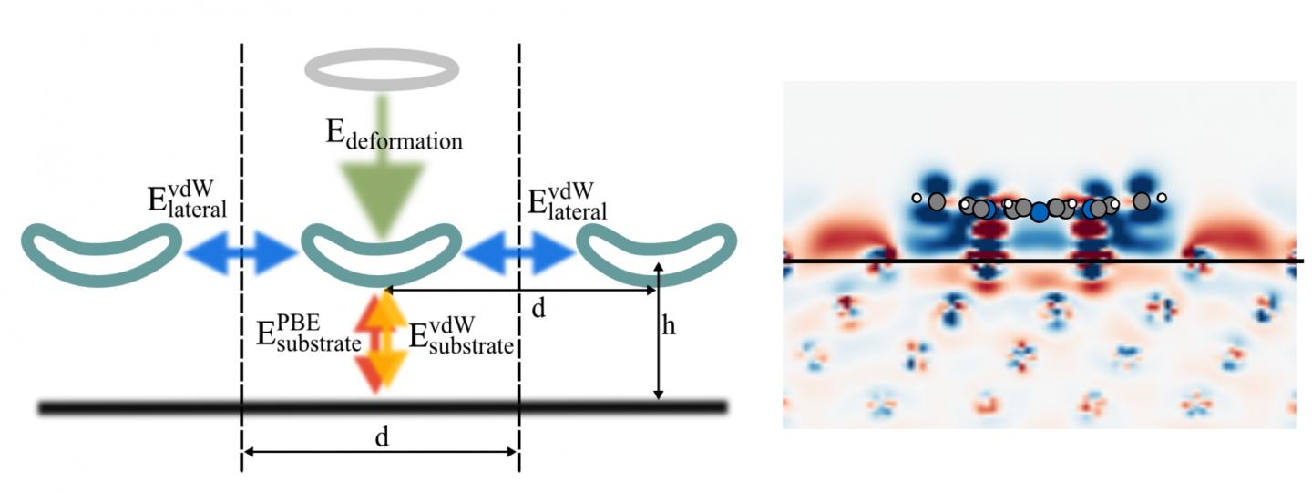
Finally, the researchers have made this illustrative diagram titled ‘Energy’ available,

Caption: Schematic depiction of different energy terms contributing to the adsorption energy, and charge density difference of 2H-P after adsorption onto Cu(111) at 12.8 Angstrom separation. Credit: M. Müller/TU Munich

Caption: Schematic depiction of different energy terms contributing to the adsorption energy, and charge density difference of 2H-P after adsorption onto Cu(111) at 12.8 Angstrom separation. Credit: M. Müller/TU Munich

Hybrid bacterial genes and virus shell combined to create ‘nano reactor’ for hydrogen biofuel

Turning water into fuel may seem like an almost biblical project (e.g., Jesus turning water to wine in the New Testament) but scientists at Indiana University are hopeful they are halfway to their goal. From a Jan. 4, 2016 news item on ScienceDaily,

Scientists at Indiana University have created a highly efficient biomaterial that catalyzes the formation of hydrogen — one half of the “holy grail” of splitting H2O to make hydrogen and oxygen for fueling cheap and efficient cars that run on water.

A Jan. 4, 2016 Indiana University (IU) news release (also on EurekAlert*), which originated the news item, explains further (Note: Links have been removed),

A modified enzyme that gains strength from being protected within the protein shell — or “capsid” — of a bacterial virus, this new material is 150 times more efficient than the unaltered form of the enzyme.

“Essentially, we’ve taken a virus’s ability to self-assemble myriad genetic building blocks and incorporated a very fragile and sensitive enzyme with the remarkable property of taking in protons and spitting out hydrogen gas,” said Trevor Douglas, the Earl Blough Professor of Chemistry in the IU Bloomington College of Arts and Sciences’ Department of Chemistry, who led the study. “The end result is a virus-like particle that behaves the same as a highly sophisticated material that catalyzes the production of hydrogen.”

The genetic material used to create the enzyme, hydrogenase, is produced by two genes from the common bacteria Escherichia coli, inserted inside the protective capsid using methods previously developed by these IU scientists. The genes, hyaA and hyaB, are two genes in E. coli that encode key subunits of the hydrogenase enzyme. The capsid comes from the bacterial virus known as bacteriophage P22.

The resulting biomaterial, called “P22-Hyd,” is not only more efficient than the unaltered enzyme but also is produced through a simple fermentation process at room temperature.

The material is potentially far less expensive and more environmentally friendly to produce than other materials currently used to create fuel cells. The costly and rare metal platinum, for example, is commonly used to catalyze hydrogen as fuel in products such as high-end concept cars.

“This material is comparable to platinum, except it’s truly renewable,” Douglas said. “You don’t need to mine it; you can create it at room temperature on a massive scale using fermentation technology; it’s biodegradable. It’s a very green process to make a very high-end sustainable material.”

In addition, P22-Hyd both breaks the chemical bonds of water to create hydrogen and also works in reverse to recombine hydrogen and oxygen to generate power. “The reaction runs both ways — it can be used either as a hydrogen production catalyst or as a fuel cell catalyst,” Douglas said.

The form of hydrogenase is one of three occurring in nature: di-iron (FeFe)-, iron-only (Fe-only)- and nitrogen-iron (NiFe)-hydrogenase. The third form was selected for the new material due to its ability to easily integrate into biomaterials and tolerate exposure to oxygen.

NiFe-hydrogenase also gains significantly greater resistance upon encapsulation to breakdown from chemicals in the environment, and it retains the ability to catalyze at room temperature. Unaltered NiFe-hydrogenase, by contrast, is highly susceptible to destruction from chemicals in the environment and breaks down at temperatures above room temperature — both of which make the unprotected enzyme a poor choice for use in manufacturing and commercial products such as cars.

These sensitivities are “some of the key reasons enzymes haven’t previously lived up to their promise in technology,” Douglas said. Another is their difficulty to produce.

“No one’s ever had a way to create a large enough amount of this hydrogenase despite its incredible potential for biofuel production. But now we’ve got a method to stabilize and produce high quantities of the material — and enormous increases in efficiency,” he said.

The development is highly significant according to Seung-Wuk Lee, professor of bioengineering at the University of California-Berkeley, who was not a part of the study.

“Douglas’ group has been leading protein- or virus-based nanomaterial development for the last two decades. This is a new pioneering work to produce green and clean fuels to tackle the real-world energy problem that we face today and make an immediate impact in our life in the near future,” said Lee, whose work has been cited in a U.S. Congressional report on the use of viruses in manufacturing.

Beyond the new study, Douglas and his colleagues continue to craft P22-Hyd into an ideal ingredient for hydrogen power by investigating ways to activate a catalytic reaction with sunlight, as opposed to introducing elections using laboratory methods.

“Incorporating this material into a solar-powered system is the next step,” Douglas said.

Here’s a link to and a citation for the paper,

Self-assembling biomolecular catalysts for hydrogen production by Paul C. Jordan, Dustin P. Patterson, Kendall N. Saboda, Ethan J. Edwards, Heini M. Miettinen, Gautam Basu, Megan C. Thielges, & Trevor Douglas. Nature Chemistry (2015) doi:10.1038/nchem.2416 Published online 21 December 2015

This paper is behind a paywall.

*(also on EurekAlert) added on Jan. 5, 2016 at 1550 PST.

Self-assembling nanofibres could help mitigate side effects from pain killers

The research itself is pretty exciting but even more so is the fact that it was conducted by an undergraduate student. From an April 3, 2015 news item on Azonano,

A Chemistry undergraduate at the University of York [UK] has helped to develop a new drug release gel, which may help avoid some of the side effects of painkillers such as ibuprofen and naproxen.

In a final year project, MChem undergraduate student Edward Howe, working in Professor David Smith’s research team in the Department of Chemistry at York looked for a way of eliminating the adverse side-effects associated pain-killing drugs, particularly in the stomach, and the problems, such as ulceration, this could cause patients.

A March 31, 2015 University of York press release, which originated the news item, describes the research in more detail,

Supervised by PhD student Babatunde Okesola, whose research is supported by The Wild Chemistry Scholars Fund, Edward hoped to create gels which could interact with drugs such as Naproxen, and release them at the slightly alkaline pH values found in the intestine rather than the acidic conditions in the stomach.  His aim was to both protect the pain-killing drugs and help limit some of the side effects they can cause.

The researchers created a new gel, based on small molecules which self-assemble into nanofibers which could interact with a variety of anti-inflammatory, painkiller drugs, including iburofen and naproxen. The research is published in Chemical Communications.

Specific interactions between the gel nanofibres and the drugs meant that high loadings could be achieved, and more importantly, the release of the drug could be precisely controlled.  The gels were able to release naproxen at pH 8 – the value found in the intestine, but not at lower pH values found elsewhere in the body.

Professor Smith said: “Although researchers have used gels before to try and improve the formulation of naproxen, this is the first time that a self-assembling system has been used for the job, with the advantages of directed interactions between the nanoscale delivery scaffold and the drug.  As such, this is the first time that such precise control has been achieved.”

Edward Howe said: “The research really fascinated me. The prospect of being involved in developing a method to reduce the pain of others filled me with great pride. Understanding the interactions between the gel and the painkillers was very interesting and improved my knowledge of supramolecular chemistry.”

The next step for Professor Smith’s team will involve stabilising the gel drug delivery systems in the very acidic, low pH conditions found in the stomach so that they can transit safely to the intestine before delivering naproxen just where it is needed.

Professor Smith added: “Perhaps this is something that one of next year’s undergraduate project students might solve. As a research-intensive institution, York is committed to its undergraduates carrying out cutting-edge research such as this.”

Here’s a link to and a citation for the paper,

Self-assembled sorbitol-derived supramolecular hydrogels for the controlled encapsulation and release of active pharmaceutical ingredients by Edward J. Howe, Babatunde O. Okesola, and David K. Smith. Chem. Commun., 2015, Advance Article DOI: 10.1039/C5CC01868D First published online 31 Mar 2015

This paper is behind a paywall.

Opals, Diana Ross, and nanophotonic hybridization

It was a bit of a stretch to include Diana Ross in a Jan. 12, 2015 news item on Nanowerk about nanophotonic research at the University of Twente’s MESA+ Institute for Nano­technology  but I’m glad they did,

Ever since the early 1900s work of Niels Bohr and Hendrik Lorentz, it is known that atoms display characteristic resonant behavior to light. The hallmark of a resonance is its characteristic peak-trough behavior of the refractive index with optical frequency. Scientists from the Dutch MESA+ Institute for Nano­technology at the University of Twente have recently infiltrated cesium atoms in a self-assembled opal to create a hybrid nanophotonic system. By tuning the opal’s forbidden gap relative to the atomic resonance, dra­matic changes are observed in reflectivity. In the most extreme case, the atomic reflection spectrum is turned upside down[1] compared to the traditional case. Since dispersion is crucial in the control of optical signal pulses, the new results offer opportunities for optical information manipulation. As atoms are exquisite storage de­vices for light quanta, the results open vistas on quantum information processing, as well as on new nanoplasmonics.

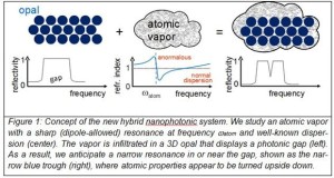
A Jan. 12, 2015 MESA+ Institute for Nano­technology at the University of Twente press release, which originated the news item, provides an illustrative diagram and a wealth of technical detail about the research,

Courtesy of the University of Twente

Courtesy of the University of Twente

While the speed of light c is proverbial, it can readily be modified by sending light through a medium with a certain refractive index n. In the medium, the speed will be decreased by the index to c/n. In any material, the refractive index depends on the frequency of the light. Usually the refractive index increases with frequency, called normal dispersion as it prevails at most frequencies in most materials such as a glass of water, a telecom fiber, or an atomic vapor. Close to the resonance frequency of the material, the index strongly decreases, called anomalous dispersion.

Dispersion is essential to control how optical bits of information – encoded as short pulses – is manipulated optical circuits. In modern optics at the nanoscale, called nanophotonics, dispersion is controlled with classes of complex nanostruc­tures that cause novel behavior to emerge. An example is a photonic crystal fiber, which does not consist of only glass like a traditional fiber, but of an intricate arrange­ment of holes and glass nanostructures.

The Twente team led by Harding devised a hybrid system consisting of an atomic vapor infiltrated in an opal photonic crystal. Photonic crystals have attracted considerable attention for their ability to radically control propagation and emission of light. These nanostructures are well-known for their ability to control the emission and propagation of light. The opals have a periodic variation of the refractive index (see Figure 1) that ensures that a certain color of light is forbidden to exist inside the opal. The light cannot enter the opal as it is reflected, which is called a gap (see Figure 1). In an analogy to semiconductors, such an effect is called a “photonic band gap”. Photonic gaps are at the basis of tiny on-chip light sources and lasers, efficient solar cells, invisibility cloaks, and devices to process optical information.

The Twente team changed the index of refraction of the voids in a photonic crystal by substituting the air by a vapor of atoms with a strong resonance, as shown in Figure 1. The contrast of the refractive index between the vapor and the opal’s silica nano­spheres was effectively used as a probe. The density of the cesium vapor was greatly varied by changing the temperature in the cell up to 420 K. At the same time, the photonic gap of the opal shifted relative to the atomic resonance due to a slow chemical reaction between the opal’s backbone material (silica) and the cesium.

On resonance, light excites an atom to a higher state and subsequently the atom reemits the light. Hence, an atom behaves like a little cavity that stores light. Simultaneously the index of refraction changes strongly for colors near resonance. For slightly longer wavelengths the index of refraction is high, on resonance it is close to one, and slightly shorter wavelengths it can even decrease below one. This effect of the cesium atoms is clearly visible in the reflectivity spectra, shown in Figure 2 [not included here], as a sharp increase and decrease of the reflectivity near the atomic resonance. Intriguingly, the characteristic peak-and-trough behavior of atoms (seen at 370 K) was turned upside down at the highest temperature (420 K), where the ce­sium reso­nance was on the red side of the opal’s stopgap.

In nanophotonics, many efforts are currently being devoted to create arrays of nanoresonators in photonic crystals, for exquisite optical signal control on a chip. Unfortunately, however, there is a major challenge in engineering high-quality pho­tonic resonators: they are all different due to inevitable fabrication variations. Hence, it is difficult to tune every resonator in sync. “Our atoms in the opal may be consid­ered as the equivalent of an carefully engineered array of nano-resonators” explains Willem Vos, “Nature takes care that all resonators are all exactly the same. Our hy­brid system solves the variability problem and could perhaps be used to make pho­tonic memories, sensors or switches that are naturally tuned.” And leading Spanish theorist Javier Garcia de Abajo (ICFO) enthuses: “This is a fine and exciting piece of work, initiating the study of atomic resonances with photonic modes in a genuinely new fashion, and suggesting many exciting possibilities, for example through the extension of this study towards combinations with metal nanoplasmonics.”

Here’s a link to and a citation for the paper published in Physical Review B,

Nanophotonic hybridization of narrow atomic cesium resonances and photonic stop gaps of opaline nanostructures by Philip J. Harding, Pepijn W. H. Pinkse, Allard P. Mosk, and Willem L. Vos. Phys. Rev. B 91, 045123 – Published 20 January 2015 DOI: http://dx.doi.org/10.1103/PhysRevB.91.045123

This paper is behind a paywall but there is an earlier iteration of the paper available on the open access arXiv.org website operated by Cornell University,

Nanophotonic hybridization of narrow atomic cesium resonances and photonic stop gaps of opaline nanostructures by Philip J. Harding, Pepijn W.H. Pinkse, Allard P. Mosk, Willem L. Vos. (Submitted on 11 Sep 2014) arXiv:1409.3417

As I understand it, the arXiv.org website is intended to open up access to research and to offer an informal peer review process.

Finally, for anyone who’s nostalgic or perhaps has never heard Diana Ross sing ‘Upside Down’,

Getting up to the size of a dust speck, the first ‘large’ self-assembling DNA crystals

An Oct. 19, 2014 news item on ScienceDaily describes the latest developments in ‘DNA nanotechnology’ research at the Wyss Institute for Biologically Inspired Engineering at Harvard University,

DNA has garnered attention for its potential as a programmable material platform that could spawn entire new and revolutionary nanodevices in computer science, microscopy, biology, and more. Researchers have been working to master the ability to coax DNA molecules to self assemble into the precise shapes and sizes needed in order to fully realize these nanotechnology dreams.

For the last 20 years, scientists have tried to design large DNA crystals with precisely prescribed depth and complex features — a design quest just fulfilled by a team at Harvard’s Wyss Institute for Biologically Inspired Engineering. The team built 32 DNA crystals with precisely-defined depth and an assortment of sophisticated three-dimensional (3D) features, an advance reported in Nature Chemistry.

It seems a bit of a misleading for the Wyss Institute to state the ‘team built’ the DNA crystals as they are self-assembling according to this Oct. 19, 2014 Wyss Institute news release (also on EurekAlert), which originated the news item,

The team used their “DNA-brick self-assembly” method, which was first unveiled in a 2012 Science publication when they created more than 100 3D complex nanostructures about the size of viruses. The newly-achieved periodic crystal structures are more than 1000 times larger than those discrete DNA brick structures, sizing up closer to a speck of dust, which is actually quite large in the world of DNA nanotechnology.

“We are very pleased that our DNA brick approach has solved this challenge,” said senior author and Wyss Institute Core Faculty member Peng Yin, Ph.D., who is also an Associate Professor of Systems Biology at Harvard Medical School, “and we were actually surprised by how well it works.”

The news release goes on to describe some of the issues with other self-assembly methods along with more details about the ‘DNA brick’ approach,

Scientists have struggled to crystallize complex 3D DNA nanostructures using more conventional self-assembly methods. The risk of error tends to increase with the complexity of the structural repeating units and the size of the DNA crystal to be assembled.

The DNA brick method uses short, synthetic strands of DNA that work like interlocking Lego® bricks to build complex structures. Structures are first designed using a computer model of a molecular cube, which becomes a master canvas. Each brick is added or removed independently from the 3D master canvas to arrive at the desired shape – and then the design is put into action: the DNA strands that would match up to achieve the desired structure are mixed together and self assemble to achieve the designed crystal structures.

“Therein lies the key distinguishing feature of our design strategy—its modularity,” said co-lead author Yonggang Ke, Ph.D., formerly a Wyss Institute Postdoctoral Fellow and now an assistant professor at the Georgia Institute of Technology and Emory University. “The ability to simply add or remove pieces from the master canvas makes it easy to create virtually any design.”

The modularity also makes it relatively easy to precisely define the crystal depth. “This is the first time anyone has demonstrated the ability to rationally design crystal depth with nanometer precision, up to 80 nm in this study,” Ke said. In contrast, previous two-dimensional DNA lattices are typically single-layer structures with only 2 nm depth.

“DNA crystals are attractive for nanotechnology applications because they are comprised of repeating structural units that provide an ideal template for scalable design features”, said co-lead author graduate student Luvena Ong.

Furthermore, as part of this study the team demonstrated the ability to position gold nanoparticles into prescribed 2D architectures less than two nanometers apart from each other along the crystal structure – a critical feature for future quantum devices and a significant technical advance for their scalable production, said co-lead author Wei Sun, Ph.D., Wyss Institute Postdoctoral Fellow.

“My preconceived notions of the limitations of DNA have been consistently shattered by our new advances in DNA nanotechnology,” said William Shih, Ph.D., who is co-author of the study and a Wyss Institute Founding Core Faculty member, as well as Associate Professor in the Department of Biological Chemistry and Molecular Pharmacology at Harvard Medical School and the Department of Cancer Biology at the Dana-Farber Cancer Institute. “DNA nanotechnology now makes it possible for us to assemble, in a programmable way, prescribed structures rivaling the complexity of many molecular machines we see in Nature.”

“Peng’s team is using the DNA-brick self-assembly method to build the foundation for the new landscape of DNA nanotechnology at an impressive pace,” said Wyss Institute Founding Director Don Ingber, M.D., Ph.D. “What have been mere visions of how the DNA molecule could be used to advance everything from the semiconductor industry to biophysics are fast becoming realities.”

Here’s a link to and a citation for the latest paper,

DNA brick crystals with prescribed depths by Yonggang Ke, Luvena L. Ong, Wei Sun, Jie Song, Mingdong Dong, William M. Shih, & Peng Yin. Nature Chemistry (2014) doi:10.1038/nchem.2083 Published online 19 October 2014

This paper is behind a paywall.

New ‘Star of David’-shaped molecule from University of Manchester

It sounds like the scientists took their inspiration from Maurits Cornelius Escher (M. C. Escher) when they created their ‘Star of David’ molecule. From a Sept. 22, 2014 news item on Nanowerk,

Scientists at The University of Manchester have generated a new star-shaped molecule made up of interlocking rings, which is the most complex of its kind ever created.

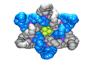
Here’s a representation of the molecule,

Atoms in the Star of David molecule. Image credit: University of Manchester

Atoms in the Star of David molecule. Image credit: University of Manchester

Here’s a ‘star’ sculpture based on Escher’s work,

Sculpture of the small stellated dodecahedron that appears in Escher's Gravitation. It can be found in front of the "Mesa+" building on the Campus of the University of Twente.

Sculpture of the small stellated dodecahedron that appears in Escher’s Gravitation. It can be found in front of the “Mesa+” building on the Campus of the University of Twente (Netherlands)

If you get a chance to see the Escher ‘star’, you’ll be able to see more plainly how the planes of the ‘star’ interlock. (I had the opportunity when visiting the University of Twente in Oct. 2012.)

Getting back to Manchester, a Sept. 22, 2014 University of Manchester press release (also on EurekAlert but dated Sept. 21, 2014), which originated the news item, describes the decades-long effort to create this molecule and provides a few technical details,

Known as a ‘Star of David’ molecule, scientists have been trying to create one for over a quarter of a century and the team’s findings are published at 1800 London time / 1300 US Eastern Time on 21 September 2014 in the journal Nature Chemistry.

Consisting of two molecular triangles, entwined about each other three times into a hexagram, the structure’s interlocked molecules are tiny – each triangle is 114 atoms in length around the perimeter. The molecular triangles are threaded around each other at the same time that the triangles are formed, by a process called ‘self-assembly’, similar to how the DNA double helix is formed in biology.

The molecule was created at The University of Manchester by PhD student Alex Stephens.

Professor David Leigh, in Manchester’s School of Chemistry, said: “It was a great day when Alex finally got it in the lab.  In nature, biology already uses molecular chainmail to make the tough, light shells of certain viruses and now we are on the path towards being able to reproduce its remarkable properties.

“It’s the next step on the road to man-made molecular chainmail, which could lead to the development of new materials which are light, flexible and very strong.  Just as chainmail was a breakthrough over heavy suits of armour in medieval times, this could be a big step towards materials created using nanotechnology. I hope this will lead to many exciting developments in the future.”

The team’s next step will be to make larger, more elaborate, interlocked structures.

Here’s a link to and a citation for the paper,

A Star of David catenane by David A. Leigh, Robin G. Pritchard, & Alexander J. Stephens. Nature Chemistry (2014) doi:10.1038/nchem.2056
Published online 21 September 2014

This paper is behind a paywall.