Tag Archives: Stanislav Gorb

Cellulose and natural nanofibres

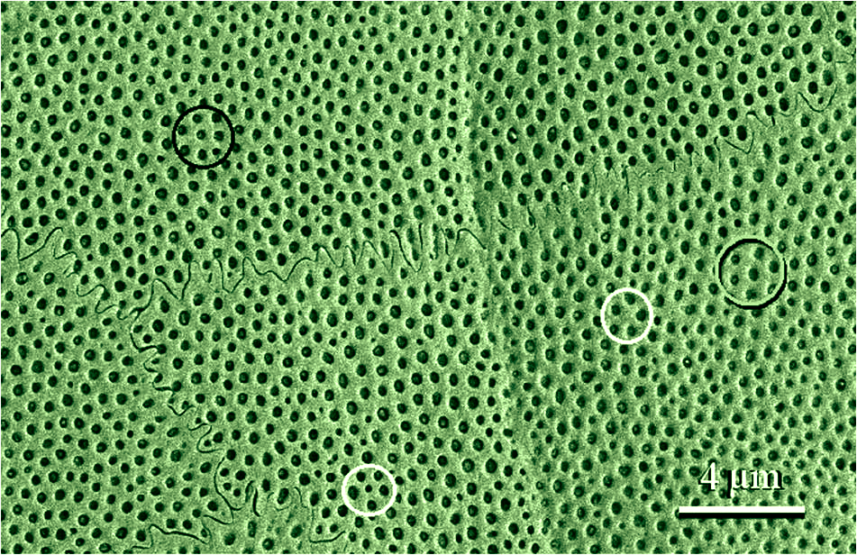
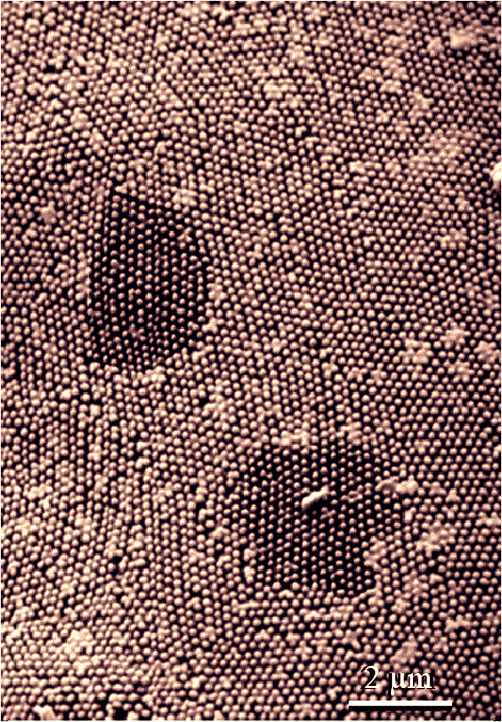
Specifically, the researchers are describing these as cellulose nanofibrils. On the left of the image, the seed look mores like an egg waiting to be fried for breakfast but the image on the right is definitely fibrous-looking,

Through contact with water, the seed of Neopallasia pectinata from the family of composite plants forms a slimy sheath. The white cellulose fibres anchor it to the seed surface. Courtesy: Kiel University (CAU)

A December 18, 2018 news item on Nanowerk describes the research into seeds and cellulose,

The seeds of some plants such as basil, watercress or plantain form a mucous envelope as soon as they come into contact with water. This cover consists of cellulose in particular, which is an important structural component of the primary cell wall of green plants, and swelling pectins, plant polysaccharides.

In order to be able to investigate its physical properties, a research team from the Zoological Institute at Kiel University (CAU) used a special drying method, which gently removes the water from the cellulosic mucous sheath. The team discovered that this method can produce extremely strong nanofibres from natural cellulose. In future, they could be especially interesting for applications in biomedicine.

A December 18, 2018 Kiel University press release, which originated the news item, offers further details about the work,

Thanks to their slippery mucous sheath, seeds can slide through the digestive tract of birds undigested. They are excreted unharmed, and can be dispersed in this way. It is presumed that the mucous layer provides protection. “In order to find out more about the function of the mucilage, we first wanted to study the structure and the physical properties of this seed envelope material,” said Zoology Professor Stanislav N. Gorb, head of the “Functional Morphology and Biomechanics” working group at the CAU. In doing so they discovered that its properties depend on the alignment of the fibres that anchor them to the seed surface

Diverse properties: From slippery to sticky

The pectins in the shell of the seeds can absorb a large quantity of water, and thus form a gel-like capsule around the seed in a few minutes. It is anchored firmly to the surface of the seed by fine cellulose fibres with a diameter of just up to 100 nanometres, similar to the microscopic adhesive elements on the surface of highly-adhesive gecko feet. So in a sense, the fibres form the stabilising backbone of the mucous sheath.

The properties of the mucous change, depending on the water concentration. “The mucous actually makes the seeds very slippery. However, if we reduce the water content, it becomes sticky and begins to stick,” said Stanislav Gorb, summarising a result from previous studies together with Dr Agnieszka Kreitschitz. The adhesive strength is also increased by the forces acting between the individual vertically-arranged nanofibres of the seed and the adhesive surface.

Specially dried

In order to be able to investigate the mucous sheath under a scanning electron microscope, the Kiel research team used a particularly gentle method, so-called critical-point drying (CPD). They dehydrated the mucous sheath of various seeds step-by-step with liquid carbon dioxide – instead of the normal method using ethanol. The advantage of this method is that evaporation of liquid carbon dioxide can be controlled under certain pressure and temperature conditions, without surface tension developing within the sheath. As a result, the research team was able to precisely remove water from the mucous, without drying out the surface of the sheath and thereby destroying the original cell structure. Through the highly-precise drying, the structural arrangement of the individual cellulose fibres remained intact.

Almost as strongly-adhesive as carbon nanotubes

The research team tested the dried cellulose fibres regarding their friction and adhesion properties, and compared them with those of synthetically-produced carbon nanotubes. Due to their outstanding properties, such as their tensile strength, electrical conductivity or their friction, these microscopic structures are interesting for numerous industrial applications of the future.

“Our tests showed that the frictional and adhesive forces of the cellulose fibres are almost as strong as with vertically-arranged carbon nanotubes,” said Dr Clemens Schaber, first author of the study. The structural dimensions of the cellulose nanofibers are similar to the vertically aligned carbon nanotubes. Through the special drying method, they can also vary the adhesive strength in a targeted manner. In Gorb’s working group, the zoologist and biomechanic examines the functioning of biological nanofibres, and the potential to imitate them with technical means. “As a natural raw material, cellulose fibres have distinct advantages over carbon nanotubes, whose health effects have not yet been fully investigated,” continued Schaber. Nanocellulose is primarily found in biodegradable polymer composites, which are used in biomedicine, cosmetics or the food industry.

Here’s a link to and a citation for the paper,

Friction-Active Surfaces Based on Free-Standing Anchored Cellulose Nanofibrils by Clemens F. Schaber, Agnieszka Kreitschitz, and Stanislav N. Gorb. ACS Appl. Mater. Interfaces, 2018, 10 (43), pp 37566–37574 DOI: 10.1021/acsami.8b05972 Publication Date (Web): September 19, 2018

Copyright © 2018 American Chemical Society

This paper is behind a paywall.

Similarities between a moth’s eye and snakeskin

Finding patterns in nature that are repeated seems to be the order of the day although there is a twist to this particular story. This time, researchers at Kiel University (also known as, University of Kiel or Christian-Albrechts University of  Kiel [Germany]) have found superficial similarities between a moth’s eye and snakeskin according to a May 4, 2016 news item on Nanowerk,

One thing is obvious: moth’s eyes and snake’s skin are entirely different. Researchers at Kiel University have taken a closer look, however, and have now brought the supposed ‘apples and oranges’ to a common denominator. They have opened up a completely new, comparative view of biological surfaces using a newly developed method, and have thus come closer to the solution of how these surfaces work. Dr. Alexander Kovalev, Dr. Alexander Filippov and Professor Stanislav Gorb from the Zoological Institute at Kiel University have published their findings in the current edition of the scientific journal Applied Physics A (“Correlation analysis of symmetry breaking in the surface nanostructure ordering: case study of the ventral scale of the snake Morelia viridis”).

A May 3, 2016 Kiel University press release, which originated the news item, describes the scientists’ first approach to the research,

One surface demonstrates reduced light reflection, the other is water repellent and resistant to abrasion. Surfaces in the animal world are evolved to adapt to their environments and give the animal they cover the greatest possible evolutionary advantage. Scientists are today still puzzled by exactly how and why these different structures develop in detail.

Current research looks right into the surface nano-structures using the latest research techniques. Normally, we would limit ourselves to comparisons within closely related species and just look thoroughly at small areas of the surface, says Gorb: “That is why we asked ourselves which structural differences can be found between completely different species. To do so, we changed biology’s typical perspective and addressed larger surface areas from various species.” These types of cross-species or cross-material studies of nanostructures are common in other technical or inorganic fields. In Biology, however, this method is completely new, Gorb continues.

They got the idea from the decorations in the hallway of their own institute, where scanning electron microscope images of moth’s eyes and snake’s skin are displayed. At some point, theoretical physicist Filippov noticed similarities between the images, which showed the surfaces at a resolution of a few millionths of a millimetre. Nipples and dimples could be seen which seemed to the human eye to follow a certain pattern. Using methods that are normally used in crystallography, the scientists were finally able to recognise the particular patterns that distinguish the two species. “The structure of moth’s eyes is perfectly organised. Nipples are highly ordered, and preferred directions are exhibited in the structural organisation”, explains Kovalev, biophysicist and main author of the study. The scientists were already aware of the eye structure’s strict symmetry. However, the fact that this goes right through to the nano-level and is repeated across the entire surface in so-called domains, is an important new finding.

So which symmetry does snake’s skin have, which at first glance appears similar, perhaps even more perfectly organised? “Compared to the structure of the moth’s eye, the structure of the snake’s skin is unorganised”, explains Kovalev. He continued: “If we concentrate on one dimple in the skin, like one nipple in the eye, we only see a diffuse cloud of further dimples in the close surroundings. Neither particular directions nor the regular arrangement can be defined. This unorganised structure continues across the entire surface.”

On concluding there were significant differences as well as similarities, the scientists took a closer look,

On their own, these findings about the organised eye structure on the one hand and the unorganised skin structure on the other hand are not especially significant. But by taking the common denominator, i.e. investigating both structures with the same degree of resolution, it is possible for the first time to compare fundamentally different structures, explains Gorb: “However, the ‘coincidental’ degree of organisation is not coincidental, but a result of evolution. That would mean that the perfect organisation gives the moth its incredible night vision, while the imperfect organisation in snake’s skin ensures the best friction properties.” That sounds logical, when you consider the laws of physics, that a symmetrical structure is necessary for good vision and good friction properties require the surface ordering in the contact with the ground to be as low as possible.

If the Kiel-based researchers had followed the usual approaches and compared snakes to snakes and moths to moths, the organisation of the elements at nano-level would have hardly been considered significant. “By comparing evolutionary distant species, we now see that the key to understanding surface functions must be right at the smallest level. Every biological surface is adapted to its environment, and these adaptations are reflected in the organisation of their smallest elements in a certain perfect or imperfect degree”, Gorb concludes.

This is the snakeskin,

Scanning electron microscopy image of the tail ventral scale in the snake Morelia viridis. The black shadowed gray circle marks a typical hexagonal arrangement of dimples, whereas both white and black circles mark five- and sevenfold symmetrical arrangement of dimples, respectively. Credit: research group Gorb

Scanning electron microscopy image of the tail ventral scale in the snake Morelia viridis. The black shadowed gray circle marks a typical hexagonal arrangement of dimples, whereas both white and black circles mark five- and sevenfold symmetrical arrangement of dimples, respectively. Credit: research group Gorb

This is the moth’s eye,


Scanning electronmicroscopy image of a single ommatidium surface of an eye in the moth Manduca sexta. Credit: research group Gorb

Scanning electronmicroscopy image of a single ommatidium surface of an eye in the moth Manduca sexta.
Credit: research group Gorb

Here’s a link to and a citation for the paper,

Correlation analysis of symmetry breaking in the surface nanostructure ordering: case study of the ventral scale of the snake Morelia viridis by A. Kovalev, A. Filippov, S. N. Gorb. Applied Physics A March 2016, 122:253 DOI:  10.1007/s00339-016-9795-2 First online: 03 March 2016

This paper is behind a paywall.