Tag Archives: structural colour

Squeezing out ‘polymer opals’ for smart clothing and more

Researchers at the University of Cambridge have developed a technology for producing ‘polymer opals’ on industrial scales according to a June 3, 2016 news item on Nanowerk (Note: A link has been removed),

Using a new method called Bend-Induced-Oscillatory-Shearing (BIOS), the researchers are now able to produce hundreds of metres of these materials, known as ‘polymer opals’, on a roll-to-roll process. The results are reported in the journal Nature Communications (“Large-scale ordering of nanoparticles using viscoelastic shear processing”).

A June 3, 2016 University of Cambridge press release (also on EurekAlert), which originated the news item, provides more detail (Note: Links have been removed),

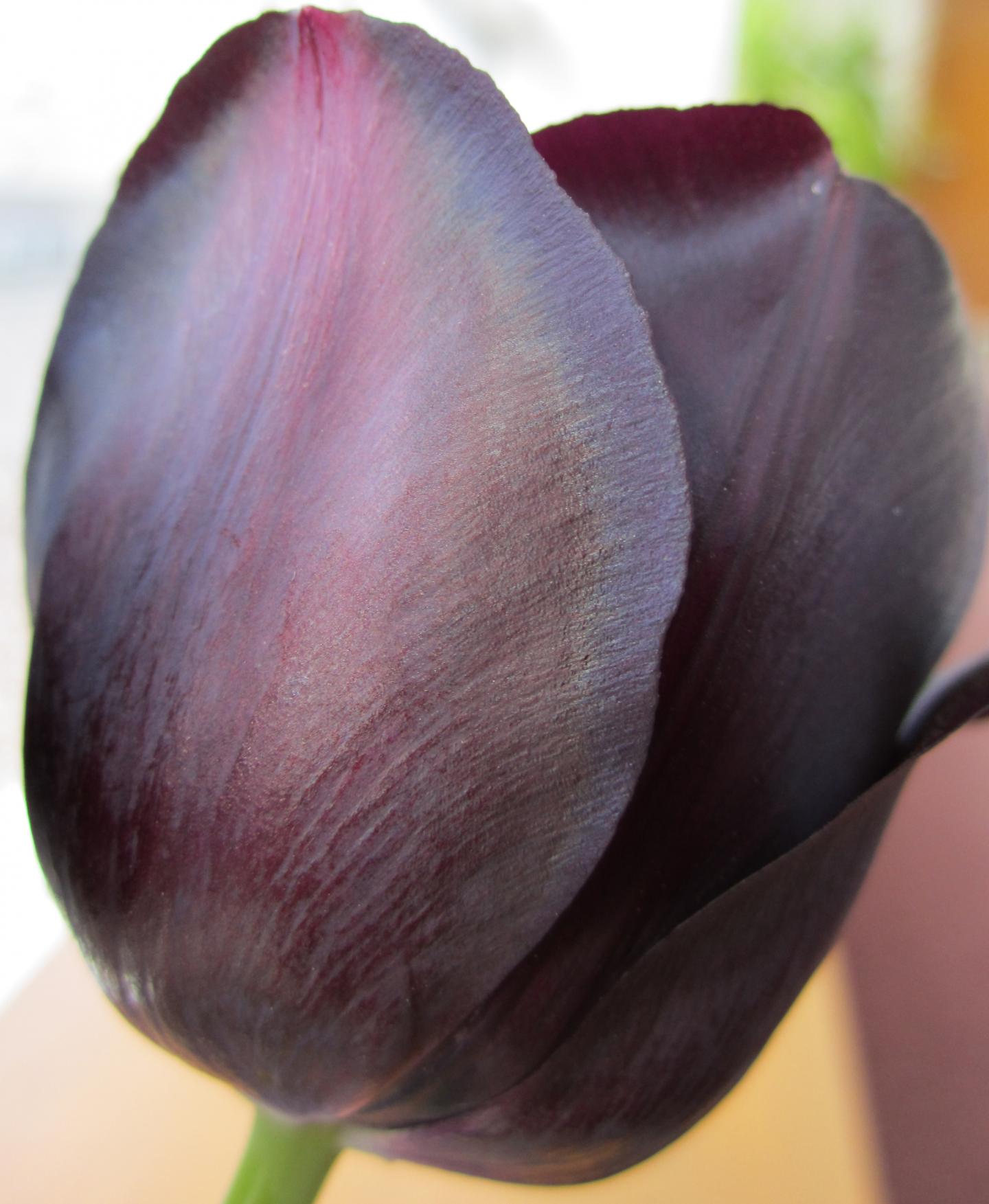
Researchers have devised a new method for stacking microscopic marbles into regular layers, producing intriguing materials which scatter light into intense colours, and which change colour when twisted or stretched.

Some of the brightest colours in nature can be found in opal gemstones, butterfly wings and beetles. These materials get their colour not from dyes or pigments, but from the systematically-ordered microstructures they contain.

The team behind the current research, based at Cambridge’s Cavendish Laboratory, have been working on methods of artificially recreating this ‘structural colour’ for several years, but to date, it has been difficult to make these materials using techniques that are cheap enough to allow their widespread use.

In order to make the polymer opals, the team starts by growing vats of transparent plastic nano-spheres. Each tiny sphere is solid in the middle but sticky on the outside. The spheres are then dried out into a congealed mass. By bending sheets containing a sandwich of these spheres around successive rollers the balls are magically forced into perfectly arranged stacks, by which stage they have intense colour.

By changing the sizes of the starting nano-spheres, different colours (or wavelengths) of light are reflected. And since the material has a rubber-like consistency, when it is twisted and stretched, the spacing between the spheres changes, causing the material to change colour. When stretched, the material shifts into the blue range of the spectrum, and when compressed, the colour shifts towards red. When released, the material returns to its original colour. Such chameleon materials could find their way into colour-changing wallpapers, or building coatings that reflect away infrared thermal radiation.

I always like it when there are quotes which seem spontaneous (from the press release),

“Finding a way to coax objects a billionth of a metre across into perfect formation over kilometre scales is a miracle [emphasis mine],” said Professor Jeremy Baumberg, the paper’s senior author. “But spheres are only the first step, as it should be applicable to more complex architectures on tiny scales.”

In order to make polymer opals in large quantities, the team first needed to understand their internal structure so that it could be replicated. Using a variety of techniques, including electron microscopy, x-ray scattering, rheology and optical spectroscopy, the researchers were able to see the three-dimensional position of the spheres within the material, measure how the spheres slide past each other, and how the colours change.

“It’s wonderful [emphasis mine] to finally understand the secrets of these attractive films,” said PhD student Qibin Zhao, the paper’s lead author.

There’s also the commercialization aspect to this work (from the press release),

Cambridge Enterprise, the University’s commercialisation arm which is helping to commercialise the material, has been contacted by more than 100 companies interested in using polymer opals, and a new spin-out Phomera Technologies has been founded. Phomera will look at ways of scaling up production of polymer opals, as well as selling the material to potential buyers. Possible applications the company is considering include coatings for buildings to reflect heat, smart clothing and footwear, or for banknote security [emphasis mine] and packaging applications.

There is a Canadian company already selling its anti-counterfeiting (banknote security) bioinspired technology. It’s called Opalux and it’s not the only bioinspired anti-counterfeiting Canadian technology company, there’s also NanoTech Security which takes its inspiration from a butterfly (Blue Morpho) wing.

Getting back to Cambridge, here’s a link to and a citation for the research team’s paper,

Large-scale ordering of nanoparticles using viscoelastic shear processing by Qibin Zhao, Chris E. Finlayson, David R. E. Snoswell, Andrew Haines, Christian Schäfer, Peter Spahn, Goetz P. Hellmann, Andrei V. Petukhov, Lars Herrmann, Pierre Burdet, Paul A. Midgley, Simon Butler, Malcolm Mackley, Qixin Guo, & Jeremy J. Baumberg. Nature Communications 7, Article number: 11661  doi:10.1038/ncomms11661 Published 03 June 2016

This paper is open access.

There is a video demonstrating the stretchability of their ‘polymer opal’ film

It was posted on YouTube three years ago when the researchers were first successful. It’s nice to see they’ve been successful at getting the technology to the commercialization stage.

How tarantulas get blue

Cobalt Blue Tarantula [downloaded from http://www.tarantulaguide.com/tarantula-pictures/cobalt-blue-tarantula-4/]

Cobalt Blue Tarantula [downloaded from http://www.tarantulaguide.com/tarantula-pictures/cobalt-blue-tarantula-4/]

That’s a stunning shade of blue on the tarantula and now scientists can explain why these and other ‘spiders’ are sometimes blue, from a Nov. 30, 2015 news item on ScienceDaily,

Scientists recently discovered that tiny, multilayer nanostructures inside a tarantula’s hair are responsible for its vibrant color. The science behind how these hair-raising spiders developed their blue hue may lead to new ways to improve computer or TV screens using biomimicry.

A Nov. 30, 2015 University of California at San Diego news release by Annie Reisewitz, which originated the news item, explains more,

Researchers from Scripps Institution of Oceanography at UC San Diego and University of Akron found that many species of tarantulas have independently evolved the ability to grow blue hair using nanostructures in their exoskeletons, rather than pigments. The study, published in the Nov. 27 issue of Science Advances, is the first to show that individual species evolved separately to make the same shade of a non-iridescent color, one that doesn’t change when viewed at different angles.

Since tarantulas’ blue color is not iridescent, the researchers suggest that the same process can be applied to make pigment replacements that never fade and help reduce glare on wide-angle viewing systems in phones, televisions, and other devices.

“There is strikingly little variety in the shade of blue produced by different species of tarantulas,” said Dimitri Deheyn, a Scripps Oceanography researcher studying marine and terrestrial biomimicry and coauthor of the study. “We see that different types of nanostructures evolved to produce the same ‘blue’ across distant branches of the tarantula family tree in a way that uniquely illustrates natural selection through convergent evolution.”

Unlike butterflies and birds that use nanostructures to produce vibrant colors to attract the attention of females during display courtship, tarantulas have poor vision and likely evolved this trait for a different reason. While the researchers still don’t understand the benefits tarantulas receive from being blue, they are now investigating how to reproduce the tarantula nanostructures in the laboratory.

The tarantula study is just one example of the biomimicry research being conducted in the Deheyn lab at Scripps Oceanography. In a cover article in the Nov. 10 of Chemistry of Materials, Deheyn and colleagues published new findings on the nanostructure of ragweed pollen, which shows interesting optical properties and has possible biomimicry applications. By transforming the pollen into a magnetic material with a specialized coating to give it more or less reflectance, the particle could adhere in a similar way that pollen does in nature while being able to adjust its visibility. The researchers suggest this design could be applied to create a new type of tagging or tracking technology.

Using a high-powered microscope, known as a hyperspectral imaging system, Deheyn is able to map a species’ color field pixel by pixel, which correlates to the shape and geometry of the nanostructures and gives them their unique color.

“This unique technology allows us to associate structure with optical property,” said Deheyn. “Our inspiration is to learn about how nature evolves unique traits that we could mimic to benefit future technologies.”

Here’s a link to and a citation for the paper,

Blue reflectance in tarantulas is evolutionarily conserved despite nanostructural diversity by Bor-Kai Hsiung, Dimitri D. Deheyn, Matthew D. Shawkey, and Todd A. Blackledge. Science Advances  27 Nov 2015: Vol. 1, no. 10, e1500709 DOI: 10.1126/sciadv.1500709

This paper appears to be open access.

Structural colo(u)r with a twist

There’s a nice essay about structural colour on the Duke University website (h/t Nanowerk). Long time readers know my favourite piece of writing on the subject is by Cristina Luiggi for The Scientist magazine which I profiled here in a Feb. 7, 2013 posting.

This latest piece seems to have been *written by Anika Radiya-Dixit* and it is very good. From the Oct. 27, 2015 Duke University blog posting titled, Iridescent Beauty: Development, function and evolution of plant nanostructures that influence animal behavior,

Iridescent wings of a Morpho butterfly

Iridescent wings of a Morpho butterfly

Creatures like the Morpho butterfly on the leaf above appear to be covered in shimmering blue and green metallic colors. This phenomenon is called “iridescence,” meaning that color appears to change as the angle changes, much like soap bubbles and sea shells.

In animals, the physical mechanisms and function of structural color have been studied significantly as a signal for recognition or mate choice.

Glover, one of the post’s authors, is a scientist who believes there may be another reason for iridescence,

On the other hand, Beverley Glover believes that such shimmering in plants can actually influence animal behavior by attracting pollinators better than their non-iridescent counterparts. Glover,Director of Cambridge University Botanic Garden,  presented her study during the Biology Seminar Series in the French Family Science Center on Monday [Oct. 26, 2015] earlier this week.

Hibiscus Trionum

Hibiscus Trionum

The metallic property of flowers like the Hibiscus Trionum above are generated by diffraction grating – similar to the way CD shines – to create color from transparent material.

In order to observe the effects of the iridescence on pollinators like bees, Glover created artificial materials with a surface structure of nanoscale ridges, similar to the microscopic view of a petal’s epidermal surface below.

Nanoscale ridges on a petal's epidermal surface.

Nanoscale ridges on a petal’s epidermal surface.

In the first set of experiments, Glover and her team marked bees with paint to follow their behavior as they set the insects to explore iridescent flowers. Some were covered in a red grating – containing a sweet solution as a reward – and others with a blue iridescent grating – containing a sour solution as deterrent. The experiment demonstrated that the bees were able to detect the iridescent signal produced by the petal’s nanoridges, and – as a result – correctly identified the rewarding flowers.

It’s worth reading the Oct. 27, 2015 Duke University blog posting to just to see the pictures used to illustrate the ideas and to find out about the second experiment.

*’written by Beverley Glover and Anika Radiya-Dixit’ has been changed to ‘Anika Radiya-Dixit’ as per information in a comment received November 27, 2021.

Self-assembly for stunning structural colour

Researchers from McGill University (Montréal, Canada) have developed a computational model which they believe explains how nature achieves structural colo(u)r as exemplified by this tulip,

 Caption: The Queen of the Night tulip displays an iridescent shimmer caused by microscopic ridges on its petals that diffract light. Credit: S. Vignolini/


Caption: The Queen of the Night tulip displays an iridescent shimmer caused by microscopic ridges on its petals that diffract light.
Credit: S. Vignolini/

A Sept. 16, 2015 news item on phys.org describes the phenomenon,

The tulip called Queen of the Night has a fitting name. Its petals are a lush, deep purple that verges on black. An iridescent shimmer dances on top of the nighttime hues, almost like moonlight glittering off regal jewels.

Certain rainforest plants in Malaysia demonstrate an even more striking color feature: Their iridescent blue leaves turn green when dunked in water.

Both the tulip’s rainbow sparkle and the Malaysian plants’ color change are examples of structural color—an optical effect that is produced by a physical structure, instead of a chemical pigment.

Now researchers have shown how plant cellulose can self-assemble [emphasis mine] into wrinkled surfaces that give rise to effects like iridescence and color change. Their findings provide a foundation to understand structural color in nature, as well as yield insights that could guide the design of devices like optical humidity sensors. …

A Sept. 15, 2015 American Institute of Physics news release on EurekAlert, which originated the news item, describes the research into cellulose and structural colour in more detail,

Cellulose is one of the most abundant organic materials on Earth. It forms a key part of the cell wall of green plants, where the cellulose fibers are found in layers. The fibers in a single layer tend to align in a single direction. However, when you move up or down a layer the axis of orientation of the fibers can shift. If you imagined an arrow pointing in the direction of the fiber alignment, it would often spin in a circle as you moved through the layers of cellulose. This twisting pattern is called a cholesteric phase, because it was first observed while studying cholesterol molecules.

Scientists think that cellulose twists mainly to provide strength. “The fibers reinforce in the direction they are oriented,” said Alejandro Rey, a chemical engineer at McGill University in Montreal, Canada. “When the orientation rotates you get multi-directional stiffness.”

Rey and his colleagues, however, weren’t primarily interested in cellulose’s mechanical properties. Instead, they wondered if the twisting structure could produce striking optical effects, as seen in plants like iridescent tulips.

The team constructed a computational model to examine the behavior of cholesteric phase cellulose. In the model, the axis of twisting runs parallel to the surface of the cellulose. The researchers found that subsurface helices naturally caused the surface to wrinkle. The tiny ridges had a height range in the nanoscale and were spaced apart on the order of microns.

The pattern of parallel ridges resembled the microscopic pattern on the petals of the Queen of the Night tulip. The ridges split white light into its many colored components and create an iridescent sheen — a process called diffraction. The effect can also be observed when light hits the microscopic grooves in a CD.

The researchers also experimented with how the amount of water in the cellulose layers affected the optical properties. More water made the layers twist less tightly, which in turn made the ridges farther apart. How tightly the cellulose helices twist is called the pitch. The team found that a surface with spatially varying pitch (in which some areas were more hydrated than others) was less iridescent and reflected a longer primary wavelength of light than surfaces with a constant pitch. The wavelength shift from around 460 nm (visible blue light) to around 520 nm (visible green light) could explain some plants’ color changing properties, Rey said.

Insights into Nature and Inspiration for New Technologies

Although proving that diffractive surfaces in nature form in the same way will require further work, the model does offer a good foundation to further explore structural color, the researchers said. They imagine the model could also guide the design of new optical devices, for example sensors that change color to indicate a change in humidity.

“The results show the optics [of cholesteric cellulose] are just as exciting as the mechanical properties,” Rey said. He said scientists tend to think of the structures as biological armor, because of their reinforcing properties. “We’ve shown this armor can also have striking colors,” he said.

Here’s a link to and a citation for the paper,

Tunable nano-wrinkling of chiral surfaces: Structure and diffraction optics by P. Rofouie, D. Pasini, and A. D. Rey. J. Chem. Phys. 143, 114701 (2015); http://dx.doi.org/10.1063/1.4929337

This is an open access paper.

Sea sapphires: now you see them, now you don’t and more about structural colour/color

The structural colour of the sea sapphire

 Scientists are studying the disappearing act of this ocean-dwelling copepod. Credit: Kaj Maney, www.liquidguru.com Courtesy: American Chemical Society


Scientists are studying the disappearing act of this ocean-dwelling copepod.
Credit: Kaj Maney, www.liquidguru.com Courtesy: American Chemical Society

Now, you’ve seen a sea sapphire. Here’s more about them and the interest they hold for experts in photonics, from a July 15, 2015 news item on ScienceDaily,

Sapphirina, or sea sapphire, has been called “the most beautiful animal you’ve never seen,” and it could be one of the most magical. Some of the tiny, little-known copepods appear to flash in and out of brilliantly colored blue, violet or red existence. Now scientists are figuring out the trick to their hues and their invisibility. The findings appear in the Journal of the American Chemical Society and could inspire the next generation of optical technologies.

A July 15, 2015 American Chemical Society (ACS) news release, which originated the news item, provides more detail,

Copepods are tiny aquatic crustaceans that live in both fresh and salt water. Some males of the ocean-dwelling Sapphirina genus display striking, iridescent colors that scientists think play a role in communication and mate recognition. The shimmering animals’ colors result when light bounces off of the thin, hexagonal crystal plates that cover their backs. These plates also help them vanish, if only fleetingly. Scientists didn’t know specifically what factors contributed to creating different shades. Scientists at the Weizmann Institute [Israel] and the Interuniversity Institute for Marine Sciences in Eilat [Israel] wanted to investigate the matter.

The researchers measured the light reflectance — which determines color — of live Sapphirina males and the spacing between crystal layers. They found that changes of reflectance depended on the thickness of the spacing. And for at least one particular species, when light hits an animal at a 45-degree angle, reflectance shifts out of the visible light range and into the ultraviolet, and it practically disappears. Their results could help inform the design of artificial photonic crystal structures, which have many potential uses in reflective coatings, optical mirrors and optical displays.

To sum this up, the colour and the invisibility properties are due to thin, hexagonal crystal plates and the spacing of these plates, in other words, structural colour, which is usually achieved at the nanoscale.

Here’s a link to and a citation for the paper,

Structural Basis for the Brilliant Colors of the Sapphirinid Copepods by Dvir Gur, Ben Leshem, Maria Pierantoni, Viviana Farstey, Dan Oron, Steve Weiner, and Lia Addadi. J. Am. Chem. Soc., 2015, 137 (26), pp 8408–8411 DOI: 10.1021/jacs.5b05289 Publication Date (Web): June 22, 2015

Copyright © 2015 American Chemical Society

This paper is behind a paywall.

For anyone who’s interested, Lynn Kimlicka has a nice explanation of structural colour in a July 22, 2015 posting on the Something About Science blog where she discusses some recent research iridescence in bird feathers and synthetic melanin. She also shares a picture of her budgie and its iridescent feathers. The ‘melanin’ research was mentioned here in a May 19, 2015 posting where I also provide a link to a great 2013 piece on structural throughout the animal and plant kingdoms by Cristina Luiggi for The Scientist.

Understanding how nanostructures can affect optical properties could be leading to new ways of managing light. A July 23, 2015 news item on ScienceDaily describes a project at the University of Delaware dedicated to “changing the color of light,”

Researchers at the University of Delaware have received a $1 million grant from the W.M. Keck Foundation to explore a new idea that could improve solar cells, medical imaging and even cancer treatments. Simply put, they want to change the color of light.

A July 23, 2015 University of Delaware (UD) news release, which originated the news item, provides more information about the proposed research,

“A ray of light contains millions and millions of individual units of light called photons,” says project leader Matthew Doty. “The energy of each photon is directly related to the color of the light — a photon of red light has less energy than a photon of blue light. You can’t simply turn a red photon into a blue one, but you can combine the energy from two or more red photons to make one blue photon.”

This process, called “photon upconversion,” isn’t new, Doty says. However, the UD team’s approach to it is.

They want to design a new kind of semiconductor nanostructure that will act like a ratchet. It will absorb two red photons, one after the other, to push an electron into an excited state when it can emit a single high-energy (blue) photon.

These nanostructures will be so teeny they can only be viewed when magnified a million times under a high-powered electron microscope.

“Think of the electrons in this structure as if they were at a water park,” Doty says. “The first red photon has only enough energy to push an electron half-way up the ladder of the water slide. The second red photon pushes it the rest of the way up. Then the electron goes down the slide, releasing all of that energy in a single process, with the emission of the blue photon. The trick is to make sure the electron doesn’t slip down the ladder before the second photon arrives. The semiconductor ratchet structure is how we trap the electron in the middle of the ladder until the second photon arrives to push it the rest of the way up.”

The UD team will develop new semiconductor structures containing multiple layers of different materials, such as aluminum arsenide and gallium bismuth arsenide, each only a few nanometers thick. This “tailored landscape” will control the flow of electrons into states with varying potential energy, turning once-wasted photons into useful energy.

The UD team has shown theoretically that their semiconductors could reach an upconversion efficiency of 86 percent, which would be a vast improvement over the 36 percent efficiency demonstrated by today’s best materials. What’s more, Doty says, the amount of light absorbed and energy emitted by the structures could be customized for a variety of applications, from lightbulbs to laser-guided surgery.

How do you even begin to make structures so tiny they can only be seen with an electron microscope? In one technique the UD team will use, called molecular beam epitaxy, nanostructures will be built by depositing layers of atoms one at a time. Each structure will be tested to see how well it absorbs and emits light, and the results will be used to tailor the structure to improve performance.

The researchers also will develop a milk-like solution filled with millions of identical individual nanoparticles, each one containing multiple layers of different materials. The multiple layers of this structure, like multiple candy shells in an M&M, will implement the photon ratchet idea. Through such work, the team envisions a future upconversion “paint” that could be easily applied to solar cells, windows and other commercial products.

Improving medical tests and treatments

While the initial focus of the three-year project will be on improving solar energy harvesting, the team also will explore biomedical applications.

A number of diagnostic tests and medical treatments, ranging from CT [computed tomography] and PET [positron emission tomography] scans to chemotherapy, rely on the release of fluorescent dyes and pharmaceutical drugs. Ideally, such payloads are delivered both at specific disease sites and at specific times, but this is hard to control in practice.

The UD team aims to develop an upconversion nanoparticle that can be triggered by light to release its payload. The goal is to achieve the controlled release of drug therapies even deep within diseased human tissue while reducing the peripheral damage to normal tissue by minimizing the laser power required.

“This is high-risk, high-reward research,” Doty says. “High-risk because we don’t yet have proof-of-concept data. High-reward because it has such a huge potential impact in renewable energy to medicine. It’s amazing to think that this same technology could be used to harvest more solar energy and to treat cancer. We’re excited to get started!”

That’s it for structural colour/color today.

World’s first full-color, flexible thin-film reflective display: a step forward for camouflage?

Caption: Dr. Chanda used an iconic National Geographic photographic of an Afghan girl to demonstrate the color-changing abilities of the nanostructured reflective display developed by his team. Credit: University of Central Florida, used with permission from National Geographic

Caption: Dr. Chanda used an iconic National Geographic photographic of an Afghan girl to demonstrate the color-changing abilities of the nanostructured reflective display developed by his team. Credit: University of Central Florida, used with permission from National Geographic

This has gotten a lot of attention. A June 25, 2015 news item on Azonano describes a couple of possible applications,

Imagine a soldier who can change the color and pattern of his camouflage uniform from woodland green to desert tan at will. Or an office worker who could do the same with his necktie. Is someone at the wedding reception wearing the same dress as you? No problem – switch yours to a different color in the blink of an eye.

A June 24, 2015 University of Central Florida news release on EurekAlert, which originated the news item, provides some insight into the research along with some technical details,

Chanda’s [Professor Debashis Chanda] research was inspired by nature. Traditional displays like those on a mobile phone require a light source, filters and a glass plates. But animals like chameleons, octopuses and squids are born with thin, flexible, color-changing displays that don’t need a light source – their skin.

“All manmade displays – LCD, LED, CRT – are rigid, brittle and bulky. But you look at an octopus, they can create color on the skin itself covering a complex body contour, and it’s stretchable and flexible,” Chanda said. “That was the motivation: Can we take some inspiration from biology and create a skin-like display?”

As detailed in the cover article of the June issue of the journal Nature Communications, Chanda is able to change the color on an ultrathin nanostructured surface by applying voltage. The new method doesn’t need its own light source. Rather, it reflects the ambient light around it.

A thin liquid crystal layer is sandwiched over a metallic nanostructure shaped like a microscopic egg carton that absorbs some light wavelengths and reflects others. The colors reflected can be controlled by the voltage applied to the liquid crystal layer. The interaction between liquid crystal molecules and plasmon waves on the nanostructured metallic surface played the key role in generating the polarization-independent, full-color tunable display.

His method is groundbreaking. It’s a leap ahead of previous research that could produce only a limited color palette. And the display is only about few microns thick, compared to a 100-micron-thick human hair. Such an ultrathin display can be applied to flexible materials like plastics and synthetic fabrics.

The research has major implications for existing electronics like televisions, computers and mobile devices that have displays considered thin by today’s standards but monstrously bulky in comparison. But the potentially bigger impact could be whole new categories of displays that have never been thought of.

“Your camouflage, your clothing, your fashion items – all of that could change,” Chanda said. “Why would I need 50 shirts in my closet if I could change the color and pattern?”

Researchers used a simple and inexpensive nano-imprinting technique that can produce the reflective nanostructured surface over a large area.

“This is a cheap way of making displays on a flexible substrate with full-color generation,” Chanda said. “That’s a unique combination.”

Here’s a link to and a citation for the paper,

Polarization-independent actively tunable colour generation on imprinted plasmonic surfaces by Daniel Franklin, Yuan Chen, Abraham Vazquez-Guardado, Sushrut Modak, Javaneh Boroumand, Daming Xu, Shin-Tson Wu & Debashis Chanda. Nature Communications 6, Article number: 7337 doi:10.1038/ncomms8337 Published 11 June 2015

This paper is open access.

Iridescent bird feathers inspire synthetic melanin for structural color/colour

I’m hoping one day they’ll be able to create textiles that rely on structure rather than pigment or dye for colour so my clothing will no longer fade with repeated washings and exposure to sunlight. There was one such textile, morphotex (named for the Blue Morpho butterfly, no longer produced by Japanese manufacturer Teijin but you can see a photo of the fabric which was fashioned into a dress by Australian designer Donna Sgro in my July 19, 2010 posting.

This particular project at the University of California at San Diego (UCSD), sadly, is not textile-oriented, but has resulted in a film according to a May 13, 2015 news item on ScienceDaily,

Inspired by the way iridescent bird feathers play with light, scientists have created thin films of material in a wide range of pure colors — from red to green — with hues determined by physical structure rather than pigments.

Structural color arises from the interaction of light with materials that have patterns on a minute scale, which bend and reflect light to amplify some wavelengths and dampen others. Melanosomes, tiny packets of melanin found in the feathers, skin and fur of many animals, can produce structural color when packed into solid layers, as they are in the feathers of some birds.

“We synthesized and assembled nanoparticles of a synthetic version of melanin to mimic the natural structures found in bird feathers,” said Nathan Gianneschi, a professor of chemistry and biochemistry at the University of California, San Diego. “We want to understand how nature uses materials like this, then to develop function that goes beyond what is possible in nature.”

A May 13, 2015 UCSD news release by Susan Brown (also on EurekAlert), which originated the news item, describes the inspiration and the work in more detail,

Gianneschi’s work focuses on nanoparticles that can sense and respond to the environment. He proposed the project after hearing Matthew Shawkey, a biology professor at the University of Akron, describe his work on the structural color in bird feathers at a conference. Gianneschi, Shawkey and colleagues at both universities report the fruits of the resulting collaboration in the journal ACS Nano, posted online May 12 [2015].

To mimic natural melanosomes, Yiwen Li, a postdoctoral fellow in Gianneschi’s lab, chemically linked a similar molecule, dopamine, into meshes. The linked, or polydopamine, balled up into spherical particles of near uniform size. Ming Xiao, a graduate student who works with Shawkey and polymer science professor Ali Dhinojwala at the University of Akron, dried different concentrations of the particles to form thin films of tightly packed polydopamine particles.

The films reflect pure colors of light; red, orange, yellow and green, with hue determined by the thickness of the polydopamine layer and how tightly the particles packed, which relates to their size, analysis by Shawkey’s group determined.

The colors are exceptionally uniform across the films, according to precise measurements by Dimitri Deheyn, a research scientist at UC San Diego’s Scripps Institution of Oceanography who studies how a wide variety of organisms use light and color to communicate. “This spatial mapping of spectra also tells you about color changes associated with changes in the size or depth of the particles,” Deheyn said.

The qualities of the material contribute to its potential application. Pure hue is a valuable trait in colorimetric sensors. And unlike pigment-based paints or dyes, structural color won’t fade. Polydopamine, like melanin, absorbs UV light, so coatings made from polydopamine could protect materials as well. Dopamine is also a biological molecule used to transmit information in our brains, for example, and therefore biodegradable.

“What has kept me fascinated for 15 years is the idea that one can generate colors across the rainbow through slight (nanometer scale) changes in structure,” said Shawkey whose interests range from the physical mechanisms that produce colors to how the structures grow in living organisms. “This idea of biomimicry can help solve practical problems but also enables us to test the mechanistic and developmental hypotheses we’ve proposed,” he said.

Natural melanosomes found in bird feathers vary in size and particularly shape, forming rods and spheres that can be solid or hollow. The next step is to vary the shapes of nanoparticles of polydopamine to mimic that variety to experimentally test how size and shape influence the particle’s interactions with light, and therefore the color of the material. Ultimately, the team hopes to generate a palette of biocompatible, structural color.

Here’s a link to and a citation for the paper,

Bio-Inspired Structural Colors Produced via Self-Assembly of Synthetic Melanin Nanoparticles by Ming Xiao, Yiwen Li, Michael C. Allen, Dimitri D. Deheyn, Xiujun Yue, Jiuzhou Zhao, Nathan C. Gianneschi, Matthew D. Shawkey, and Ali Dhinojwala. ACS Nano, Article ASAP DOI: 10.1021/acsnano.5b01298 Publication Date (Web): May 4, 2015

Copyright © 2015 American Chemical Society

This paper is behind a paywall.

For anyone who’d like to explore structural colour further, there’s this Feb. 7, 2013 posting which features excerpts from and a link to an excellent article by Cristina Luiggi for The Scientist.

Chameleon-like artificial skin

A March 12, 2015 news item on phys.org describes artificial skin inspired by chameleons,

Borrowing a trick from nature, engineers from the University of California at Berkeley have created an incredibly thin, chameleon-like material that can be made to change color—on demand—by simply applying a minute amount of force.

This new material-of-many-colors offers intriguing possibilities for an entirely new class of display technologies, color-shifting camouflage, and sensors that can detect otherwise imperceptible defects in buildings, bridges, and aircraft.

“This is the first time anybody has made a flexible chameleon-like skin that can change color simply by flexing it,” said Connie J. Chang-Hasnain, a member of the Berkeley team and co-author on a paper published today in Optica, The Optical Society’s (OSA) new journal.

A March 12, 2015 OSA news release (also on EurekAlert), which originated the news item, provides more information about this structural color project,

The colors we typically see in paints, fabrics, and other natural substances occur when white, broad spectrum light strikes their surfaces. The unique chemical composition of each surface then absorbs various bands, or wavelengths of light. Those that aren’t absorbed are reflected back, with shorter wavelengths giving objects a blue hue and longer wavelengths appearing redder and the entire rainbow of possible combinations in between. Changing the color of a surface, such as the leaves on the trees in autumn, requires a change in chemical make-up.

Recently, engineers and scientists have been exploring another approach, one that would create designer colors without the use of chemical dyes and pigments. Rather than controlling the chemical composition of a material, it’s possible to control the surface features on the tiniest of scales so they interact and reflect particular wavelengths of light. This type of “structural color” is much less common in nature, but is used by some butterflies and beetles to create a particularly iridescent display of color.

Controlling light with structures rather than traditional optics is not new. In astronomy, for example, evenly spaced slits known as diffraction gratings are routinely used to direct light and spread it into its component colors. Efforts to control color with this technique, however, have proved impractical because the optical losses are simply too great.

The authors of the Optica paper applied a similar principle, though with a radically different design, to achieve the color control they were looking for. In place of slits cut into a film they instead etched rows of ridges onto a single, thin layer of silicon. Rather than spreading the light into a complete rainbow, however, these ridges — or bars — reflect a very specific wavelength of light. By “tuning” the spaces between the bars, it’s possible to select the specific color to be reflected. Unlike the slits in a diffraction grating, however, the silicon bars were extremely efficient and readily reflected the frequency of light they were tuned to.

Fascinatingly, the reflected colors can be selected (from the news release),

Since the spacing, or period, of the bars is the key to controlling the color they reflect, the researchers realized it would be possible to subtly shift the period — and therefore the color — by flexing or bending the material.

“If you have a surface with very precise structures, spaced so they can interact with a specific wavelength of light, you can change its properties and how it interacts with light by changing its dimensions,” said Chang-Hasnain.

Earlier efforts to develop a flexible, color shifting surface fell short on a number of fronts. Metallic surfaces, which are easy to etch, were inefficient, reflecting only a portion of the light they received. Other surfaces were too thick, limiting their applications, or too rigid, preventing them from being flexed with sufficient control.

The Berkeley researchers were able to overcome both these hurdles by forming their grating bars using a semiconductor layer of silicon approximately 120 nanometers thick. Its flexibility was imparted by embedding the silicon bars into a flexible layer of silicone. As the silicone was bent or flexed, the period of the grating spacings responded in kind.

The semiconductor material also allowed the team to create a skin that was incredibly thin, perfectly flat, and easy to manufacture with the desired surface properties. This produces materials that reflect precise and very pure colors and that are highly efficient, reflecting up to 83 percent of the incoming light.

Their initial design, subjected to a change in period of a mere 25 nanometers, created brilliant colors that could be shifted from green to yellow, orange, and red – across a 39-nanometer range of wavelengths. Future designs, the researchers believe, could cover a wider range of colors and reflect light with even greater efficiency.

Here’s a link to and a citation for the paper,

Flexible photonic metastructures for tunable coloration by Li Zhu, Jonas Kapraun, James Ferrara, and Connie J. Chang-Hasnain. Optica, Vol. 2, Issue 3, pp. 255-258 (2015)
http://dx.doi.org/10.1364/OPTICA.2.000255

This paper is open access (for now at least).

Final note: I recently wrote about research into how real chameleons are able to effect colour changes in a March 16, 2015 post.

Chameleons (male panther chameleons in particular)—colour revelation

Caption: These are male panther chameleons (Furcifer pardalis) photographed in Madagascar. Credit: © Michel Milinkovitch

Caption: These are male panther chameleons (Furcifer pardalis) photographed in Madagascar.
Credit: © Michel Milinkovitch

Researchers at Switzerland’s University of Geneva/Université de Genève (UNIGE) have revealed the mechanisms (note the plural) by which chameleons change their colour. From a March 10, 2015 news item on phys.org,

Many chameleons have the remarkable ability to exhibit complex and rapid color changes during social interactions. A collaboration of scientists within the Sections of Biology and Physics of the Faculty of Science from the University of Geneva (UNIGE), Switzerland, unveils the mechanisms that regulate this phenomenon.

In a study published in Nature Communications [March 10, 2015], the team led by professors Michel Milinkovitch and Dirk van der Marel demonstrates that the changes take place via the active tuning of a lattice of nanocrystals present in a superficial layer of dermal cells called iridophores. The researchers also reveal the existence of a deeper population of iridophores with larger and less ordered crystals that reflect the infrared light. The organisation of iridophores into two superimposed layers constitutes an evolutionary novelty and it allows the chameleons to rapidly shift between efficient camouflage and spectacular display, while providing passive thermal protection.

Male chameleons are popular for their ability to change colorful adornments depending on their behaviour. If the mechanisms responsible for a transformation towards a darker skin are known, those that regulate the transition from a lively color to another vivid hue remained mysterious. Some species, such as the panther chameleon, are able to carry out such a change within one or two minutes to court a female or face a competing male.

A March 10, 2015 University of Geneva press release on EurekAlert (French language version is here on the university website), which originated the news item, explains the chameleon’s ability as being due to its ability to display structural colour,

Besides brown, red and yellow pigments, chameleons and other reptiles display so-called structural colors. «These colors are generated without pigments, via a physical phenomenon of optical interference. They result from interactions between certain wavelengths and nanoscopic structures, such as tiny crystals present in the skin of the reptiles», says Michel Milinkovitch, professor at the Department of Genetics and Evolution at UNIGE. These nanocrystals are arranged in layers that alternate with cytoplasm, within cells called iridophores. The structure thus formed allows a selective reflection of certain wavelengths, which contributes to the vivid colors of numerous reptiles.

To determine how the transition from one flashy color to another one is carried out in the panther chameleon, the researchers of two laboratories at UNIGE worked hand in hand, combining their expertise in both quantum physics and in evolutionary biology. «We discovered that the animal changes its colors via the active tuning of a lattice of nanocrystals. When the chameleon is calm, the latter are organised into a dense network and reflect the blue wavelengths. In contrast, when excited, it loosens its lattice of nanocrystals, which allows the reflection of other colors, such as yellows or reds», explain the physicist Jérémie Teyssier and the biologist Suzanne Saenko, co-first authors of the article. This constitutes a unique example of an auto-organised intracellular optical system controlled by the chameleon.

The press release goes on to note that the iridophores have another function,

The scientists also demonstrated the existence of a second deeper layer of iridophores. «These cells, which contain larger and less ordered crystals, reflect a substantial proportion of the infrared wavelengths», states Michel Milinkovitch. This forms an excellent protection against the thermal effects of high exposure to sun radiations in low-latitude regions.

The organisation of iridophores in two superimposed layers constitutes an evolutionary novelty: it allows the chameleons to rapidly shift between efficient camouflage and spectacular display, while providing passive thermal protection.

In their future research, the scientists will explore the mechanisms that explain the development of an ordered nanocrystals lattice within iridophores, as well as the molecular and cellular mechanisms that allow chameleons to control the geometry of this lattice.

Here’s a link to and a citation for the paper,

Photonic crystals cause active colour change in chameleons by Jérémie Teyssier, Suzanne V. Saenko, Dirk van der Marel, & Michel C. Milinkovitch. Nature Communications 6, Article number: 6368 doi:10.1038/ncomms7368 Published 10 March 2015

This article is open access.

Blue-striped limpets and their nanophotonic features

This is a structural colour story limpets and the Massachusetts Institute of Technology (MIT) and Harvard University. For the impatient here’s a video summary of the work courtesy of the researchers,

A Feb. 26, 2015 news item on ScienceDaily reiterates the details for those who like to read their science,

The blue-rayed limpet is a tiny mollusk that lives in kelp beds along the coasts of Norway, Iceland, the United Kingdom, Portugal, and the Canary Islands. These diminutive organisms — as small as a fingernail — might escape notice entirely, if not for a very conspicuous feature: bright blue dotted lines that run in parallel along the length of their translucent shells. Depending on the angle at which light hits, a limpet’s shell can flash brilliantly even in murky water.

Now scientists at MIT and Harvard University have identified two optical structures within the limpet’s shell that give its blue-striped appearance. The structures are configured to reflect blue light while absorbing all other wavelengths of incoming light. The researchers speculate that such patterning may have evolved to protect the limpet, as the blue lines resemble the color displays on the shells of more poisonous soft-bodied snails.

A Feb. 26, 2015 MIT news release (also on EurekAlert), which originated the news item, explains why this discovery is special,

The findings, reported this week in the journal Nature Communications, represent the first evidence of an organism using mineralized structural components to produce optical displays. While birds, butterflies, and beetles can display brilliant blues, among other colors, they do so with organic structures, such as feathers, scales, and plates. The limpet, by contrast, produces its blue stripes through an interplay of inorganic, mineral structures, arranged in such a way as to reflect only blue light.

The researchers say such natural optical structures may serve as a design guide for engineering color-selective, controllable, transparent displays that require no internal light source and could be incorporated into windows and glasses.

“Let’s imagine a window surface in a car where you obviously want to see the outside world as you’re driving, but where you also can overlay the real world with an augmented reality that could involve projecting a map and other useful information on the world that exists on the other side of the windshield,” says co-author Mathias Kolle, an assistant professor of mechanical engineering at MIT. “We believe that the limpet’s approach to displaying color patterns in a translucent shell could serve as a starting point for developing such displays.”

The news release then reveals how this research came about,

Kolle, whose research is focused on engineering bioinspired, optical materials — including color-changing, deformable fibers — started looking into the optical features of the limpet when his brother Stefan, a marine biologist now working at Harvard, brought Kolle a few of the organisms in a small container. Stefan Kolle was struck by the mollusk’s brilliant patterning, and recruited his brother, along with several others, to delve deeper into the limpet shell’s optical properties.

To do this, the team of researchers — which also included Ling Li and Christine Ortiz at MIT and James Weaver and Joanna Aizenberg at Harvard — performed a detailed structural and optical analysis of the limpet shells. They observed that the blue stripes first appear in juveniles, resembling dashed lines. The stripes grow more continuous as a limpet matures, and their shade varies from individual to individual, ranging from deep blue to turquoise.

The researchers scanned the surface of a limpet’s shell using scanning electron microscopy, and found no structural differences in areas with and without the stripes — an observation that led them to think that perhaps the stripes arose from features embedded deeper in the shell.

To get a picture of what lay beneath, the researchers used a combination of high-resolution 2-D and 3-D structural analysis to reveal the 3-D nanoarchitecture of the photonic structures embedded in the limpets’ translucent shells.

What they found was revealing: In the regions with blue stripes, the shells’ top and bottom layers were relatively uniform, with dense stacks of calcium carbonate platelets and thin organic layers, similar to the shell structure of other mollusks. However, about 30 microns beneath the shell surface the researchers noted a stark difference. In these regions, the researchers found that the regular plates of calcium carbonate morphed into two distinct structural features: a multilayered structure with regular spacing between calcium carbonate layers resembling a zigzag pattern, and beneath this, a layer of randomly dispersed, spherical particles.

The researchers measured the dimensions of the zigzagging plates, and found the spacing between them was much wider than the more uniform plates running through the shell’s unstriped sections. They then examined the potential optical roles of both the multilayer zigzagging structure and the spherical particles.

Kolle and his colleagues used optical microscopy, spectroscopy, and diffraction microscopy to quantify the blue stripe’s light-reflection properties. They then measured the zigzagging structures and their angle with respect to the shell surface, and determined that this structure is optimized to reflect blue and green light.

The researchers also determined that the disordered arrangement of spherical particles beneath the zigzag structures serves to absorb transmitted light that otherwise could de-saturate the reflected blue color.

From these results, Kolle and his team deduced that the zigzag pattern acts as a filter, reflecting only blue light. As the rest of the incoming light passes through the shell, the underlying particles absorb this light — an effect that makes a shell’s stripes appear even more brilliantly blue.

And, for those who can never get enough detail, the news release provides a bit more than the video,

The team then sought to tackle a follow-up question: What purpose do the blue stripes serve? The limpets live either concealed at the base of kelp plants, or further up in the fronds, where they are visually exposed. Those at the base grow a thicker shell with almost no stripes, while their blue-striped counterparts live higher on the plant.

Limpets generally don’t have well-developed eyes, so the researchers reasoned that the blue stripes must not serve as a communication tool, attracting one organism to another. Rather, they think that the limpet’s stripes may be a defensive mechanism: The mollusk sits largely exposed on a frond, so a plausible defense against predators may be to appear either invisible or unappetizing. The researchers determined that the latter is more likely the case, as the limpet’s blue stripes resemble the patterning of poisonous marine snails that also happen to inhabit similar kelp beds.

Kolle says the group’s work has revealed an interesting insight into the limpet’s optical properties, which may be exploited to engineer advanced transparent optical displays. The limpet, he points out, has evolved a microstructure in its shell to satisfy an optical purpose without overly compromising the shell’s mechanical integrity. Materials scientists and engineers could take inspiration from this natural balancing act.

“It’s all about multifunctional materials in nature: Every organism — no matter if it has a shell, or skin, or feathers — interacts in various ways with the environment, and the materials with which it interfaces to the outside world frequently have to fulfill multiple functions simultaneously,” Kolle says. “[Engineers] are more and more focusing on not only optimizing just one single property in a material or device, like a brighter screen or higher pixel density, but rather on satisfying several … design and performance criteria simultaneously. We can gain inspiration and insight from nature.”

Peter Vukusic, an associate professor of physics at the University of Exeter in the United Kingdom, says the researchers “have done an exquisite job” in uncovering the optical mechanism behind the limpet’s conspicuous appearance.

“By using multiple and complementary analysis techniques they have elucidated, in glorious detail, the many structural and physiological factors that have given rise to the optical signature of this highly evolved system,” says Vukusic, who was not involved in the study. “The animal’s complex morphology is highly interesting for photonics scientists and technologists interested in manipulating light and creating specialized appearances.”

Here’s a link to and a citation for the paper,

A highly conspicuous mineralized composite photonic architecture in the translucent shell of the blue-rayed limpet by Ling Li, Stefan Kolle, James C. Weaver, Christine Ortiz, Joanna Aizenberg & Mathias Kolle. Nature Communications 6, Article number: 6322 doi:10.1038/ncomms7322 Published 26 February 2015

This article is open access.