Tag Archives: surgery

Temporary tattoos mark the spot for surgery

Presumably they used markers of some kind before deciding on tattoos to mark the spot for surgeons.  Here’s more on the latest about temporary tattoos from a Dec. 21, 2016 news item on phys.org,

Tattoos aren’t just for body art. They can have medical applications, too. Doctors are using them on patients to mark an area for future treatment—particularly for non-melanoma skin cancer such as basal cell carcinoma—but the inks can cause problems. Now scientists have developed a better solution. In the journal ACS Nano, they report a new ink that glows only under certain light conditions and can disappear altogether after a period of time.

A Dec.21, 2016 American Chemical Society news release (also on EurekAlert), which originated the news item, describes the research and the reason for it in more detail,

Patients diagnosed with skin cancer typically have to wait up to three months between a biopsy confirming their condition and treatment. Doctors can mark the spot for possible future treatment using carbon graphite, India ink or fluorescent dye. But these pigments permanently color the skin, and can require laser or surgical removal after a patient has undergone surgery. They can also cause inflammation and discomfort at the site of the tattoo. Kai Chen, Gary S. Chuang, Hsian-Rong Tseng and colleagues wanted to develop a safer, more patient-friendly option.

The researchers created a time-limited pigment by cross-linking fluorescent supramolecular nanoparticles. Under ambient lighting, the nanoparticles are invisible, which would avoid unwanted markings in a patient’s skin. But the pigment glows under light shining at a wavelength of 465 nanometers, so doctors would be able to use a special light to see the dye. Testing in mice showed that tattoos created with these nanoparticles didn’t cause inflammation and lasted for three months. This would be long enough to mark a spot from biopsy through treatment for a non-melanoma patient.

The researchers have provided an image illustrating their work,

Temporary tattoo. Courtesy: ACS Nano

Here’s a link to and a citation for the paper,

Cross-Linked Fluorescent Supramolecular Nanoparticles as Finite Tattoo Pigments with Controllable Intradermal Retention Times by Jin-sil Choi, Yazhen Zhu, Hongsheng Li, Parham Peyda, Thuy Tien Nguyen, Mo Yuan Shen, Yang Michael Yang, Jingyi Zhu, Mei Liu, Mandy M. Lee, Shih-Sheng Sun, Yang Yang, Hsiao-hua Yu, Kai Chen, Gary S. Chuang, and Hsian-Rong Tseng. ACS Nano, Article ASAP DOI: 10.1021/acsnano.6b06200 Publication Date (Web): November 30, 2016.

Copyright © 2016 American Chemical Society

This paper is behind a paywall.

Gluing a broken heart back together

The Jan. 8, 2014 news item on ScienceDaily doesn’t identify which creature(s) may have inspired the heart glue developed by researchers from Brigham and Women’s Hospital (BWH), Boston Children’s Hospital, and Massachusetts Institute of Technology (MIT),

When a child is born with a heart defect such as a hole in the heart, the highly invasive therapies are challenging due to an inability to quickly and safely secure devices inside the heart. Sutures take too much time to stitch and can cause stress on fragile heart tissue, and currently available clinical adhesives are either too toxic or tend to lose their sticking power in the presence of blood or under dynamic conditions, such as in a beating heart.

“About 40,000 babies are born with congenital heart defects in the United States annually, and those that require treatment are plagued with multiple surgeries to deliver or replace non-degradable implants that do not grow with young patients,” says Jeffrey Karp, PhD, Division of Biomedical Engineering, BWH Department of Medicine, co-senior study author of a new study that may improve how surgeons treat congenital heart defects.

In the preclinical study, researchers from Boston Children’s Hospital, BWH and Massachusetts Institute of Technology (MIT) developed a bio-inspired adhesive that could rapidly attach biodegradable patches inside a beating heart — in the exact place where congenital holes in the heart occur, such as with ventricular heart defects.

The Jan. 8, 2014 BWH news release on EurekAlert, which originated the news item, discusses the use of adhesives for repair in the body and some of the specifics of this particular application,

Recognizing that many creatures in nature have secretions that are viscous and repel water, enabling them to attach under wet and dynamic conditions, the researchers developed a material with these properties that also is biodegradable, elastic and biocompatible. According to the study authors, the degradable patches secured with the glue remained attached even at increased heart rates and blood pressure.

“This adhesive platform addresses all of the drawbacks of previous systems in that it works in the presence of blood and moving structures,” says Pedro del Nido, MD, Chief of Cardiac Surgery, Boston Children’s Hospital, co-senior study author. “It should provide the physician with a completely new, much simpler technology and a new paradigm for tissue reconstruction to improve the quality of life of patients following surgical procedures.”

Unlike current surgical adhesives, this new adhesive maintains very strong sticking power when in the presence of blood, and even in active environments.

“This study demonstrated that the adhesive was strong enough to hold tissue and patches onto the heart equivalent to suturing,” says the study’s co-first author Nora Lang, MD, Department of Cardiac Surgery, Boston Children’s Hospital. “Also, the adhesive patch is biodegradable and biocompatible, so nothing foreign or toxic stays in the bodies of these patients.”

Importantly, its adhesive abilities are activated with ultraviolent (UV) light, providing an on-demand, anti-bleeding seal within five seconds of UV light application when applied to high-pressure large blood vessels and cardiac wall defects.

“When we attached patches coated with our adhesive to the walls of a beating heart, the patches remained despite the high pressures of blood flowing through the heart and blood vessels,” says Maria N. Pereira, PhD, Division of Biomedical Engineering, BWH Department of Medicine, co-first study author.

The researchers note that their waterproof, light-activated adhesive will be useful in reducing the invasiveness of surgical procedures, as well as operating times, in addition to improving heart surgery outcomes.

As to which creature(s) may have inspired the glue, perhaps this offers a hint,

The adhesive technology (and other related platforms) has been licensed to a start-up company, Gecko Biomedical, based in Paris. [emphasis mine] The company has raised 8 million Euros in their recently announced Series A financing round and expects to bring the adhesive to the market within two to three years.

The last time geckos and adhesives were mentioned here was in a Jan. 2, 2014 posting titled: Simon Fraser University’s (Canada) gecko-type robots and the European Space Agency.

Getting back to the heart glue, here’s an image illustrating the researchers’ work,

Caption: The waterproof, light-activated glue developed by researchers at Brigham and Women's Hospital, Boston Children's Hospital and Massachusetts Institute of Technology can successfully secure biodegradable patches to seal holes in a beating heart. Credit: Karp Laboratory

Caption: The waterproof, light-activated glue developed by researchers at Brigham and Women’s Hospital, Boston Children’s Hospital and Massachusetts Institute of Technology can successfully secure biodegradable patches to seal holes in a beating heart.
Credit: Karp Laboratory

For the interested, here’s a link to and a citation for the paper,

A Blood-Resistant Surgical Glue for Minimally Invasive Repair of Vessels and Heart Defects by Nora Lang, Maria J. Pereira, Yuhan Lee, Ingeborg Friehs, Nikolay V. Vasilyev, Eric N. Feins, Klemens Ablasser, Eoin D. O’Cearbhaill, Chenjie Xu, Assunta Fabozzo, Robert Padera, Steve Wasserman, Franz Freudenthal, Lino S. Ferreira, Robert Langer, Jeffrey M. Karp, and Pedro J. del Nido. Sci Transl Med 8 January 2014: Vol. 6, Issue 218, p. 218ra6 Sci. Transl. Med. DOI: 10.1126/scitranslmed.3006557

This paper is behind a paywall.

Surgery with fingertip control

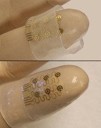
In the future, ‘surgery at your fingertips’ could be literally true. Researchers at the University of Illinois at Urbana-Champaign have created a silicon nanomembrane that can be fitted onto the fingertips and could, one day, be used in surgical procedures. From the Aug. 9, 2012 news item on ScienceDaily,

The intricate properties of the fingertips have been mimicked and recreated using semiconductor devices in what researchers hope will lead to the development of advanced surgical gloves.

The devices, shown to be capable of responding with high precision to the stresses and strains associated with touch and finger movement, are a step towards the creation of surgical gloves for use in medical procedures such as local ablations [excising or removing tissue] and ultrasound scans.

Researchers from the University of Illinois at Urbana-Champaign, Northwestern University and Dalian University of Technology have published their study August 10, in IOP [Institute of Physics] Publishing’s journal Nanotechnology.

The Aug. 10,2012 posting on the IOP website  offers this detail about the research,

The electronic circuit on the ‘skin’ is made of patterns of gold conductive lines and ultrathin sheets of silicon, integrated onto a flexible polymer called polyimide. The sheet is then etched into an open mesh geometry and transferred to a thin sheet of silicone rubber moulded into the precise shape of a finger.

This electronic ‘skin’, or finger cuff, was designed to measure the stresses and strains at the fingertip by measuring the change in capacitance – the ability to store electrical charge – of pairs of microelectrodes in the circuit.  Applied forces decreased the spacing in the skin which, in turn, increased the capacitance.

The fingertip device could also be fitted with sensors for measuring motion and temperature, with small-scale heaters as actuators for ablation and other related operations

The researchers experimented with having the electronics on the inside of the device, in contact with wearer’s skin, and also on the outside. They believe that because the device exploits materials and fabrication techniques adopted from the established semiconductor industry, the processes can be scaled for realistic use at reasonable cost.

“Perhaps the most important result is that we are able to incorporate multifunctional, silicon semiconductor device technologies into the form of soft, three-dimensional, form-fitting skins, suitable for integration not only with the fingertips but also other parts of the body,” continued Professor Rogers [John Rogers, co-author of the study].

Here’s what an image of these e-fingertips,

Virtual touch. Electronic fingertips could one day allow us to feel virtual sensations. Credit: John Rogers/University of Illinois at Urbana-Champaign

Krystnell A offers a more detailed description of the e-fingetips in an Aug. 9, 2012 story for Science NOW,

Hoping to create circuits with the flexibility of skin, materials scientist John Rogers of the University of Illinois, Urbana-Champaign, and colleagues cut up nanometer-sized strips of silicon; implanted thin, wavy strips of gold to conduct electricity; and mounted the entire circuit in a stretchable, spider web-type mesh of polymer as a support. They then embedded the circuit-polyimide structure onto a hollow tube of silicone that had been fashioned in the shape of a finger. Just like turning a sock inside out, the researchers flipped the structure so that the circuit, which was once on the outside of the tube, was on the inside where it could touch a finger placed against it.

To test the electronic fingers, the researchers put them on and pressed flat objects, such as the top of their desks. The pressure created electric currents that were transferred to the skin, which the researchers felt as mild tingling. That’s a first step in creating electrical signals that could be sent to the fingers, which could virtually recreate sensations such as heat, pressure, and texture, the team reports online today in Nanotechnology.

Rogers says another application of the technology is to custom fit the “electronic skin” around entire organs, allowing doctors to remotely monitor changes in temperature and blood flow. Electronic skin could also restore sensation to people who have lost their natural skin, he says, such as burn victims or amputees.

Here’s a link to the article which is freely accessible for 30 days after publication, from the Aug. 9, 2012 news item on ScienceDaily,

Ming Ying, Andrew P Bonifas, Nanshu Lu, Yewang Su, Rui Li, Huanyu Cheng, Abid Ameen, Yonggang Huang, John A Rogers. Silicon nanomembranes for fingertip electronics. Nanotechnology, 2012; 23 (34): 344004 DOI: 10.1088/0957-4484/23/34/344004

My best guess is that free access will no longer be available by Sept. 7 (or so) , 2012. I last wrote about John Rogers’ work in an Aug. 12, 2011 posting about electronic tattoos.

More than the “Emperor’s New Clothes” insight

Happy 2010 to all! I’ve taken some time out as I have moved locations and it’s taken longer to settle down that I hoped. (sigh) I still have loads to do but can get back to posting regularly (I hope).

New Year’s Eve I came across a very interesting article about how scientists think thanks to a reference on the Foresight Institute website. The article, Accept Defeat: The Neuroscience of Screwing Up, by Jonah Lehrer for Wired Magazine uses a story about a couple of astronomers and their investigative frustrations to illustrate research on how scientists (and the rest of us, as it turns out) think.

Before going on about the article I’m going to arbitrarily divide beliefs about scientific thinking/processes into two schools. In the first there’s the scientific method with its belief in objectivity and incontrovertible truths waiting to be discovered and validated. Later in university I was introduced to the 2nd belief about scientific thinking with the notion that scientific facts are social creations and that objectivity does not exist. From the outside it appears that scientists tend to belong to the first school and social scientists to the second but, as the Wired article points out, things are a little more amorphous than that when you dig down into the neuroscience of it all.

From the article,

The reason we’re so resistant to anomalous information — the real reason researchers automatically assume that every unexpected result is a stupid mistake — is rooted in the way the human brain works. Over the past few decades, psychologists [and other social scientists] have dismantled the myth of objectivity. The fact is, we carefully edit our reality, searching for evidence that confirms what we already believe. Although we pretend we’re empiricists — our views dictated by nothing but the facts — we’re actually blinkered, especially when it comes to information that contradicts our theories. The problem with science, then, isn’t that most experiments fail — it’s that most failures are ignored.

The DLPFC [dorsolateral prefrontal cortex] is constantly censoring the world, erasing facts from our experience. If the ACC  [anterior cingulate cortex, typically associated with errors and contradictions]] is the “Oh shit!” circuit, the DLPFC is the Delete key. When the ACC and DLPFC “turn on together, people aren’t just noticing that something doesn’t look right,” [Kevin] Dunbar says. “They’re also inhibiting that information.”

Disregarding evidence is something I’ve noticed (in others more easily than in myself) and have wondered about the implications. As noted in the article, ignoring scientific failure stymies research and ultimately more effective applications for the research. For example, there’s been a lot of interest in a new surgical procedure (still being tested) for patients with multiple sclerosis (MS). The procedure was developed by an Italian surgeon who (after his wife was stricken with the disease) reviewed literature on the disease going back 100 years and found a line of research that wasn’t being pursued actively and was a radical departure from current accepted beliefs about the nature of MS. (You can read more about the MS work here in the Globe and Mail story or here in the CBC story.) Btw, there are a couple of happy endings. The surgeon’s wife is much better and a promising new procedure is being examined.

Innovation and new research can be so difficult to pursue it’s amazing that anyone ever succeeds. Kevin Dunbar, the researcher mentioned previously, arrived at a rather interesting conclusion in his investigation on how scientists think and how they get around the ACC/DLFPC action: other people.  He tells a story about two lab groups who each had a meeting,

Dunbar watched how each of these labs dealt with their protein problem. The E. coli group took a brute-force approach, spending several weeks methodically testing various fixes. “It was extremely inefficient,” Dunbar says. “They eventually solved it, but they wasted a lot of valuable time.”The diverse lab, in contrast, mulled the problem at a group meeting. None of the scientists were protein experts, so they began a wide-ranging discussion of possible solutions. At first, the conversation seemed rather useless. But then, as the chemists traded ideas with the biologists and the biologists bounced ideas off the med students, potential answers began to emerge. “After another 10 minutes of talking, the protein problem was solved,” Dunbar says. “They made it look easy.”

When Dunbar reviewed the transcripts of the meeting, he found that the intellectual mix generated a distinct type of interaction in which the scientists were forced to rely on metaphors and analogies [my emphasis] to express themselves. (That’s because, unlike the E. coli group, the second lab lacked a specialized language that everyone could understand.) These abstractions proved essential for problem-solving, as they encouraged the scientists to reconsider their assumptions. Having to explain the problem to someone else forced them to think, if only for a moment, like an intellectual on the margins, filled with self-skepticism.

As Dunbar notes, we usually need more than an outsider to experience a Eureka moment (the story about Italian surgeon notwithstanding and it should be noted that he was an MS outsider); we need metaphors and analogies. (I’ve taken it a bit further than Dunbar likely would but I am a writer, after all.)

If you are interested in Dunbar’s work, he’s at the University of Toronto with more information here.