Tag Archives: Tal Dvir

First 3D heart printed using patient’s biological materials

This is very exciting news and it’s likely be at least 10 years before this technology could be made available to the public.
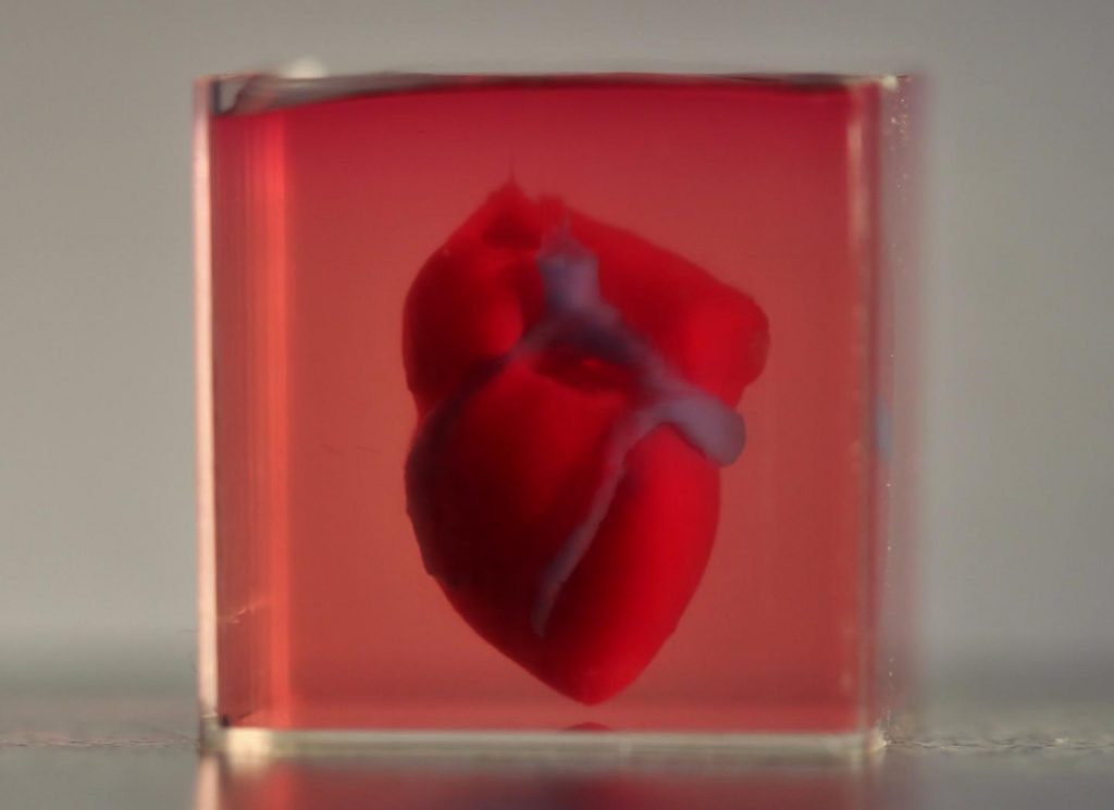
Caption: A 3D-printed, small-scaled human heart engineered from the patient’s own materials and cells. Credit: Advanced Science. © 2019 The Authors.

An April 15, 2019 news item on ScienceDaily makes a remarkable announcement,

In a major medical breakthrough, Tel Aviv University researchers have “printed” the world’s first 3D vascularised engineered heart using a patient’s own cells and biological materials. Their findings were published on April 15 [2019] in a study in Advanced Science.

Until now, scientists in regenerative medicine — a field positioned at the crossroads of biology and technology — have been successful in printing only simple tissues without blood vessels.

“This is the first time anyone anywhere has successfully engineered and printed an entire heart replete with cells, blood vessels, ventricles and chambers,” says Prof. Tal Dvir of TAU’s School of Molecular Cell Biology and Biotechnology, Department of Materials Science and Engineering, Center for Nanoscience and Nanotechnology and Sagol Center for Regenerative Biotechnology, who led the research for the study.

An April 15, 2019 Amricna Friends of Tel Aviv University (TAU) news release (also on EurekAlert), which originated the news item, provides more detail,

Heart disease is the leading cause of death among both men and women in the United States. Heart transplantation is currently the only treatment available to patients with end-stage heart failure. Given the dire shortage of heart donors, the need to develop new approaches to regenerate the diseased heart is urgent.

“This heart is made from human cells and patient-specific biological materials. In our process these materials serve as the bioinks, substances made of sugars and proteins that can be used for 3D printing of complex tissue models,” Prof. Dvir says. “People have managed to 3D-print the structure of a heart in the past, but not with cells or with blood vessels. Our results demonstrate the potential of our approach for engineering personalized tissue and organ replacement in the future.

Research for the study was conducted jointly by Prof. Dvir, Dr. Assaf Shapira of TAU’s Faculty of Life Sciences and Nadav Moor, a doctoral student in Prof. Dvir’s lab.

“At this stage, our 3D heart is small, the size of a rabbit’s heart, [emphasis mine] ” explains Prof. Dvir. “But larger human hearts require the same technology.”

For the research, a biopsy of fatty tissue was taken from patients. The cellular and a-cellular materials of the tissue were then separated. While the cells were reprogrammed to become pluripotent stem cells, the extracellular matrix (ECM), a three-dimensional network of extracellular macromolecules such as collagen and glycoproteins, were processed into a personalized hydrogel that served as the printing “ink.”

After being mixed with the hydrogel, the cells were efficiently differentiated to cardiac or endothelial cells to create patient-specific, immune-compatible cardiac patches with blood vessels and, subsequently, an entire heart.

According to Prof. Dvir, the use of “native” patient-specific materials is crucial to successfully engineering tissues and organs.

“The biocompatibility of engineered materials is crucial to eliminating the risk of implant rejection, which jeopardizes the success of such treatments,” Prof. Dvir says. “Ideally, the biomaterial should possess the same biochemical, mechanical and topographical properties of the patient’s own tissues. Here, we can report a simple approach to 3D-printed thick, vascularized and perfusable cardiac tissues that completely match the immunological, cellular, biochemical and anatomical properties of the patient.”

The researchers are now planning on culturing the printed hearts in the lab and “teaching them to behave” like hearts, Prof. Dvir says. They then plan to transplant the 3D-printed heart in animal models.

“We need to develop the printed heart further,” he concludes. “The cells need to form a pumping ability; they can currently contract, but we need them to work together. Our hope is that we will succeed and prove our method’s efficacy and usefulness.

“Maybe, in ten years, there will be organ printers in the finest hospitals around the world, and these procedures will be conducted routinely.”

Growing the heart to human size and getting the cells to work together so the heart will pump makes it seem like the 10 years Dvir imagines as the future date when there will be organ printers in hospitals routinely printing up hearts seems a bit optimistic. Regardless, I hope he’s right. Bravo to these Israeli researchers!

Here’s a link to and a citation for the paper,

3D Printing of Personalized Thick and Perfusable Cardiac Patches and Hearts by Nadav Noor, Assaf Shapira, Reuven Edri, Idan Gal, Lior Wertheim, Tal Dvir. Advanced Science DOI: https://doi.org/10.1002/advs.201900344 First published: 15 April 2019

This paper is open access.

Cardiac patch that’s both organic and engineered: a cyborg heart patch

A March 15, 2016 article by Michael Grothaus for Fast Company breaks the news about the ‘cyborg’ heart patch (Note: A link has been removed),

Researchers at Tel Aviv University’s Department of Biotechnology, Department of Materials Science and Engineering, and Center for Nanoscience and Nanotechnology have created a “cyborg heart patch” that may “single-handedly change the field of cardiac research,” reports EurekAlert. …

The researchers have made an illustration of the cyborg heart patch available,


Caption: A remotely regulated living bionic heart is pictured. The engineered tissue is comprised of living cardiac cells, polymers, and a complex nanoelectronic system. This integrated electronic system provides enhanced capabilities, such as online sensing of heart contraction, and pacing when needed. In addition, the electronics can control the release of growth factors and drugs, for stem cell recruitment and to decrease inflammation after transplantation. Credit: Tel Aviv University

A March 14, 2016 American Friends of Tel Aviv University news release (also on EurekAlert) expands on the theme,

More than 25% of the people on the national US waiting list for a heart will die before receiving one. Despite this discouraging figure, heart transplants are still on the rise. There just hasn’t been an alternative. Until now.

The “cyborg heart patch,” a new engineering innovation from Tel Aviv University, may single-handedly change the field of cardiac research. The bionic heart patch combines organic and engineered parts. In fact, its capabilities surpass those of human tissue alone. The patch contracts and expands like human heart tissue but regulates itself like a machine.

The invention is the brainchild of Prof. Tal Dvir and PhD student Ron Feiner of TAU’s Department of Biotechnology, Department of Materials Science and Engineering, and Center for Nanoscience and Nanotechnology. Their study was published today in the journal Nature Materials.

Science fiction becomes science fact

“With this heart patch, we have integrated electronics and living tissue,” Dr. Dvir said. “It’s very science fiction, but it’s already here, and we expect it to move cardiac research forward in a big way.

“Until now, we could only engineer organic cardiac tissue, with mixed results. Now we have produced viable bionic tissue, which ensures that the heart tissue will function properly.”

Prof. Dvir’s Tissue Engineering and Regenerative Medicine Lab at TAU has been at the forefront of cardiac research for the last five years, harnessing sophisticated nanotechnological tools to develop functional substitutes for tissue permanently damaged by heart attacks and cardiac disease. The new cyborg cardiac patch not only replaces organic tissue but also ensures its sound functioning through remote monitoring.

“We first ensured that the cells would contract in the patch, which explains the need for organic material,” said Dr. Dvir. “But, just as importantly, we needed to verify what was happening in the patch and regulate its function. We also wanted to be able to release drugs from the patch directly onto the heart to improve its integration with the host body.”

For the new bionic patch, Dr. Dvir and his team engineered thick bionic tissue suitable for transplantation. The engineered tissue features electronics that sense tissue function and accordingly provide electrical stimulation. In addition, electroactive polymers are integrated with the electronics. Upon activation, these polymers are able to release medication, such as growth factors or small molecules on demand.

Cardiac therapy in real time

“Imagine that a patient is just sitting at home, not feeling well,” Dr. Dvir said. “His physician will be able to log onto his computer and this patient’s file — in real time. He can view data sent remotely from sensors embedded in the engineered tissue and assess exactly how his patient is doing. He can intervene to properly pace the heart and activate drugs to regenerate tissue from afar.

“The longer-term goal is for the cardiac patch to be able to regulate its own welfare. In other words, if it senses inflammation, it will release an anti-inflammatory drug. If it senses a lack of oxygen, it will release molecules that recruit blood-vessel-forming cells to the heart.”

Dr. Dvir is currently examining how his proof of concept could apply to the brain and spinal cord to treat neurological conditions.

“This is a breakthrough, to be sure,” Dr. Dvir said. “But I would not suggest binging on cheeseburgers or quitting sports just yet. The practical realization of the technology may take some time. Meanwhile, a healthy lifestyle is still the best way to keep your heart healthy.”

It’s exciting news but this is at the proof-of-concept stage and there has been no testing, which (as Dvir seems to be hinting) means it could be several years before clinical trials.

Getting back to the heart of the matter (wordplay intended), here’s a link to and a citation for the paper,

Engineered hybrid cardiac patches with multifunctional electronics for online monitoring and regulation of tissue function by Ron Feiner, Leeya Engel, Sharon Fleischer, Maayan Malki, Idan Gal, Assaf Shapira, Yosi Shacham-Diamand & Tal Dvir. Nature Materials (2016) doi:10.1038/nmat4590 Published online 14 March 2016

This paper is behind a paywall.

The body as an electronic device—adding electronics to biological tissue

What makes this particular combination of electronic s  and living tissue special is t that it was achieved in 3-D rather than 2-D.  From the Boston Children’s Hospital Aug. 26, 2012 news release on EurekAlert,

A multi-institutional research team has developed a method for embedding networks of biocompatible nanoscale wires within engineered tissues. These networks—which mark the first time that electronics and tissue have been truly merged in 3D—allow direct tissue sensing and potentially stimulation, a potential boon for development of engineered tissues that incorporate capabilities for monitoring and stimulation, and of devices for screening new drugs.

The Aug. 27, 2012 news item on Nanowerk provides more detail about integration of the cells and electronics,

Until now, the only cellular platforms that incorporated electronic sensors consisted of flat layers of cells grown on planar metal electrodes or transistors. Those two-dimensional systems do not accurately replicate natural tissue, so the research team set out to design a 3-D scaffold that could monitor electrical activity, allowing them to see how cells inside the structure would respond to specific drugs.

The researchers built their new scaffold out of epoxy, a nontoxic material that can take on a porous, 3-D structure. Silicon nanowires embedded in the scaffold carry electrical signals to and from cells grown within the structure.

“The scaffold is not just a mechanical support for cells, it contains multiple sensors. We seed cells into the scaffold and eventually it becomes a 3-D engineered tissue,” Tian says [Bozhi Tian, a former postdoc at MIT {Massachusetts Institute of Technology} and Children’s Hospital and a lead author of the paper ].

The team chose silicon nanowires for electronic sensors because they are small, stable, can be safely implanted into living tissue and are more electrically sensitive than metal electrodes. The nanowires, which range in diameter from 30 to 80 nanometers (about 1,000 times smaller than a human hair), can detect voltages less than one-thousandth of a watt, which is the level of electricity that might be seen in a cell.

Here’s more about why the researchers want to integrate living tissue and electronics, from the Harvard University Aug. 26, 2012 news release on EurekAlert,

“The current methods we have for monitoring or interacting with living systems are limited,” said Lieber [Charles M. Lieber, the Mark Hyman, Jr. Professor of Chemistry at Harvard and one of the study’s team leaders]. “We can use electrodes to measure activity in cells or tissue, but that damages them. With this technology, for the first time, we can work at the same scale as the unit of biological system without interrupting it. Ultimately, this is about merging tissue with electronics in a way that it becomes difficult to determine where the tissue ends and the electronics begin.”

The research addresses a concern that has long been associated with work on bioengineered tissue – how to create systems capable of sensing chemical or electrical changes in the tissue after it has been grown and implanted. The system might also represent a solution to researchers’ struggles in developing methods to directly stimulate engineered tissues and measure cellular reactions.

“In the body, the autonomic nervous system keeps track of pH, chemistry, oxygen and other factors, and triggers responses as needed,” Kohane [Daniel Kohane, a Harvard Medical School professor in the Department of Anesthesia at Children’s Hospital Boston and a team leader] explained. “We need to be able to mimic the kind of intrinsic feedback loops the body has evolved in order to maintain fine control at the cellular and tissue level.”

Here’s a citation and a link to the paper (which is behind a paywall),

Macroporous nanowire nanoelectronic scaffolds for synthetic tissues by Bozhi Tian, Jia Lin, Tal Dvir, Lihua Jin, Jonathan H. Tsui, Quan  Qing, Zhigang Suo, Robert Langer, Daniel S. Kohane, and Charles M. Lieber in Nature Materials (2012) doi:10.1038/nmat3404 Published onlin26 August 2012.

This is the image MIT included with its Aug 27, 2012 news release (which originated the news item on Nanowerk),

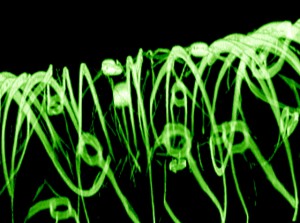
A 3-D reconstructed confocal fluorescence micrograph of a tissue scaffold.
Image: Charles M. Lieber and Daniel S. Kohane.

At this point they’re discussing therapeutic possibilities but I expect that ‘enhancement’ is also being considered although not mentioned for public consumption.