Tag Archives: Tanja Lahtinen

Novel self-assembly at 102 atoms

A Jan. 13, 2017 news item on ScienceDaily announces a discovery about self-assembly of 102-atom gold nanoclusters,

Self-assembly of matter is one of the fundamental principles of nature, directing the growth of larger ordered and functional systems from smaller building blocks. Self-assembly can be observed in all length scales from molecules to galaxies. Now, researchers at the Nanoscience Centre of the University of Jyväskylä and the HYBER Centre of Excellence of Aalto University in Finland report a novel discovery of self-assembling two- and three-dimensional materials that are formed by tiny gold nanoclusters of just a couple of nanometres in size, each having 102 gold atoms and a surface layer of 44 thiol molecules. The study, conducted with funding from the Academy of Finland and the European Research Council, has been published in Angewandte Chemie.

A Jan. 13, 2017 Academy of Finland press release, which originated the news item, provides more technical information about the work,

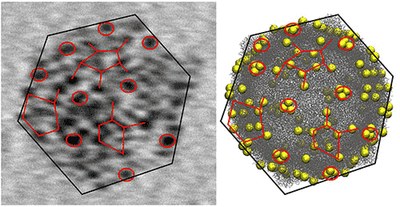
The atomic structure of the 102-atom gold nanocluster was first resolved by the group of Roger D Kornberg at Stanford University in 2007 (2). Since then, several further studies of its properties have been conducted in the Jyväskylä Nanoscience Centre, where it has also been used for electron microscopy imaging of virus structures (3). The thiol surface of the nanocluster has a large number of acidic groups that can form directed hydrogen bonds to neighbouring nanoclusters and initiate directed self-assembly.

The self-assembly of gold nanoclusters took place in a water-methanol mixture and produced two distinctly different superstructures that were imaged in a high-resolution electron microscope at Aalto University. In one of the structures, two-dimensional hexagonally ordered layers of gold nanoclusters were stacked together, each layer being just one nanocluster thick. Modifying the synthesis conditions, also three-dimensional spherical, hollow capsid structures were observed, where the thickness of the capsid wall corresponds again to just one nanocluster size (see figure).

While the details of the formation mechanisms of these superstructures warrant further systemic investigations, the initial observations open several new views into synthetically made self-assembling nanomaterials.

“Today, we know of several tens of different types of atomistically precise gold nanoclusters, and I believe they can exhibit a wide variety of self-assembling growth patterns that could produce a range of new meta-materials,” said Academy Professor Hannu Häkkinen, who coordinated the research at the Nanoscience Centre. “In biology, typical examples of self-assembling functional systems are viruses and vesicles. Biological self-assembled structures can also be de-assembled by gentle changes in the surrounding biochemical conditions. It’ll be of great interest to see whether these gold-based materials can be de-assembled and then re-assembled to different structures by changing something in the chemistry of the surrounding solvent.”

“The free-standing two-dimensional nanosheets will bring opportunities towards new-generation functional materials, and the hollow capsids will pave the way for highly lightweight colloidal framework materials,” Postdoctoral Researcher Nonappa (Aalto University) said.

Professor Olli Ikkala of Aalto University said: “In a broader framework, it has remained as a grand challenge to master the self-assemblies through all length scales to tune the functional properties of materials in a rational way. So far, it has been commonly considered sufficient to achieve sufficiently narrow size distributions of the constituent nanoscale structural units to achieve well-defined structures. The present findings suggest a paradigm change to pursue strictly defined nanoscale units for self-assemblies.”

References:

(1)    Nonappa, T. Lahtinen, J.S. Haataja, T.-R. Tero, H. Häkkinen and O. Ikkala, “Template-Free Supracolloidal Self-Assembly of Atomically Precise Gold Nanoclusters: From 2D Colloidal Crystals to Spherical Capsids”, Angewandte Chemie International Edition, published online 23 November 2016, DOI: 10.1002/anie.201609036

(2)    P. Jadzinsky et al., “Structure of a thiol-monolayer protected gold nanoparticle at 1.1Å resolution”, Science 318, 430 (2007)

(3)    V. Marjomäki et al., “Site-specific targeting of enterovirus capsid by functionalized monodispersed gold nanoclusters”, PNAS 111, 1277 (2014)

Here’s the figure mentioned in the news release,

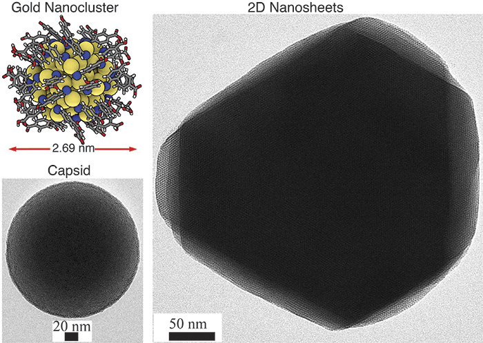
Figure: 2D hexagonal sheet-like and 3D capsid structures based on atomically precise gold nanoclusters as guided by hydrogen bonding between the ligands. The inset in the top left corner shows the atomic structure of one gold nanocluster.

Here’s a link to and a citation for the paper,

Template-Free Supracolloidal Self-Assembly of Atomically Precise Gold Nanoclusters: From 2D Colloidal Crystals to Spherical Capsids by Dr. Nonappa, Dr. Tanja Lahtinen, M. Sc. Johannes. S. Haataja, Dr. Tiia-Riikka Tero, Prof. Hannu Häkkinen, and Prof. Olli Ikkala. Angewandte Chemie International Edition Volume 55, Issue 52, pages 16035–16038, December 23, 2016 Version of Record online: 23 NOV 2016 DOI: 10.1002/anie.201609036

© 2016 Wiley-VCH Verlag GmbH & Co. KGaA, Weinheim

This paper is behind a paywall.

Gold atoms: sometimes they’re a metal and sometimes they’re a molecule

Fascinating work out of Finland shows that a minor change in the number of gold atoms in your gold nanoparticle can mean the difference between a metal and a molecule (coincidentally, this phenomenon is alluded to in my April 14, 2015 post (Nature’s patterns reflected in gold nanoparticles); more about that at the end of this piece. Getting back to Finland and when gold is metal and when it’s a molecule, here’s more from an April 10, 2015 news item on ScienceDaily,

Researchers at the Nanoscience Center at the University of Jyväskylä, Finland, have shown that dramatic changes in the electronic properties of nanometre-sized chunks of gold occur in well-defined size range. Small gold nanoclusters could be used, for instance, in short-term storage of energy or electric charge in the field of molecular electronics. Funded by the Academy of Finland, the researchers have been able to obtain new information which is important, among other things, in developing bioimaging and sensing based on metal-like clusters.

An April 10, 2015 news release (also on EurekAlert) on the Academy of Finland (Suomen Akatemia) website, which originated the news item, describes the work in more detail,

Two recent papers by the researchers at Jyväskylä (1, 2) demonstrate that the electronic properties of two different but still quite similar gold nanoclusters can be drastically different. The clusters were synthesised by chemical methods incorporating a stabilising ligand layer on their surface. The researchers found that the smaller cluster, with up to 102 gold atoms, behaves like a giant molecule while the larger one, with at least 144 gold atoms, already behaves, in principle, like a macroscopic chunk of metal, but in nanosize.

The fundamentally different behaviour of these two differently sized gold nanoclusters was demonstrated by shining a laser light onto solution samples containing the clusters and by monitoring how energy dissipates from the clusters into the surrounding solvent.

“Molecules behave drastically different from metals,” said Professor Mika Pettersson, the principal investigator of the team conducting the experiments. “The additional energy from light, absorbed by the metal-like clusters, transfers to the environment extremely rapidly, in about one hundred billionth of a second, while a molecule-like cluster is excited to a higher energy state and dissipates the energy into the environment with a rate that is at least 100 times slower. This is exactly what we saw: the 102-gold atom cluster is a giant molecule showing even a transient magnetic state while the 144-gold atom cluster is already a metal. We’ve thus managed to bracket an important size region where this fundamentally interesting change in the behaviour takes place.”

“These experimental results go together very well with what our team has seen from computational simulations on these systems,” said Professor Hannu Häkkinen, a co-author of the studies and the scientific director of the nanoscience centre. “My team predicted this kind of behaviour back in 2008-2009 when we saw big differences in the electronic structure of exactly these nanoclusters. It’s wonderful that robust spectroscopic experiments have now proved these phenomena. In fact, the metal-like 144-atom cluster is even more interesting, since we just published a theoretical paper where we saw a big enhancement of the metallic properties of just a few copper atoms mixed with gold.” (3)

Here are links to and citation for the papers,

Ultrafast Electronic Relaxation and Vibrational Cooling Dynamics of Au144(SC2H4Ph)60 Nanocluster Probed by Transient Mid-IR Spectroscopy by Satu Mustalahti, Pasi Myllyperkiö, Tanja Lahtinen, Kirsi Salorinne, Sami Malola, Jaakko Koivisto, Hannu Häkkinen, and Mika Pettersson. J. Phys. Chem. C, 2014, 118 (31), pp 18233–18239 DOI: 10.1021/jp505464z Publication Date (Web): July 3, 2014

Copyright © 2014 American Chemical Society

Copper Induces a Core Plasmon in Intermetallic Au(144,145)–xCux(SR)60 Nanoclusters by Sami Malola, Michael J. Hartmann, and Hannu Häkkinen. J. Phys. Chem. Lett., 2015, 6 (3), pp 515–520 DOI: 10.1021/jz502637b Publication Date (Web): January 22, 2015

Copyright © 2015 American Chemical Society

Molecule-like Photodynamics of Au102(pMBA)44 Nanocluster by Satu Mustalahti, Pasi Myllyperkiö, Sami Malola, Tanja Lahtinen, Kirsi Salorinne, Jaakko Koivisto, Hannu Häkkinen, and Mika Pettersson. ACS Nano, 2015, 9 (3), pp 2328–2335 DOI: 10.1021/nn506711a Publication Date (Web): February 22, 2015

Copyright © 2015 American Chemical Society

These papers are behind paywalls.

As for my April 14, 2015 post (Nature’s patterns reflected in gold nanoparticles), researchers at Carnegie Mellon University were researching patterns in different sized gold nanoparticles when this was noted in passing,

… Normally, gold is one of the best conductors of electrical current, but the size of Au133 is so small that the particle hasn’t yet become metallic. …

Getting new information on trafficking viruses with gold nanoparticles

Finnish researchers have developed a new technique for studying viruses according to a Jan. 15, 2014 news release on EurekAlert,

Researchers at the Nanoscience Center (NSC) of University of Jyväskylä in Finland have developed a novel method to study enterovirus structures and their functions. The method will help to obtain new information on trafficking of viruses in cells and tissues as well as on the mechanisms of virus opening inside cells.

The news release explains enteroviruses and describes the technique in more detail,

Enteroviruses are pathogenic viruses infecting humans. This group consists of polioviruses, coxsackieviruses, echoviruses and rhinoviruses. Enteroviruses are the most common causes of flu, but they also cause serious symptoms such as heart muscle infections and paralysis. Recently, enteroviruses have been linked with chronic diseases such as diabetes (2).

The infection mechanisms and infectious pathways of enteroviruses are still rather poorly known. Previous studies in the group of Dr. Varpu Marjomäki at the NSC have focused on the cellular factors that are important for the infection caused by selected enteroviruses (3). The mechanistic understanding of virus opening and the release of the viral genome in cellular structures for starting new virus production is still largely lacking. Furthermore, the knowledge of infectious processes in tissues is hampered by the lack of reliable tools for detecting virus infection.

The newly developed method involves a chemical modification of a known thiol-stabilized gold nanoparticle, the so-called Au102 cluster that was first synthesized and structurally solved by the group of Roger D Kornberg in 2007 (4) and later characterized at NSC by the groups of prof. Hannu Häkkinen and prof. Mika Pettersson in collaboration with Kornberg. (5) The organic thiol surface of the Au102 particles is modified by attaching linker molecules that make a chemical bond to sulfur-containing cysteine residues that are part of the surface structure of the virus. Several tens of gold particles can bind to a single virus, and the binding pattern shows up as dark tags reflecting the overall shape and structure of the virus (see the figure). The gold particles allow for studies on the structural changes of the viruses during their lifespan.

The study showed also that the infectivity of the viruses is not compromised by the attached gold particles which indicates that the labeling method does not interfere with the normal biological functions of viruses inside cells. This facilitates new investigations on the virus structures from samples taken from inside cells during the various phases of the virus infection, and gives possibilities to obtain new information on the mechanisms of virus uncoating (opening and release of the genome). The new method allows also for tracking studies of virus pathways in tissues. This is important for further understanding of acute and chronic symptoms caused by viruses. Finally, the method is expected to be useful for developing of new antiviral vaccines that are based on virus-like particles.

The method was developed at the NSC as a wide cross-disciplinary collaboration between chemists, physicists and biologists.

Here’s an image provided by the researchers, which illustrates their work,

Left: transmission electron microscopy (TEM) image of a single CVB3 virus showing tens of gold nanoparticles attached to its surface. The particles form a distinct "tagging pattern" that reflects the shape and the structure of the virus. The TEM image can be correlated to the model of the virus (right), where the yellow spheres mark the possible binding sites of the gold particles. The diameter of the virus is about 35 nanometers (nanometer = one billionth of a millimeter). The figure is taken from the publication. Courtesy: University of Jyväskylä

Left: transmission electron microscopy (TEM) image of a single CVB3 virus showing tens of gold nanoparticles attached to its surface. The particles form a distinct “tagging pattern” that reflects the shape and the structure of the virus. The TEM image can be correlated to the model of the virus (right), where the yellow spheres mark the possible binding sites of the gold particles. The diameter of the virus is about 35 nanometers (nanometer = one billionth of a millimeter). The figure is taken from the publication. Courtesy: University of Jyväskylä

Unfortunately, the researchers have published in the Proceedings f the National Academy of Sciences (PNAS). I noted in a previous posting that this publisher has developed a time-consuming process for getting access to a paper and payment options for reading it. I can provide a link to and a citation to the abstract for this paper but I’m not willing to spend several minutes trying to bypass the block they’ve placed on accessing papers and their payment options,

Site-specific targeting of enterovirus capsid by functionalized monodisperse gold nanoclusters by Varpu Marjomäki, Tanja Lahtinen, Mari Martikainen, Jaakko Koivisto, Sami Malola, Kirsi Salorinne, Mika Pettersson, and Hannu Häkkinenb. Proc. Natl. Acad. Sci. USA (2014), www.pnas.org/cgi/doi/10.1073/pnas.1310973111.

The University of Jyväskylä Jan. ??, 2014 news release about this work provides references (scroll down) to previous papers published on this work.