Tag Archives: Technical University of Berlin

Electrode-filled elastic fiber for wearable electronics and robots

This work comes out of Switzerland. A May 25, 2018 École Polytechnique Fédérale de Lausanne (EPFL) press release (also on EurekAlert) announces their fibers,

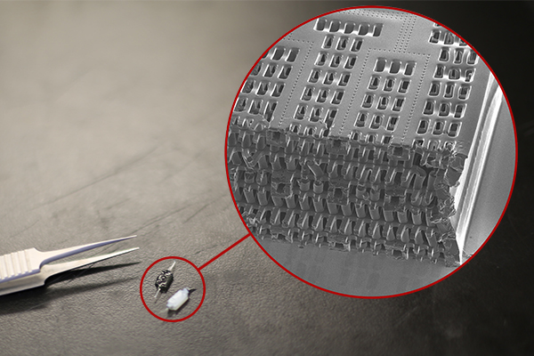
EPFL scientists have found a fast and simple way to make super-elastic, multi-material, high-performance fibers. Their fibers have already been used as sensors on robotic fingers and in clothing. This breakthrough method opens the door to new kinds of smart textiles and medical implants.

It’s a whole new way of thinking about sensors. The tiny fibers developed at EPFL are made of elastomer and can incorporate materials like electrodes and nanocomposite polymers. The fibers can detect even the slightest pressure and strain and can withstand deformation of close to 500% before recovering their initial shape. All that makes them perfect for applications in smart clothing and prostheses, and for creating artificial nerves for robots.

The fibers were developed at EPFL’s Laboratory of Photonic Materials and Fiber Devices (FIMAP), headed by Fabien Sorin at the School of Engineering. The scientists came up with a fast and easy method for embedding different kinds of microstructures in super-elastic fibers. For instance, by adding electrodes at strategic locations, they turned the fibers into ultra-sensitive sensors. What’s more, their method can be used to produce hundreds of meters of fiber in a short amount of time. Their research has just been published in Advanced Materials.

Heat, then stretch
To make their fibers, the scientists used a thermal drawing process, which is the standard process for optical-fiber manufacturing. They started by creating a macroscopic preform with the various fiber components arranged in a carefully designed 3D pattern. They then heated the preform and stretched it out, like melted plastic, to make fibers of a few hundreds microns in diameter. And while this process stretched out the pattern of components lengthwise, it also contracted it crosswise, meaning the components’ relative positions stayed the same. The end result was a set of fibers with an extremely complicated microarchitecture and advanced properties.

Until now, thermal drawing could be used to make only rigid fibers. But Sorin and his team used it to make elastic fibers. With the help of a new criterion for selecting materials, they were able to identify some thermoplastic elastomers that have a high viscosity when heated. After the fibers are drawn, they can be stretched and deformed but they always return to their original shape.

Rigid materials like nanocomposite polymers, metals and thermoplastics can be introduced into the fibers, as well as liquid metals that can be easily deformed. “For instance, we can add three strings of electrodes at the top of the fibers and one at the bottom. Different electrodes will come into contact depending on how the pressure is applied to the fibers. This will cause the electrodes to transmit a signal, which can then be read to determine exactly what type of stress the fiber is exposed to – such as compression or shear stress, for example,” says Sorin.

Artificial nerves for robots

Working in association with Professor Dr. Oliver Brock (Robotics and Biology Laboratory, Technical University of Berlin), the scientists integrated their fibers into robotic fingers as artificial nerves. Whenever the fingers touch something, electrodes in the fibers transmit information about the robot’s tactile interaction with its environment. The research team also tested adding their fibers to large-mesh clothing to detect compression and stretching. “Our technology could be used to develop a touch keyboard that’s integrated directly into clothing, for instance” says Sorin.

The researchers see many other potential applications. Especially since the thermal drawing process can be easily tweaked for large-scale production. This is a real plus for the manufacturing sector. The textile sector has already expressed interest in the new technology, and patents have been filed.

There’s a video of the lead researcher discussing the work as he offers some visual aids,

Here’s a link to and a citation for the paper,

Superelastic Multimaterial Electronic and Photonic Fibers and Devices via Thermal Drawing by Yunpeng Qu, Tung Nguyen‐Dang, Alexis Gérald Page, Wei Yan, Tapajyoti Das Gupta, Gelu Marius Rotaru, René M. Rossi, Valentine Dominique Favrod, Nicola Bartolomei, Fabien Sorin. Advanced Materials First published: 25 May 2018 https://doi.org/10.1002/adma.201707251

This paper is behind a paywall.

Mathematicians get illustrative

Frank A. Farris, an associate Professor of Mathematics at Santa Clara University (US), writes about the latest in mathematicians and data visualization in an April 4, 2017 essay on The Conversation (Note: Links have been removed),

Today, digital tools like 3-D printing, animation and virtual reality are more affordable than ever, allowing mathematicians to investigate and illustrate their work at the same time. Instead of drawing a complicated surface on a chalkboard, we can now hand students a physical model to feel or invite them to fly over it in virtual reality.

Last year, a workshop called “Illustrating Mathematics” at the Institute for Computational and Experimental Research in Mathematics (ICERM) brought together an eclectic group of mathematicians and digital art practitioners to celebrate what seems to be a golden age of mathematical visualization. Of course, visualization has been central to mathematics since Pythagoras, but this seems to be the first time it had a workshop of its own.

Visualization plays a growing role in mathematical research. According to John Sullivan at the Technical University of Berlin, mathematical thinking styles can be roughly categorized into three groups: “the philosopher,” who thinks purely in abstract concepts; “the analyst,” who thinks in formulas; and “the geometer,” who thinks in pictures.

Mathematical research is stimulated by collaboration between all three types of thinkers. Many practitioners believe teaching should be calibrated to connect with different thinking styles.

Borromean Rings, the logo of the International Mathematical Union. John Sullivan

Sullivan’s own work has benefited from images. He studies geometric knot theory, which involves finding “best” configurations. For example, consider his Borromean rings, which won the logo contest of the International Mathematical Union several years ago. The rings are linked together, but if one of them is cut, the others fall apart, which makes it a nice symbol of unity.

Apparently this new ability to think mathematics visually has influenced mathematicians in some unexpected ways,

Take mathematician Fabienne Serrière, who raised US$124,306 through Kickstarter in 2015 to buy an industrial knitting machine. Her dream was to make custom-knit scarves that demonstrate cellular automata, mathematical models of cells on a grid. To realize her algorithmic design instructions, Serrière hacked the code that controls the machine. She now works full-time on custom textiles from a Seattle studio.

In this sculpture by Edmund Harriss, the drill traces are programmed to go perpendicular to the growth rings of the tree. This makes the finished sculpture a depiction of a concept mathematicians know as ‘paths of steepest descent.’ Edmund Harriss, Author provided

Edmund Harriss of the University of Arkansas hacked an architectural drilling machine, which he now uses to make mathematical sculptures from wood. The control process involves some deep ideas from differential geometry. Since his ideas are basically about controlling a robot arm, they have wide application beyond art. According to his website, Harriss is “driven by a passion to communicate the beauty and utility of mathematical thinking.”

Mathematical algorithms power the products made by Nervous System, a studio in Massachusetts that was founded in 2007 by Jessica Rosenkrantz, a biologist and architect, and Jess Louis-Rosenberg, a mathematician. Many of their designs, for things like custom jewelry and lampshades, look like naturally occurring structures from biology or geology.

Farris’ essay is a fascinating look at mathematics and data visualization.

University of Toronto (Canada) researchers and lab-grown heart and liver tissue (person-on-a-chip)

Usually called ‘human-on-a-chip’, a team at the University of Toronto have developed a two-organ ‘person on a chip’ according to a March 7, 2016 news item on phys.org (Note: Links have been removed),

Researchers at U of T [University of Toronto] Engineering have developed a new way of growing realistic human tissues outside the body. Their “person-on-a-chip” technology, called AngioChip, is a powerful platform for discovering and testing new drugs, and could eventually be used to repair or replace damaged organs.

Professor Milica Radisic (IBBME, ChemE), graduate student Boyang Zhang and the rest of the team are among those research groups around the world racing to find ways to grow human tissues in the lab, under conditions that mimic a real person’s body. They have developed unique methods for manufacturing small, intricate scaffolds for individual cells to grow on. These artificial environments produce cells and tissues that resemble the real thing more closely than those grown lying flat in a petri dish.

The team’s recent creations have included BiowireTM—an innovative method of growing heart cells around a silk suture—as well as a scaffold for heart cells that snaps together like sheets of Velcro. But AngioChip takes tissue engineering to a whole new level. “It’s a fully three-dimensional structure complete with internal blood vessels,” says Radisic. “It behaves just like vasculature, and around it there is a lattice for other cells to attach and grow.” …

A March 7, 2016 University of Toronto news release (also on EurekAlert), which originated the news item, provides more detail about the AngioChip,

Zhang built the scaffold out of POMaC, a polymer that is both biodegradable and biocompatible. The scaffold is built out of a series of thin layers, stamped with a pattern of channels that are each about 50 to 100 micrometres wide. The layers, which resemble the computer microchips, are then stacked into a 3D structure of synthetic blood vessels. As each layer is added, UV light is used to cross-link the polymer and bond it to the layer below.

When the structure is finished, it is bathed in a liquid containing living cells. The cells quickly attach to the inside and outside of the channels and begin growing just as they would in the human body.

“Previously, people could only do this using devices that squish the cells between sheets of silicone and glass,” says Radisic. “You needed several pumps and vacuum lines to run just one chip. Our system runs in a normal cell culture dish, and there are no pumps; we use pressure heads to perfuse media through the vasculature. The wells are open, so you can easily access the tissue.”

Using the platform, the team has built model versions of both heart and liver tissues that function like the real thing. “Our liver actually produced urea and metabolized drugs,” says Radisic. They can connect the blood vessels of the two artificial organs, thereby modelling not just the organs themselves, but the interactions between them. They’ve even injected white blood cells into the vessels and watched as they squeezed through gaps in the vessel wall to reach the tissue on the other side, just as they do in the human body.

The news release also mentions potential markets and the work that needs to be accomplished before AngioChip is available for purchase,

AngioChip has great potential in the field of pharmaceutical testing. Current drug-testing methods, such as animal testing and controlled clinical trials, are costly and fraught with ethical concerns. Testing on lab-grown human tissues would provide a realistic model at a fraction of the cost, but this area of research is still in its infancy. “In the last few years, it has become possible to order cultures of human cells for testing, but they’re grown on a plate, a two-dimensional environment,” says Radisic. “They don’t capture all the functional hallmarks of a real heart muscle, for example.”

A more realistic platform like AngioChip could enable drug companies to detect dangerous side effects and interactions between organ compartments long before their products reach the market, saving countless lives. It could also be used to understand and validate the effectiveness of current drugs and even to screen libraries of chemical compounds to discover new drugs. Through TARA Biosystems Inc., a spin-off company co-founded by Radisic, the team is already working on commercializing the technology.

In future, Radisic envisions her lab-grown tissues being implanted into the body to repair organs damaged by disease. Because the cells used to seed the platform can come from anyone, the new tissues could be genetically identical to the intended host, reducing the risk of organ rejection. Even in its current form, the team has shown that the AngioChip can be implanted into a living animal, its artificial blood vessels connected to a real circulatory system. The polymer scaffolding itself simply biodegrades after several months.

The team still has much work to do. Each AngioChip is currently made by hand; if the platform is to be used industrially, the team will need to develop high-throughput manufacturing methods to create many copies at once. Still, the potential is obvious. “It really is multifunctional, and solves many problems in the tissue engineering space,” says Radisic. “It’s truly next-generation.”

Here’s a link to and a citation for the paper,

Biodegradable scaffold with built-in vasculature for organ-on-a-chip engineering and direct surgical anastomosis by Boyang Zhang, Miles Montgomery, M. Dean Chamberlain, Shinichiro Ogawa, Anastasia Korolj, Aric Pahnke, Laura A. Wells, Stéphane Massé, Jihye Kim, Lewis Reis, Abdul Momen, Sara S. Nunes, Aaron R. Wheeler, Kumaraswamy Nanthakumar, Gordon Keller, Michael V. Sefton, & Milica Radisic. Nature Materials (2016) doi:10.1038/nmat4570 Published online 07 March 2016

This paper is behind a paywall.

The researchers have made two images illustrating their work available. There’s this still image,

These tiny polymer scaffolds contain channels that are about 100 micrometres wide, about the same diameter as a human hair. When seeded with cells, the channels act as artificial blood vessels. By mimicking tissues in the human heart and other organs, these scaffolds provide a new way to test drugs for potentially dangerous side effects. (Image: Tyler Irving/Boyang Zhang/Kevin Soobrian)

These tiny polymer scaffolds contain channels that are about 100 micrometres wide, about the same diameter as a human hair. When seeded with cells, the channels act as artificial blood vessels. By mimicking tissues in the human heart and other organs, these scaffolds provide a new way to test drugs for potentially dangerous side effects. (Image: Tyler Irving/Boyang Zhang/Kevin Soobrian)

Perhaps more intriguing is this one,

UofT_AngioChipMoving

When seeded with heart cells, the flexible polymer scaffold contracts with a regular rhythm, just like real heart tissue. (Image: Boyang Zhang)

I have mentioned ‘human-on-a-chip’ projects many times here and as the news release writer notes, there is an international race. My July 1, 2015 posting (cross-posted from the June 30, 2015 posting [Testing times: the future of animal alternatives] on the International Innovation blog [a CORDIS-listed project dissemination partner for FP7 and H2020 projects]) notes a couple of those projects,

Organ-on-a-chip projects use stem cells to create human tissues that replicate the functions of human organs. Discussions about human-on-a-chip activities – a phrase used to describe 10 interlinked organ chips – were a highlight of the 9th World Congress on Alternatives to Animal Testing held in Prague, Czech Republic, last year. One project highlighted at the event was a joint US National Institutes of Health (NIH), US Food and Drug Administration (FDA) and US Defense Advanced Research Projects Agency (DARPA) project led by Dan Tagle that claimed it would develop functioning human-on-a-chip by 2017. However, he and his team were surprisingly close-mouthed and provided few details making it difficult to assess how close they are to achieving their goal.

By contrast, Uwe Marx – Leader of the ‘Multi-Organ-Chip’ programme in the Institute of Biotechnology at the Technical University of Berlin and Scientific Founder of TissUse, a human-on-a-chip start-up company – claims to have sold two-organ chips. He also claims to have successfully developed a four-organ chip and that he is on his way to building a human-on-a-chip. Though these chips remain to be seen, if they are, they will integrate microfluidics, cultured cells and materials patterned at the nanoscale to mimic various organs, and will allow chemical testing in an environment that somewhat mirrors a human.

As for where the University of Toronto efforts fit into the race, I don’t know for sure. It’s the first time I’ve come across a reference to liver tissue producing urea but I believe there’s at least one other team in China which has achieved a three-dimensional, more lifelike aspect for liver tissue in my Jan. 29, 2016 posting ‘Constructing a liver’.