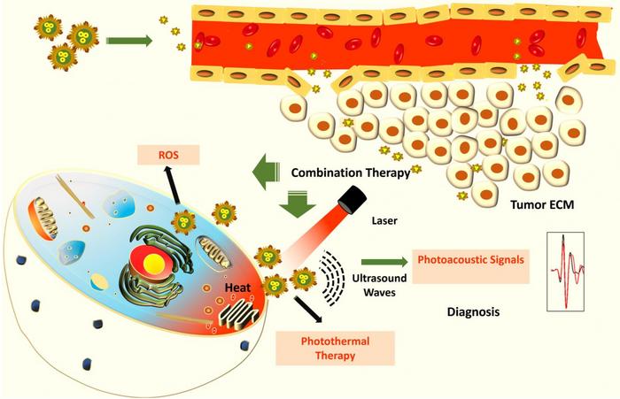
Scientists at the Indian Institute of Science (IISc) have developed a new approach to potentially detect and kill cancer cells, especially those that form a solid tumor mass. They have created hybrid nanoparticles made of gold and copper sulfide that can kill cancer cells using heat and enable their detection using sound waves, according to a study published in ACS Applied Nano Materials.
Early detection and treatment are key in the battle against cancer. Copper sulphide nanoparticles have previously received attention for their application in cancer diagnosis, while gold nanoparticles, which can be chemically modified to target cancer cells, have shown anticancer effects. In the current study, the IISc team decided to combine these two into hybrid nanoparticles.
“These particles have photothermal, oxidative stress, and photoacoustic properties,” says Jaya Prakash, Assistant Professor at the Department of Instrumentation and Applied Physics (IAP), IISc, and one of the corresponding authors of the paper. PhD students Madhavi Tripathi and Swathi Padmanabhan are co-first authors.
When light is shined on these hybrid nanoparticles, they absorb the light and generate heat, which can kill cancer cells. These nanoparticles also produce singlet oxygen atoms that are toxic for the cells. “We want both these mechanisms to kill the cancer cell,” Jaya Prakash explains.
The researchers say that the nanoparticles can also help diagnose certain cancers. Existing methods such as standalone CT and MRI scans require trained radiology professionals to decipher the images. The photoacoustic property of the nanoparticles allows them to absorb light and generate ultrasound waves, which can be used to detect cancer cells with high contrast once the particles reach them. The ultrasound waves generated from the particles allow for a more accurate image resolution as sound waves scatter less when they pass through tissues compared to light. Scans created from the generated ultrasound waves can also provide better clarity and can be used to measure the oxygen saturation in the tumour, boosting their detection.
“You can integrate this with existing systems of detection or treatment,” says Ashok M Raichur, Professor at the Department of Materials Engineering, and another corresponding author. For example, the nanoparticles can be triggered to produce heat by shining a light on them using an endoscope that is typically used for cancer screening.
Previously developed nanoparticles have limited applications because of their large size. The IISc team used a novel reduction method to deposit tiny seeds of gold onto the copper sulphide surface. The resulting hybrid nanoparticles – less than 8 nm in size – can potentially travel inside tissues easily and reach tumours. The researchers believe that the nanoparticles’ small size would also allow them to leave the human body naturally without accumulating, although extensive studies have to be carried out to determine if they are safe to use inside the human body.
In the current study, the researchers have tested their nanoparticles on lung cancer and cervical cancer cell lines in the lab. They now plan to take the results forward for clinical development.
Last week (specifically, Tuesday, March 3, 2020), someone moved away from me during a class. I’d sneezed.
The irony of the situation is that of the two of us, with my lung issues I’d be the one most at risk of getting very ill and/or dying from COVID-19. ([1] Yes, I confirmed that was the reason she’d moved. [2] The therapeutic nanoparticles news item is coming later) Here are the risk factors to take into account (from the US Centers for Disease Control’s People at Risk for Serious Illness from COVID-19 webpage,
Older adults [Note: In one report the age range was stated as ‘people over 70’]
People who have serious chronic medical conditions like:
Heart disease
Diabetes
Lung disease
I’m not suggesting that all precautions be abandoned but it would seem that panic might not be called for. Jeremy Samuel Faust, an emergency medicine physician at Brigham and Women’s Hospital in Boston, faculty in its division of health policy and public health, and an instructor at Harvard Medical School, has written a calming March 4, 2020 article (COVID-19 Isn’t As Deadly As We Think; Don’t hoard masks and food. Figure out how to help seniors and the immunosuppressed stay healthy.) for Slate.com (Note: Links have been removed],
There are many compelling reasons to conclude that SARS-CoV-2, the virus that causes COVID-19, is not nearly as deadly as is currently feared. But COVID-19 panic has set in nonetheless. You can’t find hand sanitizer in stores, and N95 face masks are being sold online for exorbitant prices, never mind that neither is the best way to protect against the virus (yes, just wash your hands). The public is behaving as if this epidemic is the next Spanish flu, which is frankly understandable given that initial reports have staked COVID-19 mortality at about 2–3 percent, quite similar to the 1918 pandemic that killed tens of millions of people.
Allow me to be the bearer of good news. These frightening numbers are unlikely to hold. The true case fatality rate, known as CFR, of this virus is likely to be far lower than current reports suggest. Even some lower estimates, such as the 1 percent death rate recently mentioned by the directors of the National Institutes of Health and the Centers for Disease Control and Prevention, likely substantially overstate the case. [emphases mine]
…
But the most straightforward and compelling evidence that the true case fatality rate of SARS-CoV-2 is well under 1 percent comes not from statistical trends and methodological massage, but from data from the Diamond Princess cruise outbreak and subsequent quarantine off the coast of Japan.
A quarantined boat is an ideal—if unfortunate—natural laboratory to study a virus. Many variables normally impossible to control are controlled. We know that all but one patient boarded the boat without the virus. We know that the other passengers were healthy enough to travel. We know their whereabouts and exposures. While the numbers coming out of China are scary, we don’t know how many of those patients were already ill for other reasons. How many were already hospitalized for another life-threatening illness and then caught the virus? How many were completely healthy, caught the virus, and developed a critical illness? In the real world, we just don’t know.
Here’s the problem with looking at mortality numbers in a general setting: In China, 9 million people die per year, which comes out to 25,000 people every single day, or around 1.5 million people over the past two months alone. A significant fraction of these deaths results from diseases like emphysema/COPD, lower respiratory infections, and cancers of the lung and airway whose symptoms are clinically indistinguishable from the nonspecific symptoms seen in severe COVID-19 cases. And, perhaps unsurprisingly, the death rate from COVID-19 in China spiked precisely among the same age groups in which these chronic diseases first become common. During the peak of the outbreak in China in January and early February, around 25 patients per day were dying with SARS-CoV-2. Most were older patients in whom the chronic diseases listed above are prevalent. Most deaths occurred in Hubei province, an area in which lung cancer and emphysema/COPD are significantly higher than national averages in China, a country where half of all men smoke. How were doctors supposed to sort out which of those 25 out of 25,000 daily deaths were solely due to coronavirus, and which were more complicated? What we need to know is how many excess deaths this virus causes.
…
This all suggests that COVID-19 is a relatively benign disease for most young people, and a potentially devastating one for the old and chronically ill, albeit not nearly as risky as reported. Given the low mortality rate among younger patients with coronavirus—zero in children 10 or younger among hundreds of cases in China, and 0.2-0.4 percent in most healthy nongeriatric adults (and this is still before accounting for what is likely to be a high number of undetected asymptomatic cases)—we need to divert our focus away from worrying about preventing systemic spread among healthy people—which is likely either inevitable, or out of our control—and commit most if not all of our resources toward protecting those truly at risk of developing critical illness and even death: everyone over 70, and people who are already at higher risk from this kind of virus.
This still largely comes down to hygiene and isolation. But in particular, we need to focus on the right people and the right places. Nursing homes, not schools. Hospitals, not planes. We need to up the hygienic and isolation ante primarily around the subset of people who can’t simply contract SARS-CoV-2 and ride it out the way healthy people should be able to.
…
Curtis Kim of Vancouver, Canada, has created a website dedicated to tracking the statistics and information about COVID-19 in Canada and around the world. Here’s more about Kim and the website from a March 8, 2020 article by Megan Devlin for the Daily Hive,
Curtis Kim, who studied Computer Systems Technology at the British Columbia Institute of Technology [BCIT], launched the site this week after getting frustrated he was spending so much time on various websites looking for daily coronavirus updates.
…
The site breaks down the number of cases in Canada, the number of deaths (zero in Canada so far), and the number of people who have recovered. Further down, it provides the same stats for global COVID-19 cases.
There’s also a colour-coded map showing where cases are distributed, and a feed of latest news articles about the virus. Kim also included information about symptoms and how to contact Canadian public health services.
…
Kim is looking for work and given what I’ve seen of his COVID-19 website, he should have no difficulty. Although I think it might be an idea for him to explain how the ‘lethality’ rate on his website has been obtained since Faust who seems to have more directly relevant experience suggests in his article that the numbers are highly problematic,
My name is Curtis, recently graduated from BCIT. I thought it would be a serious worldwide issue considering the speed of the spread of this virus ever since this COVID-19 occurred. I frequently googled to check up the current status by going through many websites and felt I was wasting time repeatedly searching with same keywords and for sure I wasn’t the only one feeling this way. That’s why I started creating this application. It provides up-to-date information on the COVID-19 broken by province and country around the world, key contact information, and latest news. I like to help people, and want them to understand this situation easily using this application. Hopefully this situation improves soon.
If you have any further inquries about the information on this web application, Please reach me at curtisk808@gmail.com
At about 11:45 am (PT) on March 9, 2020, Kim’s COVID-19 website was updated to include one death in Canada. As you might expect, ti was a resident in a long term care home. Wanyee Li’s March 9, 2020 article for The Star presents the news,
A resident at a long-term care home experiencing a COVID-19 outbreak in North Vancouver has died after contracting the virus, B.C. health officials confirmed Monday [March 9, 2020].
It is the first reported death in Canada linked to the virus.
The outbreak at the Lynn Valley Care Centre has so far been linked to three community transmission cases of the virus.
Provincial Health Officer Dr. Bonnie Henry confirmed five new cases of COVID-19 in B.C. on Monday [March 9, 2020], putting the total in the province at 32.
The five new cases include one health-care worker, two people who are close contacts of an existing case, one person who recently returned from travel to Iran and another who was in Italy recently.
Officials are conducting an investigation into the three community transmission cases at the long-term care home to determine how a health care worker contracted the virus.
…
I looked up the population figures for the province of British Columbia (BC; Wikipedia entry for Demographics of British Columbia). As of the 2016 census, there were 4,648,055 people in the province. Assuming that population number holds, 67 cases in all of Canada (with 27 cases in BC) of COVID-19 don’t seem like big numbers.
We should definitely take precautions and be careful but there’s no need to panic.
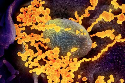
There is no vaccine or specific treatment for COVID-19, the disease caused by the severe acute respiratory syndrome coronavirus 2, or SARS-CoV-2.
Since the outbreak began in late 2019, researchers have been racing to learn more about SARS-CoV-2, which is a strain from a family of viruses known as coronavirus for their crown-like shape.
Northeastern Ûniversity] chemical engineer Thomas Webster, who specializes in developing nano-scale medicine and technology to treat diseases, is part of a contingency of scientists that are contributing ideas and technology to the Centers for Disease Control and Prevention to fight the COVID-19 outbreak.
The idea of using nanoparticles, Webster says, is that the virus behind COVID-19 consists of a structure of a similar scale as his nanoparticles. At that scale, matter is ultra-small, about ten thousand times smaller than the width of a single strand of hair.
..
This scanning electron microscope image shows SARS-CoV-2 (round gold objects) emerging from the surface of cells cultured in the lab. SARS-CoV-2, also known as 2019-nCoV, is the virus that causes COVID-19. The virus shown was isolated from a patient in the U.S. (Image: NIAID-RML)
Webster is proposing particles of similar sizes that could attach to SARS-CoV-2 viruses, disrupting their structure with a combination of infrared light treatment. That structural change would then halt the ability of the virus to survive and reproduce in the body.
“You have to think in this size range,” says Webster, Art Zafiropoulo Chair of chemical engineering at Northeastern. “In the nanoscale size range, if you want to detect viruses, if you want to deactivate them.”
Finding and neutralizing viruses with nanomedicine is at the core of what Webster and other researchers call theranostics, which focuses on combining therapy and diagnosis. Using that approach, his lab has specialized in nanoparticles to fight the microbes that cause influenza and tuberculosis.
“It’s not just having one approach to detect whether you have a virus and another approach to use it as a therapy,” he says, “but having the same particle, the same approach, for both your detection and therapy.”
…
I wish Webster good luck. As for the rest us, let’s wash our hands and keep calm.
It seems researchers at the Toronto-based (Canada), Princess Margaret Cancer Centre, have developed a new theranostic tool made of microbubbles used for imaging that are then burst into nanoparticles delivering therapeutics. From a March 30, 2015 news item on phys.org,
Biomedical researchers led by Dr. Gang Zheng at Princess Margaret Cancer Centre have successfully converted microbubble technology already used in diagnostic imaging into nanoparticles that stay trapped in tumours to potentially deliver targeted, therapeutic payloads.
The discovery, published online today [March 30, 2015] in Nature Nanotechnology, details how Dr. Zheng and his research team created a new type of microbubble using a compound called porphyrin – a naturally occurring pigment in nature that harvests light.
In the lab in pre-clinical experiments, the team used low-frequency ultrasound to burst the porphyrin containing bubbles and observed that they fragmented into nanoparticles. Most importantly, the nanoparticles stayed within the tumour and could be tracked using imaging.
“Our work provides the first evidence that the microbubble reforms into nanoparticles after bursting and that it also retains its intrinsic imaging properties. We have identified a new mechanism for the delivery of nanoparticles to tumours, potentially overcoming one of the biggest translational challenges of cancer nanotechnology. In addition, we have demonstrated that imaging can be used to validate and track the delivery mechanism,” says Dr. Zheng, Senior Scientist at the Princess Margaret and also Professor of Medical Biophysics at the University of Toronto.
Conventional microbubbles, on the other hand, lose all intrinsic imaging and therapeutic properties once they burst, he says, in a blink-of-an-eye process that takes only a minute or so after bubbles are infused into the bloodstream.
“So for clinicians, harnessing microbubble to nanoparticle conversion may be a powerful new tool that enhances drug delivery to tumours, prolongs tumour visualization and enables them to treat cancerous tumours with greater precision.”
For the past decade, Dr. Zheng’s research focus has been on finding novel ways to use heat, light and sound to advance multi-modality imaging and create unique, organic nanoparticle delivery platforms capable of transporting cancer therapeutics directly to tumours.
Interesting development, although I suspect there are many challenges yet to be met such as ensuring the microbubbles consistently arrive at their intended destination in sufficient mass to be effective both for imaging purposes and, later, as nanoparticles for drug delivery purposes.
This paper is behind a paywall but a free preview is available via ReadCube Access.
This is one of those times where I’m including the funding agencies and the ‘About’ portions of the news release,
The research published today was funded by the Canadian Institutes of Health Research (CIHR) Frederick Banting and Charles Best Canada Graduate Scholarship, the Emerging Team Grant on Regenerative Medicine and Nanomedicine co-funded by the CIHR and the Canadian Space Agency, the Natural Sciences and Engineering Research Council of Canada, the Ontario Institute for Cancer Research, the International Collaborative R&D Project of the Ministry of Knowledge Economy, South Korea, the Joey and Toby Tanenbaum/Brazilian Ball Chair in Prostate Cancer Research, the Canada Foundation for Innovation and The Princess Margaret Cancer Foundation.
About Princess Margaret Cancer Centre, University Health Network
The Princess Margaret Cancer Centre has achieved an international reputation as a global leader in the fight against cancer and delivering personalized cancer medicine. The Princess Margaret, one of the top five international cancer research centres, is a member of the University Health Network, which also includes Toronto General Hospital, Toronto Western Hospital and Toronto Rehabilitation Institute. All are research hospitals affiliated with the University of Toronto. For more information, go to http://www.theprincessmargaret.ca or http://www.uhn.ca .
I was not expecting to see South Korea or Brazil mentioned in the funding. Generally, when multiple countries are funding research, their own research institutions are also involved. As for the Princess Margaret Cancer Centre being one of the top five such centres internationally, I wonder how these rankings are determined.
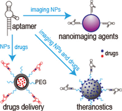
A popular concept in some circles, theranostics (sometimes called theragnostics) is a conflation of the words ‘therapeutics’ and ‘diagnostics’. A Feb. 17, 2015 news item on Nanowerk features the use of aptamers as theranostic agents,
Aptamers are composed of short RNA or single-stranded DNA sequences that, when folded into their unique 3D conformation, can bind to their targets with high specifi city and affinity. Although functionally similar to protein antibodies, oligonucleotide aptamers offer several advantages over protein antibodies in biomedical and clinical applications.
Through the enhanced permeability and retention effect, nanomedicines can improve the therapeutic index of a treatment and reduce side effects by enhancing accumulation at the disease site. However, this targets tumors passively and, thus, may not be ideal for targeted therapy.
To construct ligand-directed “active targeting” nanobased delivery systems, aptamer-equipped nanomedicines have been tested for in vitro diagnosis, in vivo imaging, targeted cancer therapy, theranostic approaches, sub-cellular molecule detection, food safety, and environmental monitoring.
There’s a very intriguing nanomedicine project in Tel Aviv, Israel. Called Nanomedicines for Personalized Theranostics, the project combines diagnostics and therapeutics for a personalized medical experience. From the Oct. 19, 2012 news item on Nanowerk (Note: I have removed a link),
Tel Aviv University [TAU] has been appointed by the Israel National Nanotechnology Initiative (INNI) to lead a consortium on “Nanomedicines for Personalized Theranostics”, a combined system of diagnostics and therapeutic treatments. This consortium of 11 laboratories will be dedicated to developing nano-sized drug delivery systems for the detection and treatment of various diseases. Eight of the labs are TAU-led, with additional participation from Hebrew University Jerusalem, Bar-Ilan University and Ben Gurion University of the Negev.
The ultimate goal is to design a new class of drugs that can destroy faulty proteins in angiogenesis-dependent diseases that involve the growth of new blood vessels from existing vessels — including cancer, infectious diseases and heart diseases — and deliver these drugs safely into the body. Beyond the academic realm, the group aims to create spin-off companies based on licensed technologies they develop, creating the basis for a thriving biotechnology industry within Israel.
The news item provides some insight into the situation in Israel,
Although considered a beacon of research and development, the field of biotechnology in Israel has suffered drawbacks, both in academia and industry. Higher salaries lure the best minds abroad, and international companies have more private capital with which to sustain businesses.
“Israel has amazing intellectual resources, but we are constantly combating budget constraints. With this project, the idea is to create future technologies built on Israeli creativity that also allow us to bring in the brightest people and better funding,” says Prof. Peer [Scientific Director Prof. Dan Peer]. While many great biotechnology ideas were born in Israel, the economic situation stymied the establishment of many more successful companies within the country, he observes. “We want to maintain the advantages that we have in the life sciences while boosting this lagging industry. Our research as part of the FTA [the Focal Technology Area within the INNI] will be a starting engine.”
Prof. Peer hopes that in two years, researchers will be able to start translating their research into practical applications.
The INNI is also working to combat “brain drain” in the academic world by giving TAU and other institutions the means to attract outstanding young researchers back home to Israel, both with funding and with the prestige of the project.
Is there a country in the world that isn’t concerned about ‘brain drain’?