Tag Archives: Thomas Fennel

Nano-fireworks

Fig. 1: Nano-fireworks in an argon nanoparticle are ignited by a moderately intense and invisible XUV laser pulse. A subsequent visible laser pulse heats the nanoparticle very efficiently, resulting in its explosion. Electrons and ions move in different directions and send out fluorescence light in various colors. Without the XUV pulse the nanoparticle would remain intact. Courtesy: Max Born Institute in Berlin (Germany)

Fig. 1: Nano-fireworks in an argon nanoparticle are ignited by a moderately intense and invisible XUV laser pulse. A subsequent visible laser pulse heats the nanoparticle very efficiently, resulting in its explosion. Electrons and ions move in different directions and send out fluorescence light in various colors. Without the XUV pulse the nanoparticle would remain intact. Courtesy: Max Born Institute in Berlin [Germany]

You can see why these have been called nano-fireworks by the researchers. Here’s more from a Jan. 23, 2016 news item on Nanowerk,

A team of researchers from the Max Born Institute in Berlin and the University of Rostock demonstrated a new way to turn initially transparent nanoparticles suddenly into strong absorbers for intense laser light and let them explode.

Intense laser pulses can transform transparent material into a plasma that captures energy of the incoming light very efficiently. Scientists from Berlin and Rostock discovered a trick to start and control this process in a way that is so efficient that it could advance methods in nanofabrication and medicine. The light-matter encounter was studied by a team of physicists from the Max Born Institute for Nonlinear Optics and Short Pulse Spectroscopy (MBI) in Berlin and from the Institute of Physics of the University of Rostock [Germany].

A Jan. 19, 2016 MBI press release, which originated the news item, offers more detail,

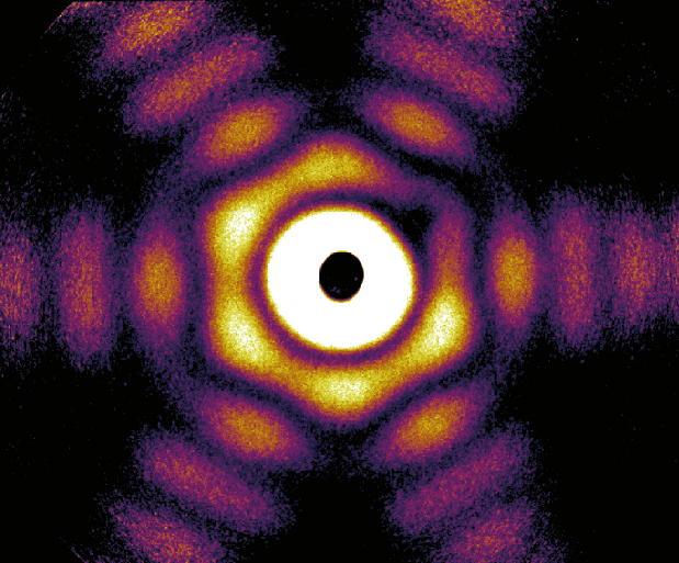
The researchers studied the interaction of intense near-infrared (NIR) laser pulses with tiny, nanometer-sized particles that contain only a few thousand Argon atoms – so-called atomic nanoclusters. The visible NIR light pulse alone can only generate a plasma if its electromagnetic waves are so strong that they rip individual atoms apart into electrons and ions. The scientists could outsmart this so-called ignition threshold by illuminating the clusters with an additional weak extreme-ultraviolet (XUV) laser pulse that is invisible to the human eye and lasts only a few femtoseconds (a femtosecond is a millionth of a billionth of a second). With this trick the researchers could “switch on” the energy transfer from the near-infrared light to the particle at unexpectedly low NIR intensities and created nano-fireworks, during which electrons, ions and colourful fluorescence light are sent out from the clusters in different directions .. . …

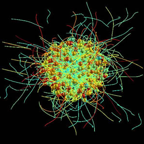
The experiments were carried out at the Max Born Institute at a 12 meter long high-harmonic generation (HHG) beamline. “The observation that argon clusters were strongly ionized even at moderate NIR laser intensities was very surprising”, explains Dr. Bernd Schütte from MBI, who conceived and performed the experiments. “Even though the additional XUV laser pulse is weak, its presence is crucial: without the XUV ignition pulse, the nanoparticles remained unaffected and transparent for the NIR light … .” Theorists around Prof. Thomas Fennel from the University of Rostock modelled the light-matter processes with numerical simulations and uncovered the origin of the observed synergy of the two laser pulses. They found that only a few seed electrons created by the ionizing radiation of the XUV pulse are sufficient to start a process similar to a snow avalanche in the mountains. The seed electrons are heated in the NIR laser light and kick out even more electrons. “In this avalanching process, the number of free electrons in the nanoparticle increases exponentially”, explains Prof. Fennel. “Eventually, the nanoscale plasma in the particles can be heated so strongly that highly charged ions are created.”

The novel concept of starting ionization avalanching with XUV light makes it possible to spatially and temporally control the strong-field ionization of nanoparticles and solids. Using HHG pulses paves the way for monitoring and controlling the ionization of nanoparticles on attosecond time scales, which is incredibly fast. One attosecond compares to a second as one second to the age of the universe. Moreover, the ignition method is expected to be applicable also to dielectric solids. This makes the concept very interesting for applications, in which intense laser pulses are used for the fabrication of nanostructures. By applying XUV pulses, a smaller focus size and therefore a higher precision could be achieved. At the same time, the overall efficiency can be improved, as NIR pulses with a much lower intensity compared to current methods could be used. In this way, novel nanolithography and nanosurgery applications may become possible in the future.

Here’s a link to and a citation for the paper,

Ionization Avalanching in Clusters Ignited by Extreme-Ultraviolet Driven Seed Electrons by Bernd Schütte, Mathias Arbeiter, Alexandre Mermillod-Blondin, Marc J. J. Vrakking, Arnaud Rouzée, and Thomas Fennel.
Phys. Rev. Lett. 116, 033001 – Published 19 January 2016 DOI:http://dx.doi.org/10.1103/PhysRevLett.116.033001

© 2016 American Physical Society

This paper is behind a paywall.

Nanoparticles in 3D courtesy of x-rays

A Feb. 4, 2015 Deutsches Elektronen-Synchrotron (DESY) press release (also on EurekAlert) announces a 3D first,

For the first time, a German-American research team has determined the three-dimensional shape of free-flying silver nanoparticles, using DESY’s X-ray laser FLASH. The tiny particles, hundreds of times smaller than the width of a human hair, were found to exhibit an unexpected variety of shapes, as the physicists from the Technical University (TU) Berlin, the University of Rostock, the SLAC National Accelerator Laboratory in the United States and from DESY report in the scientific journal Nature Communications. Besides this surprise, the results open up new scientific routes, such as direct observation of rapid changes in nanoparticles.

The press release goes on to describe the work in more detail,

“The functionality of nanoparticles is linked to their geometric form, which is often very difficult to determine experimentally,” explains Dr. Ingo Barke from the University of Rostock. “This is particularly challenging when they are present as free particles, that is, in the absence of contact with a surface or a liquid.”

The nanoparticle shape can be revealed from the characteristic way how it scatters X-ray light. Therefore, X-ray sources like DESY’s FLASH enable a sort of super microscope into the nano-world. So far, the spatial structure of nanoparticles has been reconstructed from multiple two-dimensional images, which were taken from different angles. This procedure is uncritical for particles on solid substrates, as the images can be taken from many different angles to uniquely reconstruct their three-dimensional shape.

“Bringing nanoparticles into contact with a surface or a liquid can significantly alter the particles, such that you can no longer see their actual form,” says Dr. Daniela Rupp from the TU Berlin. A free particle, however, can only be measured one time in flight before it either escapes or is destroyed by the intense X-ray light. Therefore, the scientists looked for a way to record the entire structural information of a nanoparticle with a single X-ray laser pulse.

To achieve this goal, the scientists led by Prof. Thomas Möller from the TU Berlin and Prof. Karl-Heinz Meiwes-Broer and Prof. Thomas Fennel from the University of Rostock employed a trick. Instead of taking usual small-angle scattering images, the physicists recorded the scattered X-rays in a wide angular range. “This approach virtually captures the structure from many different angles simultaneously from a single laser shot,” explains Fennel.

The researchers tested this method on free silver nanoparticles with diameters of 50 to 250 nanometres (0.00005 to 0.00025 millimetres). The experiment did not only verify the feasibility of the tricky method, but also uncovered the surprising result that large nanoparticles exhibit a much greater variety of shapes than expected.

The shape of free nanoparticles is a result of different physical principles, particularly the particles’ effort to minimize their energy. Consequently, large particles composed of thousands or millions of atoms often yield predictable shapes, because the atoms can only be arranged in a particular way to obtain an energetically favourable state.

In their experiment, however, the researchers observed numerous highly symmetrical three-dimensional shapes, including several types known as Platonic and Archimedean bodies. Examples include the truncated octahedron (a body consisting of eight regular hexagons and six squares) and the icosahedron (a body made up of twenty equilateral triangles). The latter is actually only favourable for extremely small particles consisting of few atoms, and its occurrence with free particles of this size was previously unknown. “The results show that metallic nanoparticles retain a type of memory of their structure, from the early stages of growth to a yet unexplored size range,” emphasizes Barke.

Due to the large variety of shapes, it was especially important to use a fast computational method so that the researchers were capable of mapping the shape of each individual particle. The scientists used a two-step process: the rough shape was determined first and then refined using more complex simulations on a super computer. This approach turned out to be so efficient that it could not only determine various shapes reliably, but could also differentiate between varying orientations of the same shape.

This new method for determining the three-dimensional shape and orientation of nanoparticles with a single X-ray laser shot opens up a wide spectrum of new research directions. In future projects, particles could be directly “filmed” in three dimensions during growth or during phase changes. “The ability to directly film the reaction of a nanoparticle to an intense flash of X-ray light has been a dream for many physicists – this dream could now come true, even in 3D!,” emphasises Rupp.

The researchers have provided an image showing their work,

Caption: This is a wide-angle X-ray diffraction image of a truncated twinned tetrahedra nanoparticle. Credit: Hannes Hartmann/University of Rostock

Caption: This is a wide-angle X-ray diffraction image of a truncated twinned tetrahedra nanoparticle.
Credit: Hannes Hartmann/University of Rostock

Here’s a link to and a citation for the paper,

The 3D-architecture of individual free ​silver nanoparticles captured by X-ray scattering by Ingo Barke, Hannes Hartmann, Daniela Rupp, Leonie Flückiger, Mario Sauppe, Marcus Adolph, Sebastian Schorb, Christoph Bostedt, Rolf Treusch, Christian Peltz, Stephan Bartling, Thomas Fennel, Karl-Heinz Meiwes-Broer, & Thomas Möller. Nature Communications 6, Article number: 6187 doi:10.1038/ncomms7187 Published 04 February 2015

This article is open access.