A revolutionary new type of smart window could cut window-cleaning costs in tall buildings while reducing heating bills and boosting worker productivity. Developed by University College London (UCL) with support from EPSRC, prototype samples confirm that the glass can deliver three key benefits:
Self-cleaning: The window is ultra-resistant to water, so rain hitting the outside forms spherical droplets that roll easily over the surface – picking up dirt, dust and other contaminants and carrying them away. This is due to the pencil-like, conical design of nanostructures engraved onto the glass, trapping air and ensuring only a tiny amount of water comes into contact with the surface. This is different from normal glass, where raindrops cling to the surface, slide down more slowly and leave marks behind.
Energy-saving: The glass is coated with a very thin (5-10nm) film of vanadium dioxide which during cold periods stops thermal radiation escaping and so prevents heat loss; during hot periods it prevents infrared radiation from the sun entering the building. Vanadium dioxide is a cheap and abundant material, combining with the thinness of the coating to offer real cost and sustainability advantages over silver/gold-based and other coatings used by current energy-saving windows.
Anti-glare: The design of the nanostructures also gives the windows the same anti-reflective properties found in the eyes of moths and other creatures that have evolved to hide from predators. It cuts the amount of light reflected internally in a room to less than 5 per cent – compared with the 20-30 per cent achieved by other prototype vanadium dioxide coated, energy-saving windows – with this reduction in ‘glare’ providing a big boost to occupant comfort.
This is the first time that a nanostructure has been combined with a thermochromic coating. The bio-inspired nanostructure amplifies the thermochromics properties of the coating and the net result is a self-cleaning, highly performing smart window, said Dr Ioannis Papakonstantinou of UCL.
The UCL team calculate that the windows could result in a reduction in heating bills of up to 40 per cent, with the precise amount in any particular case depending on the exact latitude of the building where they are incorporated. Windows made of the ground-breaking glass could be especially well-suited to use in high-rise office buildings.
Dr Ioannis Papakonstantinou of UCL, project leader, explains: It’s currently estimated that, because of the obvious difficulties involved, the cost of cleaning a skyscraper’s windows in its first 5 years is the same as the original cost of installing them. Our glass could drastically cut this expenditure, quite apart from the appeal of lower energy bills and improved occupant productivity thanks to less glare. As the trend in architecture continues towards the inclusion of more glass, it’s vital that windows are as low-maintenance as possible.
So, when can I buy these windows? (from the press release; Note: Links have been removed)
Discussions are now under way with UK glass manufacturers with a view to driving this new window concept towards commercialisation. The key is to develop ways of scaling up the nano-manufacturing methods that the UCL team have specially developed to produce the glass, as well as scaling up the vanadium dioxide coating process. Smart windows could begin to reach the market within around 3-5 years [emphasis mine], depending on the team’s success in securing industrial interest.
Dr Papakonstantinou says: We also hope to develop a ‘smart’ film that incorporates our nanostructures and can easily be added to conventional domestic, office, factory and other windows on a DIY [do-it-yourself] basis to deliver the triple benefit of lower energy use, less light reflection and self-cleaning, without significantly affecting aesthetics.
Professor Philip Nelson, Chief Executive of EPSRC said: This project is an example of how investing in excellent research drives innovation to produce tangible benefits. In this case the new technique could deliver both energy savings and cost reductions.
A 5-year European Research Council (ERC) starting grant (IntelGlazing) has been awarded to fabricate smart windows on a large scale and test them under realistic, outdoor environmental conditions.
…
The UCL team that developed the prototype smart window includes Mr Alaric Taylor, a PhD student in Dr Papakonstantinou’s group, and Professor Ivan Parkin from UCL’s Department of Chemistry.
I wish them good luck.
One last note, these new windows are the outcome of a 2.5 year EPSRC funded project: Biologically Inspired Nanostructures for Smart Windows with Antireflection and Self-Cleaning Properties, which ended in Sept. 2015.
This research is courtesy of the University College London (UCL) according to a Dec. 9, 2015 news item on Nanowerk,
A new test for detecting multiple explosives simultaneously has been developed by UCL scientists. The proof-of-concept sensor is designed to quickly identify and quantify five commonly used explosives in solution to help track toxic contamination in waste water and improve the safety of public spaces.
Lead researcher, Dr William Peveler (UCL Chemistry), said: “This is the first time multiple explosives have been detected using a single sensor before, demonstrating proof-of-concept for this approach. Our sensor changes colour within 10 seconds to give information about how much and what explosives are present in a sample. Following further development, we hope it will be used to quickly analyse the nature of threats and inform tailored responses.”
Dr Peveler, added: “We analysed explosives which are commonly used for industrial and military purposes to create a useful tool for environmental and security monitoring. For example, DNT is a breakdown product from landmines, and RDX and PETN have been used in terror plots in recent years as they can be hard to detect using sniffer dogs. Our test can quickly identify these compounds so we see it having a variety of applications from monitoring the waste water of munitions factories and military ranges to finding evidence of illicit activities.”
The sensor is made of quantum dots, which are tiny light-emitting particles or nanomaterials, to which explosive targeting receptors are attached. As each explosive binds to the quantum dot, it quenches the light being emitted to a different degree. The distinct changes in colour are analysed computationally in a variety of conditions to give a unique fingerprint for each compound, allowing multiple explosives to be detected with a single test.
Senior author, Professor Ivan Parkin (UCL Chemistry), said: “Our sensor is a significant step forward for multiple explosives detection. Current methods can be laborious and require expensive equipment but our test is designed to be inexpensive, fast and use a much smaller volume of sample than previously possible. Although all of these factors are important, speed and accuracy are crucial when identifying explosive compounds.”
The team plan to take it from the laboratory into the field by blind testing it with contaminated waste water samples. They also hope to improve the sensitivity of the test by tailoring the surfaces of the quantum dots. Currently, its limit is less than one part per million which the team hope to increase into the part per billion range.
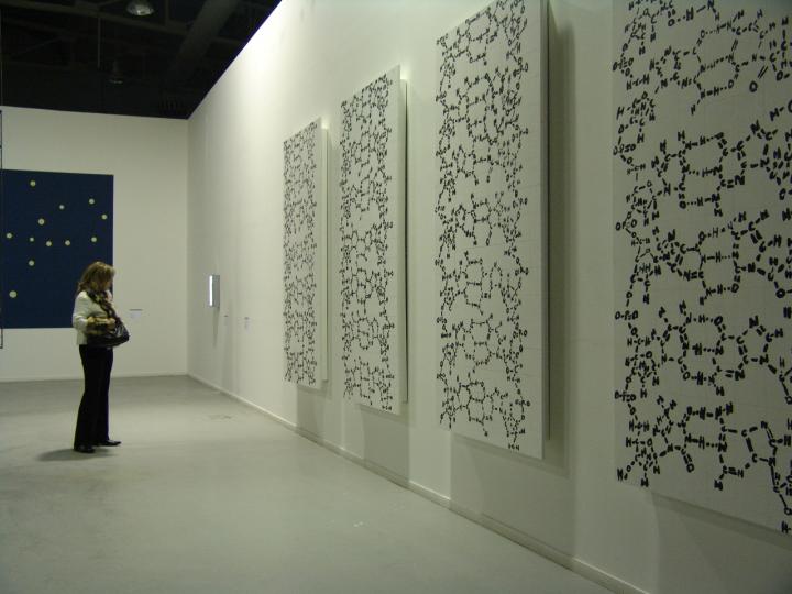
There is a fascinating set of stories about bioart designed to whet your appetite for more (*) in a Nov. 23, 2015 Cell Press news release on EurekAlert (Note: A link has been removed),
Joe Davis is an artist who works not only with paints or pastels, but also with genes and bacteria. In 1986, he collaborated with geneticist Dan Boyd to encode a symbol for life and femininity into an E. coli bacterium. The piece, called Microvenus, was the first artwork to use the tools and techniques of molecular biology. Since then, bioart has become one of several contemporary art forms (including reclamation art and nanoart) that apply scientific methods and technology to explore living systems as artistic subjects. A review of the field, published November 23, can be found in Trends in Biotechnology.
Bioart ranges from bacterial manipulation to glowing rabbits, cellular sculptures, and–in the case of Australian-British artist Nina Sellars–documentation of an ear prosthetic that was implanted onto fellow artist Stelarc’s arm. In the pursuit of creating art, practitioners have generated tools and techniques that have aided researchers, while sometimes crossing into controversy, such as by releasing invasive species into the environment, blurring the lines between art and modern biology, raising philosophical, societal, and environmental issues that challenge scientific thinking.
“Most people don’t know that bioart exists, but it can enable scientists to produce new ideas and give us opportunities to look differently at problems,” says author Ali K. Yetisen, who works at Harvard Medical School and the Wellman Center for Photomedicine, Massachusetts General Hospital. “At the same time there’s been a lot of ethical and safety concerns happening around bioart and artists who wanted to get involved in the past have made mistakes.”
Here’s a sample of Joe Davis’s work,
This photograph shows polyptich paintings by Joe Davis of his 28-mer Microvenus DNA molecule (2006 Exhibition in Greece at Athens School of Fine Arts). Credit: Courtesy of Joe Davis
The news release goes on to recount a brief history of bioart, which stretches back to 1928 and then further back into the 19th and 18th centuries,
In between experiments, Alexander Fleming would paint stick figures and landscapes on paper and in Petri dishes using bacteria. In 1928, after taking a brief hiatus from the lab, he noticed that portions of his “germ paintings,” had been killed. The culprit was a fungus, penicillin–a discovery that would revolutionize medicine for decades to come.
In 1938, photographer Edward Steichen used a chemical to genetically alter and produce interesting variations in flowering delphiniums. This chemical, colchicine, would later be used by horticulturalists to produce desirable mutations in crops and ornamental plants.
In the late 18th and early 19th centuries, the arts and sciences moved away from traditionally shared interests and formed secular divisions that persisted well into the 20th century. “Appearance of environmental art in the 1970s brought about renewed awareness of special relationships between art and the natural world,” Yetisen says.
To demonstrate how we change landscapes, American sculptor Robert Smithsonian paved a hillside with asphalt, while Bulgarian artist Christo Javacheffa (of Christo and Jeanne-Claude) surrounded resurfaced barrier islands with bright pink plastic.
These pieces could sometimes be destructive, however, such as in Ten Turtles Set Free by German-born Hans Haacke. To draw attention to the excesses of the pet trade, he released what he thought were endangered tortoises back to their natural habitat in France, but he inadvertently released the wrong subspecies, thus compromising the genetic lineages of the endangered tortoises as the two varieties began to mate.
By the late 1900s, technological advances began to draw artists’ attention to biology, and by the 2000s, it began to take shape as an artistic identity. Following Joe Davis’ transgenic Microvenus came a miniaturized leather jacket made of skin cells, part of the Tissue Culture & Art Project (initiated in 1996) by duo Oran Catts and Ionat Zurr. Other examples of bioart include: the use of mutant cacti to simulate appearance of human hair in the place of cactus spines by Laura Cinti of University College London’s C-Lab; modification of butterfly wings for artistic purposes by Marta de Menezes of Portugal; and photographs of amphibian deformation by American Brandon Ballengée.
“Bioart encourages discussions about societal, philosophical, and environmental issues and can help enhance public understanding of advances in biotechnology and genetic engineering,” says co-author Ahmet F. Coskun, who works in the Division of Chemistry and Chemical Engineering at California Institute of Technology.
Life as a Bioartist
Today, Joe Davis is a research affiliate at MIT Biology and “Artist-Scientist” at the George Church Laboratory at Harvard–a place that fosters creativity and technological development around genetic engineering and synthetic biology. “It’s Oz, pure and simple,” Davis says. “The total amount of resources in this environment and the minds that are accessible, it’s like I come to the city of Oz every day.”
But it’s not a one-way street. “My particular lab depends on thinking outside the box and not dismissing things because they sound like science fiction,” says [George M.] Church, who is also part of the Wyss Institute for Biologically Inspired Engineering. “Joe is terrific at keeping us flexible and nimble in that regard.”
For example, Davis is working with several members of the Church lab to perform metagenomics analyses of the dust that accumulates at the bottom of money-counting machines. Another project involves genetically engineering silk worms to spin metallic gold–an homage to the fairy tale of Rumpelstiltskin.
“I collaborate with many colleagues on projects that don’t necessarily have direct scientific results, but they’re excited to pursue these avenues of inquiry that they might not or would not look into ordinarily–they might try to hide it, but a lot of scientists have poetic souls,” Davis says. “Art, like science, has to describe the whole word and you can’t describe something you’re basically clueless about. The most exciting part of these activities is satiating overwhelming curiosity about everything around you.”
The number of bioartists is still small, Davis says, partly because of a lack of federal funding of the arts in general. Accessibility to the types of equipment bioartists want to experiment with can also be an issue. While Davis has partnered with labs over the past few decades, other artists affiliate themselves with community access laboratories that are run by do-it-yourself biologists. One way that universities can help is to create departmental-wide positions for bioartists to collaborate with scientists.
“In the past, there have been artists affiliated with departments in a very utilitarian way to produce figures or illustrations,” Church says. “Having someone like Joe stimulates our lab to come together in new ways and if we had more bioartists, I think thinking out of the box would be a more common thing.”
“In the era of genetic engineering, bioart will gain new meanings and annotations in social and scientific contexts,” says Yetisen. “Bioartists will surely take up new roles in science laboratories, but this will be subject to ethical criticism and controversy as a matter of course.”
Here’s a link to and a citation for the paper,
Bioart by Ali K. Yetisen, Joe Davis, Ahmet F. Coskun, George M. Church, Seok Hyun. Trends in Biotechnology, DOI: http://dx.doi.org/10.1016/j.tibtech.2015.09.011 Published Online: November 23, 2015
This paper appears to be open access.
*Removed the word ‘featured’ on Dec. 1, 2015 at 1030 hours PDT.
The story of science in the Muslim world is extraordinary, influencing science to this day, and is not well known even within its own community. The days when Muslim or Islamic scientists led the world are long gone and that is cause for concern. An Oct. 29, 2015 Malaysian Industry-Government Group for High Technology press release on EurekAlert argues that universities in Muslim countries must reinvent themselves to transform society and achieve scientific excellence,
A Task Force of international experts, formed by the Muslim World Science Initiative, today released a report [Science at Universities of the Muslim World] on the state of science at universities of the Muslim world.
…
To assess the state of science at universities of the Muslim world, the Task Force reviewed the rankings of Muslim-world’s universities globally, scientific production (number of papers published and citations), the level of spending on research and development (R&D), female participation in the scientific workforce, and other indicators.
The results were compared to those of countries deemed comparable in terms of gross domestic product (GDP) per capita, e.g. Brazil, Israel, Spain, South Africa, and South Korea.
The Task Force noted recent improvements in scientific publishing across a number of countries and a relatively healthy gender ratio among university students, even though the overall state of science in the Muslim World remains ‘poor,’ as depicted by
the disproportionately small number of Nobel Laureates
the small number of universities in top global rankings
the low spending on R&D, and
the abysmal performance of pre-university students on math and science tests
Seeking to assess if universities were the ‘main culprits’ in this sorry state of affairs, the Task Force highlighted significant challenges at the Universities of the Muslim World.
In particular, the Task Force lamented the fact that science education in most Organization of Islamic Cooperation (OIC) member countries was extremely narrow in focus and did little to enable students to think critically, especially beyond their respective domains of specialty.
The Task Force calls for broad liberal education for scientists and engineers to enable them to function effectively in addressing complex multi-disciplinary challenges that the world faces today.
The Task Force also noted that self-censorship was often practiced in the selection of topics to be taught, particularly regarding controversial subjects such as the theory of evolution.
The Task Force called for the introduction and systematic study of philosophy of science and history of the sciences of the Muslim ‘Golden Age’ and beyond for students to navigate and develop a perspective on these difficult disciplinary boundaries and overlaps. The language of instruction also created significant challenges.
Faculty members were also ill-trained to teach using cutting-edge methods such as inquiry-based science education and had little autonomy to innovate.
While the Task Force called for greater autonomy for the universities, it also emphasized that they must become meritocracies and aspire for true scientific excellence rather than playing for temporary gains in numbers or rankings. It also calls for zero tolerance on plagiarism and other forms of academic misconduct.
The Report of the Task Force includes: a foreword by the Chair, Tan Sri Zakri Abdul Hamid, the main assessment and recommendations, and individual essays written by the Task Force members on issues, including
Science, Society & the University
Are universities of the Muslim world helping spread a culture of science through society?
Should Religion Be Kept Out of the Science Classroom?
STEM Education and the Muslim Gender Divide and
The Need of Liberal Education for Science and Engineering
The Task Force is putting out an open call for universities across the Muslim world to join a voluntary Network of Excellence of Universities for Science (NEXUS), to be launched early next year.
This peer group will be managed by the task force and housed in Tan Sri Zakri’s office. NEXUS will run summer schools for university administrators, monitor the progress of reforms at participating universities, and issue a peer report card that will assess the performance of the universities in meeting milestones, thus recognizing and inspiring further improvements. True transformation will require much broader action from ministries, regulators and funding agencies, and these may be the most resistant to change.
Releasing the Report of the Task Force, Tan Sri Zakri Abdul Hamid stressed that “universities must reinvent themselves to lead the scientific reforms in the Muslim World, and as they do so they must embrace key ideas of merit and transparency, engagement with society, and pedagogical and curricular innovation.”
Professor Nidhal Guessoum, the Task Force’s Convenor, noted that “Task Force members strongly believe that the most appropriate venue for action on our recommendations is the university itself. The most essential ingredient in creating excellence in science and science teaching at a university is a realization, within a university’s highest leadership and its faculty, of the need to give up the old and dated ways, renew the purpose, and re-write the genetic code of their university.
Dr. Athar Osama, the Director of the Project noted that “the purpose of Muslim World Science Initiative is to jumpstart a dialogue within the society on critical issues at the intersection of science, society, and Islam. The Task Force has done a commendable job in laying the groundwork for a very important conversation about our universities.”
The divide between science/technology/engineering/mathematics (STEM) education and other fields of interest such as social sciences, the arts, and the humanities may be larger in the Islamic world (and to some extent reversed with humanities looking down on science) but it is a problem elsewhere, often expressed as a form of snobbery, as I alluded to in my Aug. 7, 2015 posting titled: Science snobbery and the problem of accessibility.
An Oct. 28, 2015 Nature essay about Islam, science, and the report by Nidhal Guessou and Athar Osama (two members of the Task Force; Note: Links have been removed) provides more context,
The Islamic civilization lays claim to the world’s oldest continually operational university. The University of Qarawiyyin was founded in Fes, Morocco, in ad 859, at the beginning of an Islamic Golden Age. Despite such auspicious beginnings, universities in the region are now in dire straits, as demonstrated by a report we have authored, released this week (see go.nature.com/korli3).
The 57 countries of the Muslim world — those with a Muslim-majority population, and part of the Organisation of Islamic Cooperation (OIC) — are home to nearly 25% of the world’s people. But as of 2012, they had contributed only 1.6% of the world’s patents, 6% of its academic publications, and 2.4% of the global research expenditure1, 2.
The authors note problems and at least one success with regard to curriculum (from the Nature essay; Note: Links have been removed),
Science classes themselves have serious problems. The textbooks used in OIC universities are often imported from the United States or Europe. Although the content is of a high standard, they assume a Western experience and use English or French as the language of instruction. This disadvantages many students, and creates a disconnect between their education and culture. To encourage the production of higher-quality, local textbooks and other academic material, universities need to reward staff for producing these at least as much as they do for research publication.
Some basic facts are seen as controversial, and marginalized. Evolution, for example, is usually taught only to biology students, often as “a theory”, and is rarely connected to the rest of the body of knowledge. One ongoing study has found, for example, that most Malaysian physicians and medical students reject evolution (see go.nature.com/38cswo). Evolution needs to be taught widely and shown to be compatible with Islam and its culture6. Teaching the philosophy and history of science would help, too.
The global consensus is that enquiry-based science education fosters the deepest understanding of scientific concepts and laws. But in most OIC universities, lecture-based teaching still prevails. Exceptions are rare. One is the Petroleum Institute, an engineering university in Abu Dhabi, UAE, where the faculty has created a hands-on experience with positive results on student interest and enrolment, particularly of women.
For anyone interested in the full report, it can be requested from the Muslim Science website.
One final comment, here’s the list of task force members in the Oct. 29, 2015 news release which includes someone from Mauritius (my father was born there),
Tan Sri Zakri Abdul Hamid, Science Advisor to Prime Minister of Malaysia, Chair of the Task Force on Science at the Universities of the Muslim World
Prof. Nidhal Guessoum, American University of Sharjah, UAE, Convenor of the Task Force on Science at Universities of the Muslim World
Dr. Mohammad Yusoff Sulaiman, President and CEO, MiGHT, Malaysia, Co-Convenor of the Task Force on Science at Universities of the Muslim World.
Dr. Moneef Zou’bi, Executive Director, Islamic World Academy of Science (IAS)
Prof. Adil Najam, Dean Frederick S. Pardee School of Global Studies, Boston University and former Vice Chancellor, Lahore University of Management Sciences (LUMS)
Prof. Ameenah Gurib-Fakim, Fellow of IAS, President of the Republic of Mauritius, and Professor at University of Mauritius
Prof. Mustafa El-Tayeb, President , Future University, Khartoum, Sudan
Prof. Abdur Razak Dzulkifli, President of International Association of Universities (IAU), and former Vice Chancellor USM, Malaysia
Dr. Nadia Alhasani, Dean of Student Life (formerly Dean of Women in Science and Engineering (WiSE), The Petroleum Institute, Abu Dhabi, UAE
Prof. Jamal Mimouni, Professor, University of Constantine-1, Algeria
Dr. Dato Lee Yee Cheong, Chair ISTIC Governing Board / Chair IAP SEP Global Council
Prof. Michael Reiss, Professor of Science Education, UCL Institute of Education, University College, London, Expert Advisor to the Muslim-Science.Com Task Force on Science at Universities of the Muslim World
Prof. Bruce Alberts, Professor of Biochemistry, University of California, San Francisco; President Emeritus, National Academy of Sciences, and Recipient, 2014 US Presidential Medal of Science, Expert Advisor to the Muslim-Science.Com Task Force on Science at Universities of the Muslim World
Professor Shoaib S. H. Zaidi, Professor and Dean of School of Sciences and Engineering, Habib University, Karachi
Dr. Athar Osama, Founder Muslim World Science Initiative, and Project Director of the Task Forces Project.
This show is still making its way around the world with the latest stop, as of Oct. 20, 2015, at the Library of Alexandria in Egypt.
A Jan. 21, 2010 article by Nick Higham and Margaret Ryan for BBC (British Broadcasting Corporation) news online describes some of the exhibit highlights,
From about 700 to 1700, many of history’s finest scientists and technologists were to be found in the Muslim world.
In Christian Europe the light of scientific inquiry had largely been extinguished with the collapse of the Roman empire. But it survived, and indeed blazed brightly, elsewhere.
From Moorish Spain across North Africa to Damascus, Baghdad, Persia and all the way to India, scientists in the Muslim world were at the forefront of developments in medicine, astronomy, engineering, hydraulics, mathematics, chemistry, map-making and exploration.
…
Salim Al-Hassani, a former professor of engineering at Umist (University of Manchester Institute of Science and Technology) is a moving force behind the exhibition, 1001 Inventions.
…
Visitors to the exhibition will be greeted by a 20 ft high replica of a spectacular clock designed in 1206 by the inventor Al-Jazari.
It incorporates elements from many cultures, representing the different cultural and scientific traditions which combined and flowed through the Muslim world.
The clock’s base is an elephant, representing India; inside the elephant the water-driven works of the clock derive from ancient Greece.
A Chinese dragon swings down from the top of the clock to mark the hours. At the top is a phoenix, representing ancient Egypt.
Sitting astride the elephant and inside the framework of the clock are automata, or puppets, wearing Arab turbans.
Elsewhere in the exhibition are displays devoted to water power, the spread of education (one of the world’s first universities was founded by a Muslim woman, Fatima al-Fihri), Muslim architecture and its influence on the modern world and Muslim explorers and geographers.
There is a display of 10th Century surgeons’ instruments, a lifesize model of a man called Abbas ibn Firnas, allegedly the first person to have flown with wings, and a model of the vast 100 yard-long junk commanded by the Muslim Chinese navigator, Zheng He.
The description of the exhibition items is compelling.
Science and the modern world debate (Humanism and Islam)
Yasmin Khan has written up a transcript of sorts in a Nov. 6, 2015 posting on the Guardian science blogs about a science debate (which took place Wednesday, Oct. 28, 2015 in London, UK) where Humanist and Islamic perspectives were being discussed (Note: Links have been removed),
Two important figures came head-to-head at Conway Hall, to discuss Islamic versus Humanist perspectives on science and the modern world. Jim Al-Khalili made the final public appearance of his term as president of the British Humanist Association during this stimulating, and at times provoking, debate with Ziauddin Sardar, chair of the Muslim Institute.
Al-Khalili advocated the values of the European Enlightenment, arguing that ever since the “Age of Reason” took hold during the 18th century, Humanists have looked to science instead of religion to explore and comprehend the world. Sardar upheld the view that it is the combination of faith and reason that offers a fuller understanding of the world, maintaining that it was this worldview that enabled the development of science in the Islamic golden Age.
A practising Muslim, Sardar is on an independent mission to promote rational, considered thought in interpreting the Qur’an. He explained that when he came to the UK from Pakistan, he found comfort in the familiar language of mathematics, which set him on a trajectory to train as a physicist: “God doesn’t need me, I need him. It makes me a better person and a better scientist”, he said.
…
In short, Sardar’s view is that although human knowledge at times converges with the Qur’an, the text should certainly not be treated as a scientific encyclopaedia. In support of this view, Sardar lamented the emergence of the I’jaz movement, which insists the Qur’an contains descriptions of modern scientific phenomena ranging from quantum mechanics to accurate descriptions of the stages of embryology and geology. In Sardar’s opinion, this stems from insecurity and a personal need to vindicate Islam to others.
Jim Al-Khalili agreed that ascribing literal meanings to religious texts can be perilous and that these verses should be interpreted more metaphorically. Likewise, when Einstein famously said “God does not play dice” he was using a figure of speech to acknowledge that there are things we don’t yet understand but this shouldn’t stop us from trying to find out more.
Whilst Al-Khalili is a staunch atheist, he adopts what he describes as an “accommodationist” approach in his interactions with people of religious faith: “I don’t think people who believe in God are irrational, I just don’t see a need to believe there is a purpose for why things are the way they are.” Born in Bagdad, Al-Khalili grew up in Iraq. His mother was Christian and his father was Shia, but he never heard them quarrel about religion. By the time he reached his teens he felt that he had distanced himself from needing any form of spirituality and his subsequent scientific training cemented this worldview. He asserted that his core values are empathy, humility and respect, without being driven by a reward in an afterlife: “It’s not just people of religious faith that have a moral compass – morality is what makes us human.”
I encourage you to read Khan’s piece (Nov. 6, 2015 posting) in its entirety as she provides historical and contemporary context to what seems to have been a fascinating and nuanced debate. Plus, there’s a bit of a bonus at the end where Khan is described as the producer of Sindbad Sci-Fi, a website where they are Reimagining Arab Science Fiction. From the website’s About page,
Sindbad Sci-Fi is an initiative for spurring the discovery of and engagement with Arab Science Fiction through dialogue. Our aim is to sustain a growing community of interest through brokering face-to-face and online discussion, building new partnerships and project collaborations along the way.
Many of us know and love Sindbad the sailor as the fictional sailor from the Arabian Book of OneThousand and One Nights, considered as being an early composite work of proto-science fiction and fantasy. His extraordinary voyages led him to adventures in magical places whilst meeting monsters and encountering supernatural phenomena.
Sindbad Sci-Fi is reviving Sindbad’s adventurous spirit for exploration and discovery. Join us as we continue star trekking across the Middle East, North Africa, South Asia and beyond. Together, we will boldly go where no one else has gone before!
I’m pretty sure somebody associated with this site is a Star Trek fan.
Applying the concept of superposition to photosynthesis and olfaction is not the first thought that would have occurred to me on stumbling across the European Union’s PAPETS project (Phonon-Assisted Processes for Energy Transfer and Sensing). Thankfully, a July 9, 2015 news item on Nanowerk sets the record straight (Note: A link has been removed),
Quantum physics is helping researchers to better understand photosynthesis and olfaction.
Can something be for instance in two different places at the same time? According to quantum physics, it can. More precisely, in line with the principle of ‘superposition’, a particle can be described as being in two different states simultaneously.
While it may sound like voodoo to the non-expert, superposition is based on solid science. Researchers in the PAPETS project are exploring this and other phenomena on the frontier between biology and quantum physics. Their goal is to determine the role of vibrational dynamics in photosynthesis and olfaction.
A July 7, 2015 research news article on the CORDIS website, which originated the news item, further explains (Note: A link has been removed),
Quantum effects in a biological system, namely in a photosynthetic complex, were first observed by Greg Engel and collaborators in 2007, in the USA. These effects were reproduced in different laboratories at a temperature of around -193 degrees Celsius and subsequently at ambient temperature.
‘What’s surprising and exciting is that these quantum effects have been observed in biological complexes, which are large, wet and noisy systems,’ says PAPETS project coordinator, Dr. Yasser Omar, researcher at Instituto de Telecomunicações and professor at Universidade de Lisboa [Portugal]. ‘Superposition is fragile and we would expect it to be destroyed by the environment.’
Superposition contributes to more efficient energy transport. An exciton, a quantum quasi-particle carrying energy, can travel faster along the photosynthetic complex due to the fact that it can exist in two states simultaneously. When it comes to a bifurcation it need not choose left or right. It can proceed down both paths simultaneously.
‘It’s like a maze,’ says Dr. Omar. ‘Only one door leads to the exit but the exciton can probe both left and right at the same time. It’s more efficient.’
Dr. Omar and his colleagues believe that a confluence of factors help superposition to be effected and maintained, namely the dynamics of the vibrating environment, whose role is precisely what the PAPETS project aims to understand and exploit.
Theory and experimentation meet
The theories being explored by PAPETS are also tested in experiments to validate them and gain further insights. To study quantum transport in photosynthesis, for example, researchers shoot fast laser pulses into biological systems. They then observe interference along the transport network, a signature of wavelike phenomena.
‘It’s like dropping stones into a lake,’ explains Dr. Omar. ‘You can then see whether the waves that are generated grow bigger or cancel each other when they meet.’
Applications: more efficient solar cells and odour detection
While PAPETS is essentially an exploratory project, it is generating insights that could have practical applications. PAPETS’ researchers are getting a more fundamental understanding of how photosynthesis works and this could result in the design of much more efficient solar cells.
Olfaction, the capacity to recognise and distinguish different odours, is another promising area. Experiments focus on the behaviour of Drosophila flies. So far, researchers suspect that the tunnelling of electrons associated to the internal vibrations of a molecule may be a signature of odour. Dr. Omar likens this tunnelling to a ping-pong ball resting in a bowl that goes through the side of the bowl to appear outside it.
This work could have applications in the food, water, cosmetics or drugs industries. Better artificial odour sensing could be used to detect impurities or pollution, for example.
‘Unlike seeing, hearing or touching, the sense of smell is difficult to reproduce artificially with high efficacy,’ says Dr. Omar.
The PAPETS project, involving 7 partners, runs from September 2014 to August 2016 and has a budgeted EU contribution funding of EUR 1.8 million.
You can find out more about PAPETS here. In the meantime, I found the other partners in the project (in addition to Portugal), from the PAPETS Partners webpage (Note: Links have been removed),
– Controlled Quantum Dynamics Group, Universität Ulm (UULM), Germany. PI: Martin Plenio and Susana Huelga.
– Biophysics Research Group, Vrije Universiteit Amsterdam (VUA), Netherlands. PI: Rienk van Grondelle and Roberta Croce.
– Department of Chemical Sciences, Università degli Studi di Padova (UNIPD), Italy. PI: Elisabetta Collini.
– Biomedical Sciences Research Centre “Alexander Fleming” (FLEMING), Athens, Greece. PI: Luca Turin and Efthimios M. Skoulakis.
– Biological Physics and Complex Systems Group, Centre National de la Recherche Scientifique (CNRS), Orléans, France. PI: Francesco Piazza.
– Quantum Physics of Biomolecular Processes, University College London (UCL), UK. PI: Alexandra Olaya-Castro.
Access to environmental information and use of it for environmental decision making are central pillars of environmental democracy. Yet, not much attention is paid to the question of who is producing it, and for whom? By examining the history of environmental information, since NEPA in 1969, three eras can be identified: information produced by experts, for experts (1969-1992); information produced by experts, to be shared by experts and the public (1992-2011); and finally, information produced by experts and the public to be shared by experts and the public.
Underlying these are changes in access to information, rise in levels of education and rapid change due to digital technologies. The three eras and their implication to environmental decision making will be explored, with special attention to the role of geographical information and geographical information systems and to citizen science. [emphasis mine]
Tuesday, April 29th from 10:00 – 11:30am. [EST]
I hope the speaker description and the paper being distributed on the event page mean this may be a bit more interesting to those of us curious about citizen science than is immediately apparent from the event description,
Muki (Mordechai) Haklay
Muki Haklay is a Professor of Geographic Information Science in the Department of Civil, Environmental and Geomatic Engineering, University College London. He is also the Director of the UCL Extreme Citizen Science group, which is dedicated to allowing any community, regardless of their literacy, to use scientific methods and tools to collect, analyze and interpret and use information about their area and activities.
His research interests include Public access and use of Environmental Information; Human-Computer Interaction (HCI) and Usability Engineering aspects of GIS; and Societal aspects of GIS use – in particular, participatory mapping and Citizen Science.
You can RSVP from the event page if you’re planning to attend this event in Washington, DC in person, alternatively you can watch a livestream webcast by returning to the event page on April 29, 2014 at 10 am (that will be 7 am, if you’re on the West Coast),
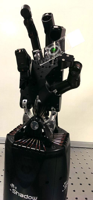
This March 27, 2014 news item on Azonano is an announcement for a new project featuring haptics and self-assembly,
NPL (UK’s National Physical Laboratory) has started a new strategic research partnership with UCL (University College of London) and MIT (Massachusetts Institute of Technology) focused on haptic-enabled sensing and micromanipulation of biological self-assembly – BioTouch.
A computer operated dexterous robotic hand holding a microscope slide with a fluorescent human cell (not to scale) embedded into a synthetic extracellular matrix. Courtesy: NPL
The news release goes on to describe the BioTouch project in more detail (Note: A link has been removed),
The project will probe sensing and application of force and related vectors specific to biological self-assembly as a means of synthetic biology and nanoscale construction. The overarching objective is to enable the re-programming of self-assembled patterns and objects by directed micro-to-nano manipulation with compliant robotic haptic control.
This joint venture, funded by the European Research Council, EPSRC and NPL’s Strategic Research Programme, is a rare blend of interdisciplinary research bringing together expertise in robotics, haptics and machine vision with synthetic and cell biology, protein design, and super- and high-resolution microscopy. The research builds on the NPL’s pioneering developments in bioengineering and imaging and world-leading haptics technologies from UCL and MIT.
Haptics is an emerging enabling tool for sensing and manipulation through touch, which holds particular promise for the development of autonomous robots that need to perform human-like functions in unstructured environments. However, the path to all such applications is hampered by the lack of a compliant interface between a predictably assembled biological system and a human user. This research will enable human directed micro-manipulation of experimental biological systems using cutting-edge robotic systems and haptic feedback.
Recently the UK government has announced ‘eight great technologies’ in which Britain is to become a world leader. Robotics, synthetic biology, regenerative medicine and advanced materials are four of these technologies for which this project serves as a merging point providing thus an excellent example of how multidisciplinary collaborative research can shape our future.
If it read this rightly, it means they’re trying to design systems where robots will work directly with materials in the labs while humans direct the robots’ actions from a remote location. My best example of this (it’s not a laboratory example) would be of a surgery where a robot actually performs the work while a human directs the robot’s actions based on haptic (touch) information the human receives from the robot. Surgeons don’t necessarily see what they’re dealing with, they may be feeling it with their fingers (haptic information). In effect, the robot’s hands become an extension of the surgeon’s hands. I imagine using a robot’s ‘hands’ would allow for less invasive procedures to be performed.
I got curious the other day about trachea transplants, a topic I first wrote about one an Aug. 22, 2011 posting featuring Andemariam Teklesenbet Beyene and wondered how things had worked out for him. For anyone who doesn’t know the story, ,
In early July 2011, there were reports of a new kind of transplant involving a body part made of a biocomposite. Andemariam Teklesenbet Beyene underwent a trachea transplant that required an artificial windpipe crafted by UK experts then flown to Sweden where Beyene’s stem cells were used to coat the windpipe before being transplanted into his body.
It is an extraordinary story not least because Beyene, a patient in a Swedish hospital planning to return to Eritrea after his PhD studies in Iceland, illustrates the international cooperation that made the transplant possible.
The scaffolding material for the artificial windpipe was developed by Professor Alex Seifalian at the University College London in a landmark piece of nanotechnology-enabled tissue engineering. Tim Harper in his July 25, 2011 posting provides more details about the scaffolding,
A team led by Professor Alexander Seifalian (UCL Division of Surgery & Interventional Science; professor of nanotechnology and regenerative medicine at University College London, UK), whose laboratories are headquartered at the Royal Free Hospital, created a glass mold of the patient’s trachea from X-ray computed tomography (CT) scans of the patient. In CT, digital geometry processing is employed to generate a 3D image of the inside of an object from a large series of 2D X-ray images taken around one single axis of rotation.
Then, they manufactured a full size y-shaped trachea scaffold at Professor Seifalian’s laboratories. The scaffold of the trachea was built using a novel nanocomposite polymer developed and patented by Professor Seifalian. Professor Seifalian worked together with Professor Paolo Macchiarini at Karolinska Institutet, Stockholm, Sweden (who also holds an Honorary appointment at UCL).
…
What I didn’t realize in 2011 was there had been some earlier transplants as Gretchen Vogel writes in her April 19, 2013 article (Trachea Transplants Test the Limits) which summarizes and critiques the work* on synthesized tracheas to date for Science magazine (the article is behind a a paywall),
More than a dozen ill people have received a bioengineered trachea seeded with stem cells during the past 5 years, but outcomes are mixed, and critics say the treatment may not do what its developers claim.
…
Although at first glance the trachea might seem like a simple tube, its thin but cartilage-reinforced walls must stand up to near-constant use as a person breathes, clears his throat, or coughs. Any transplant, therefore, has to be strong enough to withstand such pressures without collapsing. But a rigid prosthesis can rub against and damage the adjacent major blood vessels in the upper part of the chest, leaving a patient at risk for a fatal hemorrhage. At the same time, the natural blood supply for the trachea’s tissues is intricate, with vessels too small for surgeons to easily reconnect during a transplant operation. And because it is exposed to inhaled air, the wound between the implant and the remaining airway is especially vulnerable to infection.
Surgeons have tried for years to find ways around these challenges, without much success. When Castillo (Claudia Castillo, first patient to receive a trachea transplant using her own stem cells) was hospitalized in Barcelona in March 2008, Macchiarini [Paolo Macchiarini], who was then at the University of Barcelona’s Hospital Clínic, and Birchall [Martin Birchall], then at the University of Bristol in the United Kingdom, had experimented with bioengineered transplants in pigs. They would take a trachea from a pig and remove its living cells to create a so-called decellularized scaffold. They seeded this with cells from the recipient pig: bone marrow cells on the outer layer, thought to help form new cartilage, and epithelial cells on the inside, which they hoped would regrow the trachea’s lining. They allowed the cells to grow on the scaffold for several days in a bioreactor designed to provide different conditions for the two types of cells. They hoped that the decellularized scaffold would not require immunosuppressive drugs to prevent its rejection and that the seeded cells would take over the removed cells’ roles, ultimately forming a living organ.
The main difference between the 2008 Castillo operation and the 2011 Teklesenbet Beyene,operation is the scaffolding. For Castillo, they used a cadaverous** trachea where living cells were removed to create a ‘decellularized’ scaffold. For Teklesenbet Beyene, they used a nanocomposite** polymer. According to Vogel, 14 people have had the operation using either the decellularized or the nanocomposite composite polymer as the base for a new trachea. There have been some problems and deaths although Castillo who is still alive did not respond to any of Vogel’s requests for a comment . As for Teklesenbet Beyene (from the article),
His current doctor, Tomas Gudbjartsson of Landspitali University Hospital in Reykjavik, tells Science that Beyene has had several stents, but is healthy enough that he was able to complete his studies last year [2012]. The researchers have mentioned other patients in passing in several papers, but no formal reports have been published about their health, and Science has not been able to independently verify the current status of all the patients.
Both Birchall and Macchiarini have received grants for clinical trials,
In March [2013?[, Birchall received a £2.8 million ($4.3 million) grant from the United Kingdom’s Medical Research Council to conduct a trial of decellularized and stem cell–seeded upper trachea and larynx, with roughly 10 patients. Macchiarini has already completed two transplants in Russia as part of a clinical trial—funded with a $6 million grant from the Russian government—that he says should eventually enroll 20 or 25 patients. “We were allowed to do this type of transplantation only in extreme cases,” he says. “The clinical study for the first time gives us a chance to include patients who are not in such critical shape.”
Macchiarini is also the lead investigator on a 5-year, €4 million ($5.2 million) grant from the European Union to begin a clinical trial using decellularized tracheas and further develop the polymer scaffolds in large animal models. That project may need to be reorganized, however, following a legal dispute last year in Italy, where the transplants were supposed to take place—Macchiarini had a part-time position at Careggi Hospital in Florence. In September, however, Italy’s financial police accused him of attempted extortion, and briefly placed him under house arrest, for allegedly telling a patient that he could receive treatment in Germany for €150,000. Macchiarini and his lawyer say that he was simply informing the patient of possible options, not demanding payment. The main charges were soon dropped, but Macchiarini says that the charges stemmed from academic politics in Tuscany and he has severed ties with the hospital and university there. “There is no way to go back there.”
Getting back to the trachea transplants, there seems to be a major difference of opinion. While the researchers Macchiarini and Birchall have opted for human clinical trials other experts are suggesting that animal trials should be the next step for this research. I recommend reading Vogel’s article so you can fully appreciate the debate.
*’which a summary and critique of the work’ changed to ‘which summarizes and critiques the work’ for grammatical correctness on April 8, 2016.
**’pig trachea’ changed to ‘cadaverous trachea’ and ‘nanocompostie’ changed to ‘nanocomposite’ on April 19, 2016.
There are two news releases about this work which brings quantum computing a step closer to reality. I’ll start with the Nov. 15, 2013 Simon Fraser University (SFU; located in Vancouver, Canada) news release (Note: A link has been removed),,
An international team of physicists led by Simon Fraser University professor Mike Thewalt has overcome a key barrier to building practical quantum computers, taking a significant step to bringing them into the mainstream.
In their record-breaking experiment conducted on SFU’s Burnaby campus, [part of Metro Vancouver] the scientists were able to get fragile quantum states to survive in a solid material at room temperature for 39 minutes. For the average person, it may not seem like a long time, but it’s a veritable eternity to a quantum physicist.
“This opens up the possibility of truly long-term coherent information storage at room temperature,” explains Thewalt.
Quantum computers promise to significantly outperform today’s machines at certain tasks, by exploiting the strange properties of subatomic particles. Conventional computers process data stored as strings of ones or zeroes, but quantum objects are not constrained to the either/or nature of binary bits.
Instead, each quantum bit – or qubit – can be put into a superposition of both one and zero at the same time, enabling them to perform multiple calculations simultaneously. For instance, this ability to multi-task could allow quantum computers to crack seemingly secure encryption codes.
“A powerful universal quantum computer would change technology in ways that we already understand, and doubtless in ways we do not yet envisage,” says Thewalt, whose research was published in Science today.
“It would have a huge impact on security, code breaking and the transmission and storage of secure information. It would be able to solve problems which are impossible to solve on any conceivable normal computer. It would be able to model the behaviour of quantum systems, a task beyond the reach of normal computers, leading, for example, to the development of new drugs by a deeper understanding of molecular interactions.”
However, the problem with attempts to build these extraordinary number-crunchers is that superposition states are delicate structures that can collapse like a soufflé if nudged by a stray particle, such as an air molecule.
To minimize this unwanted process, physicists often cool their qubit systems to almost absolute zero (-273 C) and manipulate them in a vacuum. But such setups are finicky to maintain and, ultimately, it would be advantageous for quantum computers to operate robustly at everyday temperatures and pressures.
“Our research extends the demonstrated coherence time in a solid at room temperature by a factor of 100 – and at liquid helium temperature by a factor of 60 (from three minutes to three hours),” says Thewalt.
“These are large, significant improvements in what is possible.”
An international team including Stephanie Simmons of Oxford University report in this week’s Science a test performed as part of a project led by Mike Thewalt of Simon Fraser University, Canada, and colleagues. …
In the experiment, the team raised the temperature of a system, in which information is encoded in the nuclei of phosphorus atoms in silicon, from -269°C to 25°C and demonstrated that the superposition states survived at this balmy temperature for 39 minutes – outside of silicon the previous record for such a state’s survival at room temperature was around two seconds. [emphasis mine] The team even found that they could manipulate the qubits as the temperature of the system rose, and that they were robust enough for this information to survive being ‘refrozen’ (the optical technique used to read the qubits only works at very low temperatures).
‘Thirty-nine minutes may not seem very long but as it only takes one-hundred-thousandth of a second to flip the nuclear spin of a phosphorus ion – the type of operation used to run quantum calculations – in theory over two million operations could be applied in the time it takes for the superposition to naturally decay by 1%. Having such robust, as well as long-lived, qubits could prove very helpful for anyone trying to build a quantum computer,’ said Stephanie Simmons of Oxford University’s Department of Materials, an author of the paper.
…
The team began with a sliver of silicon doped with small amounts of other elements, including phosphorus. Quantum information was encoded in the nuclei of the phosphorus atoms: each nucleus has an intrinsic quantum property called ‘spin’, which acts like a tiny bar magnet when placed in a magnetic field. Spins can be manipulated to point up (0), down (1), or any angle in between, representing a superposition of the two other states.
The team prepared their sample at just 4°C above absolute zero (-269°C) and placed it in a magnetic field. Additional magnetic field pulses were used to tilt the direction of the nuclear spin and create the superposition states. When the sample was held at this cryogenic temperature, the nuclear spins of about 37% of the ions – a typical benchmark to measure quantum coherence – remained in their superposition state for three hours. The same fraction survived for 39 minutes when the temperature of the system was raised to 25°C.
…
There is still some work ahead before the team can carry out large-scale quantum computations. The nuclear spins of the 10 billion or so phosphorus ions used in this experiment were all placed in the same quantum state. To run calculations, however, physicists will need to place different qubits in different states. ‘To have them controllably talking to one another – that would address the last big remaining challenge,’ said Simmons.
Even for the uninitiated, going from a record of two seconds to 39 minutes has to raise an eyebrow.
ETA Nov. 18 ,2013: The University College of London has also issued a Nov. 15, 2013 news release on EurekAlert about this work. While some of this is repetitive, I think there’s enough new information to make this excerpt worthwhile,
The team even found that they could manipulate the qubits as the temperature of the system rose, and that they were robust enough for this information to survive being ‘refrozen’ (the optical technique used to read the qubits only works at very low temperatures). 39 minutes may not sound particularly long, but since it only takes a tiny fraction of a second to run quantum computations by flipping the spin of phosphorus ions (electrically charged phosphorus atoms), many millions of operations could be carried out before a system like this decays.
“This opens up the possibility of truly long-term coherent information storage at room temperature,” said Mike Thewalt (Simon Fraser University), the lead researcher in this study.
The team began with a sliver of silicon doped with small amounts of other elements, including phosphorus. They then encoded quantum information in the nuclei of the phosphorus atoms: each nucleus has an intrinsic quantum property called ‘spin’, which acts like a tiny bar magnet when placed in a magnetic field. Spins can be manipulated to point up (0), down (1), or any angle in between, representing a superposition of the two other states.
The team prepared their sample at -269 °C, just 4 degrees above absolute zero, and placed it in a magnetic field. They used additional magnetic field pulses to tilt the direction of the nuclear spin and create the superposition states. When the sample was held at this cryogenic temperature, the nuclear spins of about 37 per cent of the ions – a typical benchmark to measure quantum coherence – remained in their superposition state for three hours. The same fraction survived for 39 minutes when the temperature of the system was raised to 25 °C.
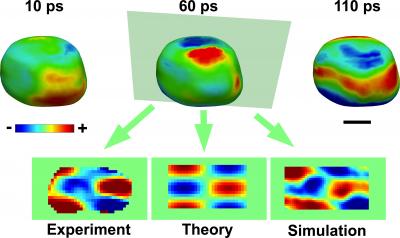
Folks at the London Centre for Nanotechnology (at the University College of London) have released a film made with a pioneering 3D imaging technique that shows how gold nanocrystals vibrate. From the May 23, 2013 news release on EurekAlert,
A billon-frames-per-second film has captured the vibrations of gold nanocrystals in stunning detail for the first time.
The film, which was made using 3D imaging pioneered at the London Centre for Nanotechnology (LCN) at UCL [University College of London], reveals important information about the composition of gold. The findings are published in the journal Science.
Jesse Clark, from the LCN and lead author of the paper said: “Just as the sound quality of a musical instrument can provide great detail about its construction, so too can the vibrations seen in materials provide important information about their composition and functions.”
“It is absolutely amazing that we are able to capture snapshots of these nanoscale motions and create movies of these processes. This information is crucial to understanding the response of materials after perturbation. “
Caption: The acoustic phonons can be visualized on the surface as regions of contraction (blue) and expansion (red). Also shown are two-dimensional images comparing the experimental results with theory and molecular dynamics simulation. The scale bar is 100 nanometers. Credit: Jesse Clark/UCL
Here are more details from the news release,
Scientists found that the vibrations were unusual because they start off at exactly the same moment everywhere inside the crystal. It was previously expected that the effects of the excitation would travel across the gold nanocrystal at the speed of sound, but they were found to be much faster, i.e., supersonic.
The new images support theoretical models for light interaction with metals, where energy is first transferred to electrons, which are able to short-circuit the much slower motion of the atoms.
The team carried out the experiments at the SLAC National Accelerator Laboratory using a revolutionary X-ray laser called the “Linac Coherent Light Source”. The pulses of X-rays are extremely short (measured in femtoseconds, or quadrillionths of a second), meaning they are able to freeze all motion of the atoms in any sample, leaving only the electrons still moving.
However, the X-ray pulses are intense enough that the team was able to take single snapshots of the vibrations of the gold nanocrystals they were examining. The vibration was started with a short pulse of infrared light.