Tag Archives: University of Antwerp

Non-invasive chemical imaging reveals the Eykian Lamb of God’s secrets

Left: color image after the 1950s treatment. The ears of the Eyckian Lamb were revealed after removal of the 16th-century overpaint obscuring the background. Right: color image after the 2019 treatment that removed all of the 16th century overpaint, revealing the face of the Eyckian Lamb. The dotted lines indicate the outline of the head before removal of 16th-century overpaint.

Fascinating, yes? More than one person has noticed that the ‘new’ lamb is “disturbingly human-like.” First, here’s more about this masterpiece and the technology used to restore it (from a July 29, 2020 University of Antwerp (Belgium) press release (Note: I do not have all of the figures (images) described in this press release embedded here),

Two non-invasive chemical imaging modalities were employed to help understand the changes made over time to the Lamb of God, the focal point of the Ghent Altarpiece (1432) by Hubert and Jan Van Eyck. Two major results were obtained: a prediction of the facial features of the Lamb of God that had been hidden beneath non-original overpaint dating from the 16th century (and later), and evidence for a smaller earlier version of the Lamb’s body with a more naturalistic build. These non-invasive imaging methods, combined with analysis of paint cross-sections and magnified examination of the paint surface, provide objective chemical evidence to understand the extent of overpaints and the state of preservation of the original Eyckian paint underneath.

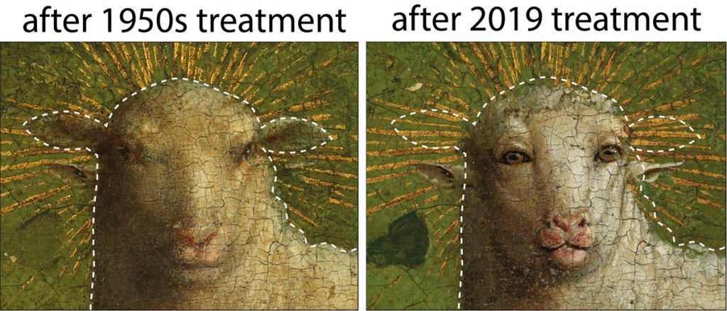
The Ghent Altarpiece is one of the founding masterpieces of Western European painting. The central panel, The Adoration of the Lamb, represents the sacrifice of Christ with a depiction of the Lamb of God standing on an altar, blood pouring into a chalice. During conservation treatment and technical analysis in the 1950s, conservators recognized the presence of overpaint on the Lamb and the surrounding area. But based on the evidence available at that time, the decision was made to remove only the overpaint obscuring the background immediately surrounding the head. As a result, the ears of the Eyckian Lamb were uncovered, leading to the surprising effect of a head with four ears (Figure 1).

Figure 1: Left: Color image after the 1950s treatment. The ears of the Eyckian Lamb were revealed after removal of the 16th century overpaint obscuring the background. (© Lukasweb.be – Art in Flanders vzw). Right: Color image after the 2019 treatment that removed all of the 16th century overpaint, revealing the face of the Eyckian Lamb. The dotted lines indicate the outline of the head before removal of 16th century overpaint. (© Lukasweb.be – Art in Flanders vzw).

During the recent conservation treatment of the central panel, chemical images collected before 16th century overpaint was removed revealed facial features that predicted aspects of the Eyckian Lamb, at that time still hidden below the overpaint. For example, the smaller, v-shaped nostrils of the Eyckian Lamb are situated higher than the 16th century nose, as revealed in the map for mercury, an element associated with the red pigment vermilion (Figure 2, red arrow). A pair of eyes that look forward, slightly lower than the 16th century eyes, can be seen in a false-color hyperspectral infrared reflectance image (Figure 2, right). This image also shows dark preparatory underdrawing lines that define pursed lips, and in conjunction with the presence of mercury in this area, suggest the Eyckian lips were more prominent. In addition, the higher, 16th century ears were painted over the gilded rays of the halo (Figure 2, yellow rays). Gilding is typically the artist’s final touch when working on a painting, which supports the conclusion that the lower set of ears is the Eyckian original. Collectively, these facial features indicate that, compared to the 16th century restorer’s overpainted face, the Eyckian Lamb has a smaller face with a distinctive expression.

Figure 2: Left: Colorized composite elemental map showing the distribution of gold (in yellow), mercury (in red), and lead (in white). The red arrow indicates the position of the Eyckian Lamb’s nostrils. (University of Antwerp). Right: Composite false-color infrared reflectance image (blue – 1000 nm, green – 1350 nm, red – 1650 nm) shows underdrawn lines indicating the position of facial features of the Eyckian Lamb, including forward-gazing eyes, the division between the lips, and the jawline. (National Gallery of Art, Washington). The dotted lines indicate the outline of the head before removal of 16th century overpaint.

The new imaging also revealed previously unrecognized revisions to the size and shape of the Lamb’s body: a more naturalistically shaped Lamb, with slightly sagging back, more rounded hindquarters and a smaller tail. The artist’s underdrawing lines used to lay out the design of the smaller shape can be seen in the false-color hyperspectral infrared reflectance image (Figure 3, lower left, white arrows). Mathematical processing of the reflectance dataset to emphasize a spectral feature associated with the pigment lead white resulted in a clearer image of the smaller Lamb (Figure 3, lower right). Differences between the paint handling of the fleece in the initial small Lamb and the revised area of the larger Lamb also were found upon reexamination of the x-radiograph and the paint surface under the microscope.

Figure 3: Upper left: Color image before removal of all 16th century overpaint. (© Lukasweb.be – Art in Flanders vzw). Upper right: Color image after removal of all 16th century overpaint. (© Lukasweb.be – Art in Flanders vzw). Lower left: False-color infrared reflectance image (blue – 1000 nm, green – 1350 nm, red – 1650 nm) reveals underdrawing lines that denote the smaller hindquarters of the initial Lamb. Lower right: Map derived from processing the infrared reflectance image cube showing the initial Lamb with a slightly sagging back, more rounded hindquarters and a smaller tail. Brighter areas of the map indicate stronger absorption from the -OH group associated with one of the forms of lead white. (National Gallery of Art, Washington).

During the conservation treatment completed in 2019, decisions were informed by well-established conservation methods (high-resolution color photography, X-radiography, infrared imaging, paint sample analysis) as well as the new chemical imaging. In this way, the conservation treatment uncovered the smaller face of the Eyckian Lamb, with forward-facing eyes that meet the viewer’s gaze. Only overpaints that could be identified as being later additions dating from the 16th century onward were carefully and safely removed. The body of the Lamb, however, has not changed. The material evidence indicates that the lead white paint layer used to define the larger squared-off hindquarters was applied prior to the 16th century restoration, but because analysis at the present time cannot definitively establish whether this was a change by the original artist(s) or a very early restoration or alteration by another artist, the enlarged contour of the Lamb was left untouched.

Chemical imaging technologies can be used to build confidence about the state of preservation of original paint and help guide the decision to remove overpaint. Combined with the conservators’ thorough optical examination, informed by years of experience and insights derived from paint cross-sections, chemical imaging methods will no doubt be central to ongoing interdisciplinary research, helping to resolve long-standing art-historical issues on the Ghent Altarpiece as well as other works of art. These findings were obtained by researchers from the University of Antwerp using macroscale X-ray fluorescence imaging and researchers at the National Gallery of Art, Washington using infrared reflectance imaging spectroscopy, interpreted in conjunction with the observations of the scientists and the conservation team from The Royal Institute for Cultural Heritage (KIK-IRPA), Brussels.

A January 22, 2020 British Broadcasting Corporation (BBC) online news item notes some of the response to the ‘new’ lamb (Note: A link has been removed),

Restorers found that the central panel of the artwork, known as the Adoration of the Mystic Lamb, had been painted over in the 16th Century.

Another artist had altered the Lamb of God, a symbol for Jesus depicted at the centre of the panel.

Now conservationists have stripped away the overpaint, revealing the lamb’s “intense gaze” and “large frontal eyes”.

Hélène Dubois, the head of the restoration project, told the Art Newspaper the original lamb had a more “intense interaction with the onlookers”.

She said the lamb’s “cartoonish” depiction, which departs from the painting’s naturalistic style, required more research.

The lamb has been described as having an “alarmingly humanoid face” with “penetrating, close-set eyes, full pink lips and flared nostrils” by the Smithsonian Magazine.

These features are “eye-catching, if not alarmingly anthropomorphic”, said the magazine, the official journal of the Smithsonian Institution.

There was also disbelief on social media, where the lamb was called “disturbing” by some and compared to an “alien creature”. Some said they felt it would have been better to not restore the lamb’s original face.

The painter of the panel, Jan Van Eyck, is considered to be one of the most technical and talented artists of his generation. However, it is widely believed that The Ghent Altarpiece was started by his brother, Hubert Van Eyck.

Taken away by the Nazis during World War Two and Napoleon’s troops in the 1700s, the altarpiece is thought to be one of the most frequently stolen artworks of all time.

If you have the time, do read the January 22, 2020 BBC news item in its entirety as it conveys more of the controversy.

Jennifer Ouellette’s July 29, 2020 article for Ars Technica delves further into the technical detail along with some history about this particular 21st Century restoration. The conservators and experts used artificial intelligence (AI) to assist.

Here’s a link to and a citation for the paper,

Dual mode standoff imaging spectroscopy documents the painting process of the Lamb of God in the Ghent Altarpiece by J. and H. Van Eyck by Geert Van der Snickt, Kathryn A. Dooley, Jana Sanyova, Hélène Dubois, John K. Delaney, E. Melanie Gifford, Stijn Legrand, Nathalie Laquiere and Koen Janssens. Science Advances 29 Jul 2020: Vol. 6, no. 31, eabb3379 DOI: 10.1126/sciadv.abb3379

This paper is open access.

Neurons and graphene carpets

I don’t entirely grasp the carpet analogy. Actually, I have no why they used a carpet analogy but here’s the June 12, 2018 ScienceDaily news item about the research,

A work led by SISSA [Scuola Internazionale Superiore di Studi Avanzati] and published on Nature Nanotechnology reports for the first time experimentally the phenomenon of ion ‘trapping’ by graphene carpets and its effect on the communication between neurons. The researchers have observed an increase in the activity of nerve cells grown on a single layer of graphene. Combining theoretical and experimental approaches they have shown that the phenomenon is due to the ability of the material to ‘trap’ several ions present in the surrounding environment on its surface, modulating its composition. Graphene is the thinnest bi-dimensional material available today, characterised by incredible properties of conductivity, flexibility and transparency. Although there are great expectations for its applications in the biomedical field, only very few works have analysed its interactions with neuronal tissue.

A June 12, 2018 SISSA press release (also on EurekAlert), which originated the news item, provides more detail,

A study conducted by SISSA – Scuola Internazionale Superiore di Studi Avanzati, in association with the University of Antwerp (Belgium), the University of Trieste and the Institute of Science and Technology of Barcelona (Spain), has analysed the behaviour of neurons grown on a single layer of graphene, observing a strengthening in their activity. Through theoretical and experimental approaches the researchers have shown that such behaviour is due to reduced ion mobility, in particular of potassium, to the neuron-graphene interface. This phenomenon is commonly called ‘ion trapping’, already known at theoretical level, but observed experimentally for the first time only now. “It is as if graphene behaves as an ultra-thin magnet on whose surface some of the potassium ions present in the extra cellular solution between the cells and the graphene remain trapped. It is this small variation that determines the increase in neuronal excitability” comments Denis Scaini, researcher at SISSA who has led the research alongside Laura Ballerini.

The study has also shown that this strengthening occurs when the graphene itself is supported by an insulator, like glass, or suspended in solution, while it disappears when lying on a conductor. “Graphene is a highly conductive material which could potentially be used to coat any surface. Understanding how its behaviour varies according to the substratum on which it is laid is essential for its future applications, above all in the neurological field” continues Scaini, “considering the unique properties of graphene it is natural to think for example about the development of innovative electrodes of cerebral stimulation or visual devices”.

It is a study with a double outcome. Laura Ballerini comments as follows: “This ‘ion trap’ effect was described only in theory. Studying the impact of the ‘technology of materials’ on biological systems, we have documented a mechanism to regulate membrane excitability, but at the same time we have also experimentally described a property of the material through the biology of neurons.”

Dexter Johnson in a June 13, 2018 posting, on his Nanoclast blog (on the IEEE [Institute of Electrical and Electronics Engineers] website), provides more context for the work (Note: Links have been removed),

While graphene has been tapped to deliver on everything from electronics to optoelectronics, it’s a bit harder to picture how it may offer a key tool for addressing neurological damage and disorders. But that’s exactly what researchers have been looking at lately because of the wonder material’s conductivity and transparency.

In the most recent development, a team from Europe has offered a deeper understanding of how graphene can be combined with neurological tissue and, in so doing, may have not only given us an additional tool for neurological medicine, but also provided a tool for gaining insights into other biological processes.

“The results demonstrate that, depending on how the interface with [single-layer graphene] is engineered, the material may tune neuronal activities by altering the ion mobility, in particular potassium, at the cell/substrate interface,” said Laura Ballerini, a researcher in neurons and nanomaterials at SISSA.

Ballerini provided some context for this most recent development by explaining that graphene-based nanomaterials have come to represent potential tools in neurology and neurosurgery.

“These materials are increasingly engineered as components of a variety of applications such as biosensors, interfaces, or drug-delivery platforms,” said Ballerini. “In particular, in neural electrode or interfaces, a precise requirement is the stable device/neuronal electrical coupling, which requires governing the interactions between the electrode surface and the cell membrane.”

This neuro-electrode hybrid is at the core of numerous studies, she explained, and graphene, thanks to its electrical properties, transparency, and flexibility represents an ideal material candidate.

In all of this work, the real challenge has been to investigate the ability of a single atomic layer to tune neuronal excitability and to demonstrate unequivocally that graphene selectively modifies membrane-associated neuronal functions.

I encourage you to read Dexter’s posting as it clarifies the work described in the SISSA press release for those of us (me) who may fail to grasp the implications.

Here’s a link to and a citation for the paper,

Single-layer graphene modulates neuronal communication and augments membrane ion currents by Niccolò Paolo Pampaloni, Martin Lottner, Michele Giugliano, Alessia Matruglio, Francesco D’Amico, Maurizio Prato, Josè Antonio Garrido, Laura Ballerini, & Denis Scaini. Nature Nanotechnology (2018) DOI: https://doi.org/10.1038/s41565-018-0163-6 Published online June 13, 2018

This paper is behind a paywall.

All this brings to mind a prediction made about the Graphene Flagship and the Human Brain Project shortly after the European Commission announced in January 2013 that each project had won funding of 1B Euros to be paid out over a period of 10 years. The prediction was that scientists would work on graphene/human brain research.

Generating power from polluted air

I have no idea how viable this concept might be but it is certainly appealing, From a May 8, 2017 news item on Nanowerk (Note: A link has been removed),

Researchers from the University of Antwerp and KU Leuven (University of Leuven), Belgium, have succeeded in developing a process that purifies air and, at the same time, generates power. The device must only be exposed to light in order to function (ChemSusChem, “Harvesting Hydrogen Gas from Air Pollutants with an Unbiased Gas Phase Photoelectrochemical Cell”).

Caption: The new device must only be exposed to light in order to purify air and generate power. Credit: UAntwerpen and KU Leuven

A May 8, 2017 University of Leuven press release (also on EurekAlert), which originated the news item, describes this nifty research in slightly more detail,

“We use a small device with two rooms separated by a membrane,” explains Professor Sammy Verbruggen (UAntwerp/KU Leuven). “Air is purified on one side, while on the other side hydrogen gas is produced from a part of the degradation products. This hydrogen gas can be stored and used later as fuel, as is already being done in some hydrogen buses, for example.”

In this way, the researchers respond to two major social needs: clean air and alternative energy production. The heart of the solution lies at the membrane level, where the researchers use specific nanomaterials. “These catalysts are capable of producing hydrogen gas and breaking down air pollution,” explains Professor Verbruggen. “In the past, these cells were mostly used to extract hydrogen from water. We have now discovered that this is also possible, and even more efficient, with polluted air.”

It seems to be a complex process, but it is not: the device must only be exposed to light. The researchers’ goal is to be able to use sunlight, as the processes underlying the technology are similar to those found in solar panels. The difference here is that electricity is not generated directly, but rather that air is purified while the generated power is stored as hydrogen gas.

“We are currently working on a scale of only a few square centimetres. At a later stage, we would like to scale up our technology to make the process industrially applicable. We are also working on improving our materials so we can use sunlight more efficiently to trigger the reactions. “

Here’s a link to and a citation for the paper,

Harvesting Hydrogen Gas from Air Pollutants with an Unbiased Gas Phase Photoelectrochemical Cell. by  Prof. Dr. Sammy W. Verbruggen, Myrthe Van Hal1, Tom Bosserez, Dr. Jan Rongé, Dr. Birger Hauchecorne, Prof. Dr. Johan A. Martens, and Prof. Dr. Silvia Lenaerts. ChemSusChem Volume 10, Issue 7, pages 1413–1418, April 10, 2017 DOI: 10.1002/cssc.201601806 Version of Record online: 6 MAR 2017

© 2017 Wiley-VCH Verlag GmbH & Co. KGaA, Weinheim

This paper is behind a paywall.

Starry gold and silica Janus particles

A Feb. 11, 2014 news item on phys.org features a joint Basque/Belgian research collaboration on a Janus-type particle useful for future biomedical applications,

Researchers from the Basque centre CIC biomaGUNE and the University of Antwerp (Belgium) have designed nanoparticles with one half formed of gold branches and the other of silicon oxide. They are a kind of Janus particle, so-called in honour of the Roman god with two faces, which could be used in phototherapy in the future to treat tumours.

The Feb. 11, 2014 Platforma SINC news release on the Alpha Galileo website, which originated the news item, elaborates on the Janus myth and on the research,

In Roman mythology, Janus was the god of gates, doors, beginnings and transitions between the past and the future. In fact, the first month of the year, January (from the Latin, ianuarĭus), bears his name. This deity was characterised by his profile of two faces, something which has inspired scientists, when naming their chemical designs with two clearly distinct components.

Now, a team of researchers from CIC biomaGUNE in San Sebastian, together with colleagues from the Belgian University of Antwerp, have created Janus particles of nanometric size. They are constituted by silicon oxide on one side and gold points on the other.

Here’s an image of the ‘starry’ particles supplied by the researchers,

Two examples of nanostars with one silicon oxide face (bluish) and another with golden branches (yellow). / Credit: Liz-Marzán et al.

Two examples of nanostars with one silicon oxide face (bluish) and another with golden branches (yellow). / Credit: Liz-Marzán et al.

The news release goes on to describe the ‘starry’ particles in more detail,

As Luis Liz-Marzán, the main author of this study published in the journal ‘Chemical Communications’, explains to SINC: “These nanostars have optical and electronic properties determined largely by their small dimensions and their morphology.”

The researchers have come up with techniques to mould the sharp gold points from nanoparticles of this metal, such that very intense electric fields can be generated on the gold points using light.

“Our research is basic science, but these fields are used in processes of ultrasensitive detection to identify negligible quantities of molecules that can be absorbed on the gold face as contaminants or biomarkers that indicate the presence of a disease,” says Liz-Marzán.

Another possible application is phototherapy, the object of which is to kill malignant cells using heat, in this case induced by lighting the gold points. The oxide face would be used to join the nanostars to specific biological receptors that would take them to the damaged cells and only to these, so that the metal part can exercise its therapeutic or diagnostic function.

These nanoparticles are produced in various stages. First, golden nanospheres are produced by the chemical reduction of a salt from the precious metal. Then, two different organic compounds are added on opposite sides of the particle in order to give them distinct affinity due to the silicon oxide. In this way, the oxide covers only one part and the other remains uncovered in order to let the golden points grow.

Here’s a link to and a citation for the research paper,

Denis Rodríguez-Fernández, Thomas Altantzis, Hamed Heidari, Sara Bals, Luis M. Liz-Marzán. “A protecting group approach toward synthesis of Au–silica Janus nanostars”. Chemical Communications 50: 79-81, 2014. DOI: 10.1039/C3CC47531J.

This article is available for free but you need to register with the website first or log in if you have already registered.

I last wrote about a Janus particle in an Aug. 13, 2009 post about research at Duke University.