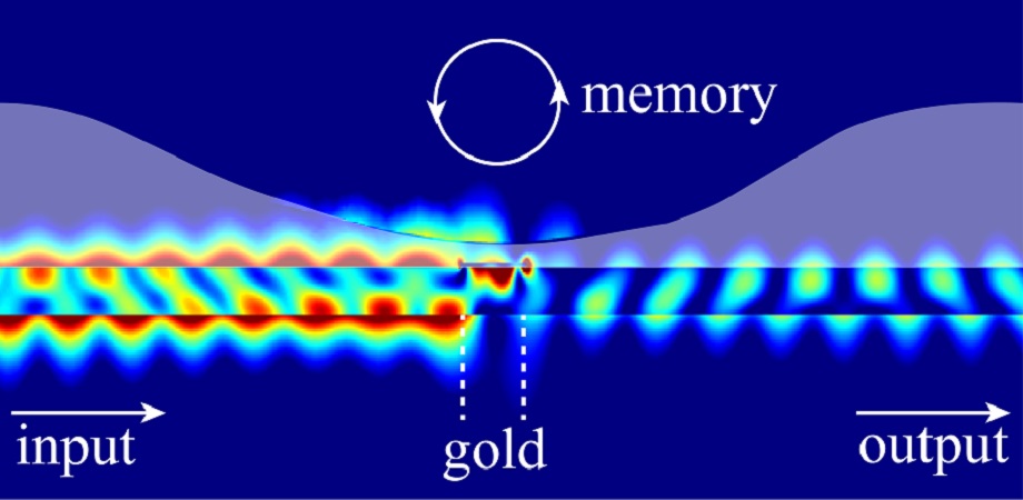
Simulation result of light affecting liquid geometry, which in turn affects reflection and transmission properties of the optical mode, thus constituting a two-way light–liquid interaction mechanism. The degree of deformation serves as an optical memory allowing to store the power magnitude of the previous optical pulse and use fluid dynamics to affect the subsequent optical pulse at the same actuation region, thus constituting an architecture where memory is part of the computation process. Credit: Gao et al., doi 10.1117/1.AP.4.4.046005
This is a fascinating approach to neuromorphic (brainlike) computing and given my recent post (August 29, 2022) about human cells being incorporated into computer chips, it’s part o my recent spate of posts about neuromorphic computing. From a July 25, 2022 news item on phys.org,
Sunlight sparkling on water evokes the rich phenomena of liquid-light interaction, spanning spatial and temporal scales. While the dynamics of liquids have fascinated researchers for decades, the rise of neuromorphic computing has sparked significant efforts to develop new, unconventional computational schemes based on recurrent neural networks, crucial to supporting wide range of modern technological applications, such as pattern recognition and autonomous driving. As biological neurons also rely on a liquid environment, a convergence may be attained by bringing nanoscale nonlinear fluid dynamics to neuromorphic computing.
Researchers from University of California San Diego recently proposed a novel paradigm where liquids, which usually do not strongly interact with light on a micro- or nanoscale, support significant nonlinear response to optical fields. As reported in Advanced Photonics, the researchers predict a substantial light–liquid interaction effect through a proposed nanoscale gold patch operating as an optical heater and generating thickness changes in a liquid film covering the waveguide.
The liquid film functions as an optical memory. Here’s how it works: Light in the waveguide affects the geometry of the liquid surface, while changes in the shape of the liquid surface affect the properties of the optical mode in the waveguide, thus constituting a mutual coupling between the optical mode and the liquid film. Importantly, as the liquid geometry changes, the properties of the optical mode undergo a nonlinear response; after the optical pulse stops, the magnitude of liquid film’s deformation indicates the power of the previous optical pulse.
Remarkably, unlike traditional computational approaches, the nonlinear response and the memory reside at the same spatial region, thus suggesting realization of a compact (beyond von-Neumann) architecture where memory and computational unit occupy the same space. The researchers demonstrate that the combination of memory and nonlinearity allow the possibility of “reservoir computing” capable of performing digital and analog tasks, such as nonlinear logic gates and handwritten image recognition.
Their model also exploits another significant liquid feature: nonlocality. This enables them to predict computation enhancement that is simply not possible in solid state material platforms with limited nonlocal spatial scale. Despite nonlocality, the model does not quite achieve the levels of modern solid-state optics-based reservoir computing systems, yet the work nonetheless presents a clear roadmap for future experimental works aiming to validate the predicted effects and explore intricate coupling mechanisms of various physical processes in a liquid environment for computation.
Using multiphysics simulations to investigate coupling between light, fluid dynamics, heat transport, and surface tension effects, the researchers predict a family of novel nonlinear and nonlocal optical effects. They go a step further by indicating how these can be used to realize versatile, nonconventional computational platforms. Taking advantage of a mature silicon photonics platform, they suggest improvements to state-of-the-art liquid-assisted computation platforms by around five orders magnitude in space and at least two orders of magnitude in speed.
This breakthrough in neuromorphic (brainlike) computing is being attributed to the pandemic (COVID-19) according to a September 3, 2021 news item on phys.org,
Isaac Newton’s groundbreaking scientific productivity while isolated from the spread of bubonic plague is legendary. University of California San Diego physicists can now claim a stake in the annals of pandemic-driven science.
A team of UC San Diego [University of California San Diego] researchers and colleagues at Purdue University have now simulated the foundation of new types of artificial intelligence computing devices that mimic brain functions, an achievement that resulted from the COVID-19 pandemic lockdown. By combining new supercomputing materials with specialized oxides, the researchers successfully demonstrated the backbone of networks of circuits and devices that mirror the connectivity of neurons and synapses in biologically based neural networks.
As bandwidth demands on today’s computers and other devices reach their technological limit, scientists are working towards a future in which new materials can be orchestrated to mimic the speed and precision of animal-like nervous systems. Neuromorphic computing based on quantum materials, which display quantum-mechanics-based properties, allow scientists the ability to move beyond the limits of traditional semiconductor materials. This advanced versatility opens the door to new-age devices that are far more flexible with lower energy demands than today’s devices. Some of these efforts are being led by Department of Physics Assistant Professor Alex Frañó and other researchers in UC San Diego’s Quantum Materials for Energy Efficient Neuromorphic Computing (Q-MEEN-C), a Department of Energy-supported Energy Frontier Research Center.
“In the past 50 years we’ve seen incredible technological achievements that resulted in computers that were progressively smaller and faster—but even these devices have limits for data storage and energy consumption,” said Frañó, who served as one of the PNAS paper’s authors, along with former UC San Diego chancellor, UC president and physicist Robert Dynes. “Neuromorphic computing is inspired by the emergent processes of the millions of neurons, axons and dendrites that are connected all over our body in an extremely complex nervous system.”
As experimental physicists, Frañó and Dynes are typically busy in their laboratories using state-of-the-art instruments to explore new materials. But with the onset of the pandemic, Frañó and his colleagues were forced into isolation with concerns about how they would keep their research moving forward. They eventually came to the realization that they could advance their science from the perspective of simulations of quantum materials.
“This is a pandemic paper,” said Frañó. “My co-authors and I decided to study this issue from a more theoretical perspective so we sat down and started having weekly (Zoom-based) meetings. Eventually the idea developed and took off.”
The researchers’ innovation was based on joining two types of quantum substances—superconducting materials based on copper oxide and metal insulator transition materials that are based on nickel oxide. They created basic “loop devices” that could be precisely controlled at the nano-scale with helium and hydrogen, reflecting the way neurons and synapses are connected. Adding more of these devices that link and exchange information with each other, the simulations showed that eventually they would allow the creation of an array of networked devices that display emergent properties like an animal’s brain.
Like the brain, neuromorphic devices are being designed to enhance connections that are more important than others, similar to the way synapses weigh more important messages than others.
“It’s surprising that when you start to put in more loops, you start to see behavior that you did not expect,” said Frañó. “From this paper we can imagine doing this with six, 20 or a hundred of these devices—then it gets exponentially rich from there. Ultimately the goal is to create a very large and complex network of these devices that will have the ability to learn and adapt.”
With eased pandemic restrictions, Frañó and his colleagues are back in the laboratory, testing the theoretical simulations described in the PNAS [Proceedings of the National Academy of Sciences] paper with real-world instruments.
The gene editing tool .CRISPR (clustered regularly interspaced short palindromic repeats) does feature in this story but only as a minor character; the real focus is on the delivery system. From a February 9, 2018 news item on Nanowerk ()Note: A link has been removed),
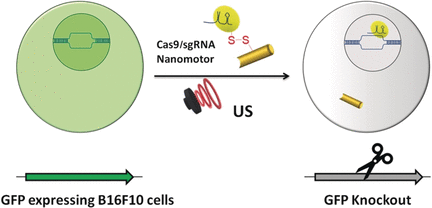
In cancer research, the “Cas-9–sgRNA” complex is an effective genomic editing tool, but its delivery across the cell membrane to the target (tumor) genome has not yet been satisfactorily solved.
American and Danish scientists have now developed an active nanomotor for the efficient transport, delivery, and release of this gene scissoring system. As detailed in their paper in the journal Angewandte Chemie (“Active Intracellular Delivery of a Cas9/sgRNA Complex Using Ultrasound-Propelled Nanomotors”), their nanovehicle is propelled towards its target by ultrasound.
The publisher (Wiley) has made this image illustrating the work available,
Genomic engineering as a promising cancer therapeutic approach has experienced a tremendous surge since the discovery of the adaptive bacterial immune defense system “CRISPR” and its potential as a gene editing tool over a decade ago. Engineered CRISPR systems for gene editing now contain two main components, a single guide RNA or sgRNA and Cas-9 nuclease. While the sgRNA guides the nuclease to the specified gene sequence, Cas-9 nuclease performs its editing with surgical efficiency. However, the delivery of the large machinery to the target genome is still problematic. The authors of the Angewandte Chemie study, Liangfang Zhang and Joseph Wang from the University of California San Diego, and their colleagues now propose ultrasound-propelled gold nanowires as an active transport/release vehicle for the Cas9-sgRNA complex over the membrane.
Gold nanowires may cross a membrane passively, but thanks to their rod- or wirelike asymmetric shape, active motion can be triggered by ultrasound. “The asymmetric shape of the gold nanowire motor, given by the fabrication process, is essential for the acoustic propulsion,” the authors remarked. They assembled the vehicle by attaching the Cas-9 protein/RNA complex to the gold nanowire through sulfide bridges. These reduceable linkages have the advantage that inside the tumor cell, the bonds would be broken by glutathione, a natural reducing compound enriched in tumor cells. The Cas9-sgRNA would be released and sent to the nucleus to do its editing work, for, example, the knockout of a gene.
As a test system, the scientists monitored the suppression of fluorescence emitted by green fluorescence protein expressing melanoma B16F10 cells. Ultrasound was applied for five minutes, which accelerated the nanomotor carrying the Cas9-sgRNA complex across the membrane, accelerating it even inside the cell, as the authors noted. Moreover, they observed their Cas9-sgRNA complex effectively suppressing fluorescence with only tiny concentrations of the complex needed.
Thus, both the effective use of an acoustic nanomotor as an active transporter and the small payload needed for efficient gene knockout are intriguing results of the study. The simplicity of the system, which uses only few and readily available components, is another remarkable achievement.
Here’s a link to and a citation for the paper,
Active Intracellular Delivery of a Cas9/sgRNA Complex Using Ultrasound-Propelled Nanomotors by Malthe Hansen-Bruhn, Dr. Berta Esteban-Fernández de Ávila, Dr. Mara Beltrán-Gastélum, Prof. Jing Zhao, Dr. Doris E. Ramírez-Herrera, Pavimol Angsantikul, Prof. Kurt Vesterager Gothelf, Prof. Liangfang Zhang, and Prof. Joseph Wang. Angewandte Chemie International Edition Vol. 57 Issue 7 DOI: 10.1002/anie.201713082 Version of Record online: 6 FEB 2018
Researcher Bor-Kai Hsiung’s work has graced this blog before but the topic was tarantulas and their structural colour. This time, it’s all about Australian peacock spiders and their structural colour according to a December 22, 2017 news item on ScienceDaily,
Even if you are arachnophobic, you probably have seen pictures or videos of Australian peacock spiders (Maratus spp.). These tiny spiders are only 1-5 mm long but are famous for their flamboyant courtship displays featuring diverse and intricate body colorations, patterns, and movements.
The spiders extremely large anterior median eyes have excellent color vision and combine with their bright colors to make peacock spiders cute enough to cure most people of their arachnophobia. But these displays aren’t just pretty to look at, they also inspire new ways for humans to produce color in technology.
One species of peacock spider — the rainbow peacock spider (Maratus robinsoni) is particularly neat, because it showcases an intense rainbow iridescent signal in males’ courtship displays to the females. This is the first known instance in nature of males using an entire rainbow of colors to entice females. Dr. Bor-Kai Hsiung led an international team of researchers from the US (UAkron, Cal Tech, UC San Diego, UNL [University of Nebraska-Lincoln]), Belgium (Ghent University), Netherlands (UGroningen), and Australia to discover how rainbow peacock spiders produce this unique multi-color iridescent signal.
Using a diverse array of research techniques, including light and electron microscopy, hyperspectral imaging, imaging scatterometry, nano 3D printing and optical modeling, the team found the origin of this intense rainbow iridescence emerged from specialized abdominal scales of the spiders. These scales have an airfoil-like microscopic 3D contour with nanoscale diffraction grating structures on the surface.
The interaction between the surface nano-diffraction grating and the microscopic curvature of the scales enables separation and isolation of light into its component wavelengths at finer angles and smaller distances than are possible with current manmade engineering technologies.
Inspiration from these super iridescent scales can be used to overcome current limitations in spectral manipulation, and to further reduce the size of optical spectrometers for applications where fine-scale spectral resolution is required in a very small package, notably instruments on space missions, or wearable chemical detection systems. And it could have a wide array of implications to fields ranging from life sciences and biotechnologies to material sciences and engineering.
Here’s a video of an Australian rainbow peacock spider,
Here’s more from the YouTube description published on April 13, 2017 by Peacockspiderman,
Scenes of Maratus robinsoni, a spider Peter Robinson discovered and David Hill and I named it after him in 2012. You can read our description on pages 36-41 in Peckhamia 103.2, which can be downloaded from the Peckhamia website http://peckhamia.com/peckhamia_number…. This is one of the two smallest species of peacock spider (2.5 mm long) and the only spider we know of in which colour changes occur every time it moves, this video was created to document this. Music: ‘Be Still’ by Johannes Bornlöf licensed through my MCN ‘Brave Bison’ from ‘Epidemic Sound’ For licensing inquiries please contact Brave Bison licensing@bravebison.io
The University of California at San Diego also published a December 22, 2017 news release about this work, which covers some of the same ground while providing a few new tidbits of information,
Brightly colored Australian peacock spiders (Maratus spp.) captivate even the most arachnophobic viewers with their flamboyant courtship displays featuring diverse and intricate body colorations, patterns, and movements – all packed into miniature bodies measuring less than five millimeters in size for many species. However, these displays are not just pretty to look at. They also inspire new ways for humans to produce color in technology.
One species of peacock spider – the rainbow peacock spider (Maratus robinsoni) – is particularly impressive, because it showcases an intense rainbow iridescent signal in males’ courtship displays to females. This is the first known instance in nature of males using an entire rainbow of colors to entice females to mate. But how do males make their rainbows? A new study published in Nature Communications looked to answer that question.
Figuring out the answers was inherently interdisciplinary so Bor-Kai Hsiung, a postdoctoral scholar at Scripps Institution of Oceanography at the University of California San Diego, assembled an international team that included biologists, physicists and engineers. Starting while he was a Ph.D. student at The University of Akron under the mentorship of Todd Blackledge and Matthew Shawkey, the team included researchers from UA, Scripps Oceanography, California Institute of Technology, and University of Nebraska-Lincoln, the University of Ghent in Belgium, University of Groningen in Netherlands, and Australia to discover how rainbow peacock spiders produce this unique iridescent signal.
The team investigated the spider’s photonic structures using techniques that included light and electron microscopy, hyperspectral imaging, imaging scatterometry and optical modeling to generate hypotheses about how the spider’s scale generate such intense rainbows. The team then used cutting-edge nano 3D printing to fabricate different prototypes to test and validate their hypotheses. In the end, they found that the intense rainbow iridescence emerged from specialized abdominal scales on the spiders. These scales combine an airfoil-like microscopic 3D contour with nanoscale diffraction grating structures on the surface. It is the interaction between the surface nano-diffraction grating and the microscopic curvature of the scales that enables separation and isolation of light into its component wavelengths at finer angles and smaller distances than are possible with current engineering technologies.
“Who knew that such a small critter would create such an intense iridescence using extremely sophisticated mechanisms that will inspire optical engineers,” said Dimitri Deheyn, Hsuing’s advisor at Scripps Oceanography and a coauthor of the study.
For Hsiung, the finding wasn’t quite so unexpected.
“One of the main questions that I wanted to address in my Ph.D. dissertation was ‘how does nature modulate iridescence?’ From a biomimicry perspective, to fully understand and address a question, one has to take extremes from both ends into consideration. I purposefully chose to study these tiny spiders with intense iridescence after having investigated the non-iridescent blue tarantulas,” said Hsiung.
The mechanism behind these tiny rainbows may inspire new color technology, but would not have been discovered without research combining basic natural history with physics and engineering, the researchers said.
“Nanoscale 3D printing allowed us to experimentally validate our models, which was really exciting,” said Shawkey. “We hope that these techniques will become common in the future.”
“As an engineer, what I found fascinating about these spider structural colors is how these long evolved complex structures can still outperform human engineering,” said Radwanul Hasan Siddique, a postdoctoral scholar at Caltech and study coauthor. “Even with high-end fabrication techniques, we could not replicate the exact structures. I wonder how the spiders assemble these fancy structural patterns in the first place!”
Inspiration from these super iridescent spider scales can be used to overcome current limitations in spectral manipulation, and to reduce the size of optical spectrometers for applications where fine-scale spectral resolution is required in a very small package, notably instruments on space missions, or wearable chemical detection systems.
In the end, peacock spiders don’t just produce nature’s smallest rainbows.They could also have implications for a wide array of fields ranging from life sciences and biotechnologies to material sciences and engineering.
Before citing the paper and providing a link, here’s a story by Robert F. Service for Science magazine about attempts to capitalize on ‘spider technology’, in this case spider silk,
The hype over spider silk has been building since 1710. That was the year François Xavier Bon de Saint Hilaire, president of the Royal Society of Sciences in Montpellier, France, wrote to his colleagues, “You will be surpriz’d to hear, that Spiders make a Silk, as beautiful, strong and glossy, as common Silk.” Modern pitches boast that spider silk is five times stronger than steel yet more flexible than rubber. If it could be made into ropes, a macroscale web would be able to snare a jetliner.
The key word is “if.” Researchers first cloned a spider silk gene in 1990, in hopes of incorporating it into other organisms to produce the silk. (Spiders can’t be farmed like silkworms because they are territorial and cannibalistic.) Today, Escherichia coli bacteria, yeasts, plants, silkworms, and even goats have been genetically engineered to churn out spider silk proteins, though the proteins are often shorter and simpler than the spiders’ own. Companies have managed to spin those proteins into enough high-strength thread to produce a few prototype garments, including a running shoe by Adidas and a lightweight parka by The North Face. But so far, companies have struggled to mass produce these supersilks.
Some executives say that may finally be about to change. One Emeryville, California-based startup, Bolt Threads, says it has perfected growing spider silk proteins in yeast and is poised to turn out tons of spider silk thread per year. In Lansing, Michigan, Kraig Biocraft Laboratories says it needs only to finalize negotiations with silkworm farms in Vietnam to produce mass quantities of a combination spider/silkworm silk, which the U.S. Army is now testing for ballistics protection. …
I encourage you to read Service’s article in its entirety if the commercialization prospects for spider silk interest you as it includes gems such as this,
Spider silk proteins are already making their retail debut—but in cosmetics and medical devices, not high-strength fibers. AMSilk grows spider silk proteins in E. coli and dries the purified protein into powders or mixes it into gels, for use as additives for personal care products, such as moisture-retaining skin lotions. The silk proteins supposedly help the lotions form a very smooth, but breathable, layer over the skin. Römer says the company now sells tons of its purified silk protein ingredients every year.
…
Finally, here’s a citation for and a link to the paper about Australian peacock spiders and nanophotonics,
Rainbow peacock spiders inspire miniature super-iridescent optics by Bor-Kai Hsiung, Radwanul Hasan Siddique, Doekele G. Stavenga, Jürgen C. Otto, Michael C. Allen, Ying Liu, Yong-Feng Lu, Dimitri D. Deheyn, Matthew D. Shawkey, & Todd A. Blackledge. Nature Communications 8, Article number: 2278 (2017) doi:10.1038/s41467-017-02451-x Published online: 22 December 2017
When was the last time you saw a six-year old or a twelve-year old attend a political candidates’ meeting or vote in an election? Sadly, most creative science outreach in Canada is aimed at children and teenagers in the misbegotten belief that adults don’t matter and ‘youth are the future’. There are three adult science outreach scenarios although they didn’t tend to be particularly creative. (1) Should scientists feel hard done by elected representatives, they reach out to other adults for support. (2) Should those other adults become disturbed by any scientific or technological ‘advance’ then scientific experts will arrive to explain why that’s wrong. (3) Should the science enterprise want money, then a call goes out (see my May 12, 2017 posting about the Canada Science and Technology Museums Corporation gala and, yes, they were a bit creative about it).
I am oversimplifying the situation but not by much especially if one considers two upcoming national Canadian science events: Science Rendezvous which is a day-long (May 13, 2017) cross country science event taking place during while the Science Odyssey holds a 10-day (May 12 – 2017) cross country science event. The two groups arranged their events separately and then decided to coordinate their efforts. Science Odyssey is a rebranding of the Canada Science and Technology Week organized by the federal government for at least two decades and which was held (until 2016) in the fall of each year. Science Rendezvous (About page) was launched in Toronto in 2008 (University of Toronto, Ryerson University, York University and the University of Ontario Institute of Technology (UOIT)).
Regardless, both events are clearly aimed at children (and families).
I’m not suggesting that exciting science outreach for children should be curtailed. Let’s expand the efforts to9 include the adult and senior populations too.
IMAGE: Philip Guo surveyed adults between the ages of 60 and 85 who were users of pythontutor.com and learning how to code. They were mix of retired, semi-retired and still working. Credit: Courtesy Philip Guo, UC San Diego
Philip Guo caught the coding bug in high school, at a fairly typical age for a Millennial. Less typical is that the UC San Diego cognitive scientist is now eager to share his passion for programming with a different demographic. And it’s not one you’re thinking of – it’s not elementary or middle school-aged kids. Guo wants to get adults age 60 and up.
In the first known study of older adults learning computer programming, Guo outlines his reasons: People are living and working longer. This is a growing segment of the population, and it’s severely underserved by learn-to-code intiatives, which usually target college students and younger. Guo wants to change that. He would like this in-demand skill to become more broadly accessible.
“Computers are everywhere, and digital literacy is becoming more and more important,” said Guo, assistant professor in the Department of Cognitive Science, who is also affiliated with UC San Diego’s Design Lab and its Department of Computer Science and Engineering. “At one time, 1,000 years ago, most people didn’t read or write – just some monks and select professionals could do it. I think in the future people will need to read and write in computer language as well. In the meantime, more could benefit from learning how to code.”
Guo’s study was recently awarded honorable mention by the world’s leading organization in human-computer interaction, ACM SIGCHI. Guo will present his findings at the group’s premier international conference, CHI, in May [2017].
When prior human-computer interaction studies have focused on older adults at all, Guo said, it has been mostly as consumers of new technology, of social networking sites like Facebook, say, or ride-sharing services. While a few have investigated the creation of content, like blogging or making digital music, these have involved the use of existing apps. None, to his knowledge, have looked at older adults as makers of entirely new software applications, so he set out to learn about their motivations, their frustrations and if these provided clues to design opportunities.
The Study
For his study, Guo surveyed users of pythontutor.com. A web-based education tool that Guo started in 2010, Python Tutor helps those learning to program visualize their work. Step by step, it displays what a computer is doing with each line of code that it runs. More than 3.5 million people in more than 180 countries have now used Python Tutor, including those around the world taking MOOCs (massive open online courses). Despite its legacy name, the tool helps people supplement their studies not only of the Python programming language but also Java, JavaScript, Ruby, C and C++, all of which are commonly used to teach programing. The users of Python Tutor represent a wide range of demographic groups.
Guo’s survey included 504 people between the ages of 60 and 85, from 52 different countries. Some were retired and semi-retired while others were still working.
What Guo discovered: Older adults are motivated to learn programming for a number of reasons. Some are age-related. They want to make up for missed opportunities during youth (22 percent) and keep their brains “challenged, fresh and sharp” as they age (19 percent). A few (5 percent) want to connect with younger family members.
Reasons not related to age include seeking continuing education for a current job (14 percent) and wanting to improve future job prospects (9 percent). A substantial group is in it just for personal enrichment: 19 percent to implement a specific hobby project idea, 15 percent for fun and entertainment, and 10 percent out of general interest.
Interestingly, 8 percent said they wanted to learn to teach others.
Topping the list of frustrations for older students of coding was bad pedagogy. It was mentioned by 21 percent of the respondents and ranged from the use of jargon to sudden spikes in difficulty levels. Lack of real-world relevance came up 6 percent of the time. A 74-year-old retired physician wrote: “Most [tutorials] are offered by people who must know how to program but don’t seem to have much training in teaching.”
Other frustrations included a perceived decline in cognitive abilities (12 percent) and no human contact with tutors and peers (10 percent).
The study’s limitations are tied in part to the instrument – self-reporting on an online survey – and in part to the survey respondents themselves. Most hailed from North America and other English-speaking nations. Most, 84 percent, identified themselves as male; this stat is consistent with other surveys of online learning, especially in math and science topics. There was a diverse array of occupations reported, but the majority of those surveyed were STEM professionals, managers and technicians. These learners, Guo said, likely represent “early adopters” and “the more technology-literate and self-motivated end of the general population.” He suggests future studies look both at in-person learning and at a broader swath of the public. But he expects the lessons learned from this group will generalize.
The Implications
Based on this first set of findings and using a learner-centered design approach, Guo proposes tailoring computer-programming tools and curricula specifically for older learners. He notes, for example, that many of his respondents seemed to take pride in their years and in their tech-savvy, so while it may be good to advertise products as targeting this age group, they should not appear patronizing. It might make sense to reframe lessons as brain-training games, like Lumosity, now popular among the older set.
Just as it’s key to understand who the learners are so is understanding where they have trouble. Repetition and frequent examples might be good to implement, as well as more in-person courses or video-chat-based workshops, Guo said, which may lead to improvements in the teaching of programming not just for older adults but across the board.
Context matters, too. Lessons are more compelling when they are put into domains that people personally care about. And Guo recommends coding curricula that enable older adults to tell their life stories or family histories, for example, or write software that organizes health information or assists care-givers.
Guo, who is currently working on studies to extend coding education to other underrepresented groups, advocates a computing future that is fully inclusive of all ages.
“There are a number of social implications when older adults have access to computer programming – not merely computer literacy,” he said. “These range from providing engaging mental stimulation to greater gainful employment from the comfort of one’s home.”
By moving the tech industry away from its current focus on youth, Guo argues, we all stand to gain. [emphasis mine]
Guo joined the UC San Diego cognitive science faculty in 2016 after two years as an assistant professor at the University of Rochester. He received his bachelor’s and master’s degrees in computer science from MIT in 2006 and his Ph.D. from Stanford in 2012. Before becoming a professor, he built online learning tools as a software engineer at Google and a research scientist at edX. He also blogs, vlogs and podcasts at http://pgbovine.net/
When was the last time you heard about a ‘coding’ camp for adults and seniors in Canada? Also,, ask yourself if after you’d reached a certain age (40? 50? more? less?) you’d feel welcome at the Science Rendezvous events (without a child in tow), Science Odyssey events (without a child in tow), or the May 17, 2017 National Science and Innovation Gala in Ottawa (from my May 12, 2017 posting “It would seem the only person over the age of 30 who’s expected to attend is the CBC host, Heather Hiscox.”)?
If I read the news release rightly (keep scrolling), this particular artificial retina does not require a device outside the body (e.g. specially developed eyeglasses) to capture an image to be transmitted to the implant. This new artificial retina captures the image directly.
The announcement of a new artificial retina is made in a March 13, 2017 news item on Nanowerk (Note: A link has been removed),
A team of engineers at the University of California San Diego and La Jolla-based startup Nanovision Biosciences Inc. have developed the nanotechnology and wireless electronics for a new type of retinal prosthesis that brings research a step closer to restoring the ability of neurons in the retina to respond to light. The researchers demonstrated this response to light in a rat retina interfacing with a prototype of the device in vitro.
They detail their work in a recent issue of the Journal of Neural Engineering (“Towards high-resolution retinal prostheses with direct optical addressing and inductive telemetry”). The technology could help tens of millions of people worldwide suffering from neurodegenerative diseases that affect eyesight, including macular degeneration, retinitis pigmentosa and loss of vision due to diabetes
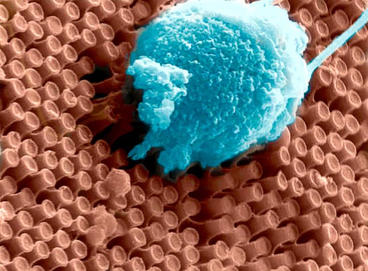
Caption: These are primary cortical neurons cultured on the surface of an array of optoelectronic nanowires. Here a neuron is pulling the nanowires, indicating the the cell is doing well on this material. Credit: UC San Diego
Despite tremendous advances in the development of retinal prostheses over the past two decades, the performance of devices currently on the market to help the blind regain functional vision is still severely limited–well under the acuity threshold of 20/200 that defines legal blindness.
“We want to create a new class of devices with drastically improved capabilities to help people with impaired vision,” said Gabriel A. Silva, one of the senior authors of the work and professor in bioengineering and ophthalmology at UC San Diego. Silva also is one of the original founders of Nanovision.
The new prosthesis relies on two groundbreaking technologies. One consists of arrays of silicon nanowires that simultaneously sense light and electrically stimulate the retina accordingly. The nanowires give the prosthesis higher resolution than anything achieved by other devices–closer to the dense spacing of photoreceptors in the human retina. The other breakthrough is a wireless device that can transmit power and data to the nanowires over the same wireless link at record speed and energy efficiency.
One of the main differences between the researchers’ prototype and existing retinal prostheses is that the new system does not require a vision sensor outside of the eye [emphasis mine] to capture a visual scene and then transform it into alternating signals to sequentially stimulate retinal neurons. Instead, the silicon nanowires mimic the retina’s light-sensing cones and rods to directly stimulate retinal cells. Nanowires are bundled into a grid of electrodes, directly activated by light and powered by a single wireless electrical signal. This direct and local translation of incident light into electrical stimulation makes for a much simpler–and scalable–architecture for the prosthesis.
The power provided to the nanowires from the single wireless electrical signal gives the light-activated electrodes their high sensitivity while also controlling the timing of stimulation.
“To restore functional vision, it is critical that the neural interface matches the resolution and sensitivity of the human retina,” said Gert Cauwenberghs, a professor of bioengineering at the Jacobs School of Engineering at UC San Diego and the paper’s senior author.
Wireless telemetry system
Power is delivered wirelessly, from outside the body to the implant, through an inductive powering telemetry system developed by a team led by Cauwenberghs.
The device is highly energy efficient because it minimizes energy losses in wireless power and data transmission and in the stimulation process, recycling electrostatic energy circulating within the inductive resonant tank, and between capacitance on the electrodes and the resonant tank. Up to 90 percent of the energy transmitted is actually delivered and used for stimulation, which means less RF wireless power emitting radiation in the transmission, and less heating of the surrounding tissue from dissipated power.
The telemetry system is capable of transmitting both power and data over a single pair of inductive coils, one emitting from outside the body, and another on the receiving side in the eye. The link can send and receive one bit of data for every two cycles of the 13.56 megahertz RF signal; other two-coil systems need at least 5 cycles for every bit transmitted.
Proof-of-concept test
For proof-of-concept, the researchers inserted the wirelessly powered nanowire array beneath a transgenic rat retina with rhodopsin P23H knock-in retinal degeneration. The degenerated retina interfaced in vitro with a microelectrode array for recording extracellular neural action potentials (electrical “spikes” from neural activity).
The horizontal and bipolar neurons fired action potentials preferentially when the prosthesis was exposed to a combination of light and electrical potential–and were silent when either light or electrical bias was absent, confirming the light-activated and voltage-controlled responsivity of the nanowire array.
The wireless nanowire array device is the result of a collaboration between a multidisciplinary team led by Cauwenberghs, Silva and William R. Freeman, director of the Jacobs Retina Center at UC San Diego, UC San Diego electrical engineering professor Yu-Hwa Lo and Nanovision Biosciences.
A path to clinical translation
Freeman, Silva and Scott Thorogood, have co-founded La Jolla-based Nanovision Biosciences, a partner in this study, to further develop and translate the technology into clinical use, with the goal of restoring functional vision in patients with severe retinal degeneration. Animal tests with the device are in progress, with clinical trials following.
“We have made rapid progress with the development of the world’s first nanoengineered retinal prosthesis as a result of the unique partnership we have developed with the team at UC San Diego,” said Thorogood, who is the CEO of Nanovision Biosciences.
An artificial blood vessel network that could lead the way to regenerating biologically-based blood vessel networks has been printed in 3D at the University of California at San Diego (UCSD) according to a March 2, 2017 news item on ScienceDaily,
Nanoengineers at the University of California San Diego have 3D printed a lifelike, functional blood vessel network that could pave the way toward artificial organs and regenerative therapies.
The new research, led by nanoengineering professor Shaochen Chen, addresses one of the biggest challenges in tissue engineering: creating lifelike tissues and organs with functioning vasculature — networks of blood vessels that can transport blood, nutrients, waste and other biological materials — and do so safely when implanted inside the body.
Researchers from other labs have used different 3D printing technologies to create artificial blood vessels. But existing technologies are slow, costly and mainly produce simple structures, such as a single blood vessel — a tube, basically. These blood vessels also are not capable of integrating with the body’s own vascular system.
“Almost all tissues and organs need blood vessels to survive and work properly. This is a big bottleneck in making organ transplants, which are in high demand but in short supply,” said Chen, who leads the Nanobiomaterials, Bioprinting, and Tissue Engineering Lab at UC San Diego. “3D bioprinting organs can help bridge this gap, and our lab has taken a big step toward that goal.”
Chen’s lab has 3D printed a vasculature network that can safely integrate with the body’s own network to circulate blood. These blood vessels branch out into many series of smaller vessels, similar to the blood vessel structures found in the body. The work was published in Biomaterials.
Chen’s team developed an innovative bioprinting technology, using their own homemade 3D printers, to rapidly produce intricate 3D microstructures that mimic the sophisticated designs and functions of biological tissues. Chen’s lab has used this technology in the past to create liver tissue and microscopic fish that can swim in the body to detect and remove toxins.
Researchers first create a 3D model of the biological structure on a computer. The computer then transfers 2D snapshots of the model to millions of microscopic-sized mirrors, which are each digitally controlled to project patterns of UV light in the form of these snapshots. The UV patterns are shined onto a solution containing live cells and light-sensitive polymers that solidify upon exposure to UV light. The structure is rapidly printed one layer at a time, in a continuous fashion, creating a 3D solid polymer scaffold encapsulating live cells that will grow and become biological tissue.
“We can directly print detailed microvasculature structures in extremely high resolution. Other 3D printing technologies produce the equivalent of ‘pixelated’ structures in comparison and usually require sacrificial materials and additional steps to create the vessels,” said Wei Zhu, a postdoctoral scholar in Chen’s lab and a lead researcher on the project.
And this entire process takes just a few seconds — a vast improvement over competing bioprinting methods, which normally take hours just to print simple structures. The process also uses materials that are inexpensive and biocompatible.
Chen’s team used medical imaging to create a digital pattern of a blood vessel network found in the body. Using their technology, they printed a structure containing endothelial cells, which are cells that form the inner lining of blood vessels.
The entire structure fits onto a small area measuring 4 millimeters × 5 millimeters, 600 micrometers thick (as thick as a stack containing 12 strands of human hair).
Researchers cultured several structures in vitro for one day, then grafted the resulting tissues into skin wounds of mice. After two weeks, the researchers examined the implants and found that they had successfully grown into and merged with the host blood vessel network, allowing blood to circulate normally.
Chen noted that the implanted blood vessels are not yet capable of other functions, such as transporting nutrients and waste. “We still have a lot of work to do to improve these materials. This is a promising step toward the future of tissue regeneration and repair,” he said.
Moving forward, Chen and his team are working on building patient-specific tissues using human induced pluripotent stem cells, which would prevent transplants from being attacked by a patient’s immune system. And since these cells are derived from a patient’s skin cells, researchers won’t need to extract any cells from inside the body to build new tissue. The team’s ultimate goal is to move their work to clinical trials. “It will take at least several years before we reach that goal,” Chen said.
A Jan. 18, 2017 news item on Nanowerk announces research into hair strength from the University of California at San Diego (UCSD or UC San Diego),
In a new study, researchers at the University of California San Diego investigate why hair is incredibly strong and resistant to breaking. The findings could lead to the development of new materials for body armor and help cosmetic manufacturers create better hair care products.
Hair has a strength to weight ratio comparable to steel. It can be stretched up to one and a half times its original length before breaking. “We wanted to understand the mechanism behind this extraordinary property,” said Yang (Daniel) Yu, a nanoengineering Ph.D. student at UC San Diego and the first author of the study.
“Nature creates a variety of interesting materials and architectures in very ingenious ways. We’re interested in understanding the correlation between the structure and the properties of biological materials to develop synthetic materials and designs — based on nature — that have better performance than existing ones,” said Marc Meyers, a professor of mechanical engineering at the UC San Diego Jacobs School of Engineering and the lead author of the study.
In a study published online in Dec. in the journal Materials Science and Engineering C, researchers examined at the nanoscale level how a strand of human hair behaves when it is deformed, or stretched. The team found that hair behaves differently depending on how fast or slow it is stretched. The faster hair is stretched, the stronger it is. “Think of a highly viscous substance like honey,” Meyers explained. “If you deform it fast it becomes stiff, but if you deform it slowly it readily pours.”
Hair consists of two main parts — the cortex, which is made up of parallel fibrils, and the matrix, which has an amorphous (random) structure. The matrix is sensitive to the speed at which hair is deformed, while the cortex is not. The combination of these two components, Yu explained, is what gives hair the ability to withstand high stress and strain.
And as hair is stretched, its structure changes in a particular way. At the nanoscale, the cortex fibrils in hair are each made up of thousands of coiled spiral-shaped chains of molecules called alpha helix chains. As hair is deformed, the alpha helix chains uncoil and become pleated sheet structures known as beta sheets. This structural change allows hair to handle a large amount deformation without breaking.
This structural transformation is partially reversible. When hair is stretched under a small amount of strain, it can recover its original shape. Stretch it further, the structural transformation becomes irreversible. “This is the first time evidence for this transformation has been discovered,” Yu said.
“Hair is such a common material with many fascinating properties,” said Bin Wang, a UC San Diego PhD alumna from the Department of Mechanical and Aerospace Engineering and co-author on the paper. Wang is now at the Shenzhen Institutes of Advanced Technology in China continuing research on hair.
The team also conducted stretching tests on hair at different humidity levels and temperatures. At higher humidity levels, hair can withstand up to 70 to 80 percent deformation before breaking (dry hair can undergo up to 50 percent deformation). Water essentially “softens” hair — it enters the matrix and breaks the sulfur bonds connecting the filaments inside a strand of hair. Researchers also found that hair starts to undergo permanent damage at 60 degrees Celsius (140 degrees Fahrenheit). Beyond this temperature, hair breaks faster at lower stress and strain.
“Since I was a child I always wondered why hair is so strong. Now I know why,” said Wen Yang, a former postdoctoral researcher in Meyers’ research group and co-author on the paper.
The team is currently conducting further studies on the effects of water on the properties of human hair. Moving forward, the team is investigating the detailed mechanism of how washing hair causes it to return to its original shape.
Here’s a link to and a citation for the paper,
Structure and mechanical behavior of human hair by Yang Yua, Wen Yang, Bin Wang, Marc André Meyers. Materials Science and Engineering: C Volume 73, 1 April 2017, Pages 152–163 http://dx.doi.org/10.1016/j.msec.2016.12.008
A Nov. 14, 2016 news item on ScienceDaily describes research that could lead to applications useful for ‘lab-on-a-chip’ operations,
A team of mechanical engineers at the University of California San Diego [UCSD] has successfully used acoustic waves to move fluids through small channels at the nanoscale. The breakthrough is a first step toward the manufacturing of small, portable devices that could be used for drug discovery and microrobotics applications. The devices could be integrated in a lab on a chip to sort cells, move liquids, manipulate particles and sense other biological components. For example, it could be used to filter a wide range of particles, such as bacteria, to conduct rapid diagnosis.
The researchers detail their findings in the Nov. 14 issue of Advanced Functional Materials. This is the first time that surface acoustic waves have been used at the nanoscale.
The field of nanofluidics has long struggled with moving fluids within channels that are 1000 times smaller than the width of a hair, said James Friend, a professor and materials science expert at the Jacobs School of Engineering at UC San Diego. Current methods require bulky and expensive equipment as well as high temperatures. Moving fluid out of a channel that’s just a few nanometers high requires pressures of 1 megaPascal, or the equivalent of 10 atmospheres.
Researchers led by Friend had tried to use acoustic waves to move the fluids along at the nano scale for several years. They also wanted to do this with a device that could be manufactured at room temperature.
After a year of experimenting, post-doctoral researcher Morteza Miansari, now at Stanford, was able to build a device made of lithium niobate with nanoscale channels where fluids can be moved by surface acoustic waves. This was made possible by a new method Miansari developed to bond the material to itself at room temperature. The fabrication method can be easily scaled up, which would lower manufacturing costs. Building one device would cost $1000 but building 100,000 would drive the price down to $1 each.
The device is compatible with biological materials, cells and molecules.
Researchers used acoustic waves with a frequency of 20 megaHertz to manipulate fluids, droplets and particles in nanoslits that are 50 to 250 nanometers tall. To fill the channels, researchers applied the acoustic waves in the same direction as the fluid moving into the channels. To drain the channels, the sound waves were applied in the opposite direction.
By changing the height of the channels, the device could be used to filter a wide range of particles, down to large biomolecules such as siRNA, which would not fit in the slits. Essentially, the acoustic waves would drive fluids containing the particles into these channels. But while the fluid would go through, the particles would be left behind and form a dry mass. This could be used for rapid diagnosis in the field.
Over millions of years, butterflies evolved sophisticated cellular mechanisms to produce brightly colored wings for mating and camouflage. iStock photo by Borut Trdina
A team of physicists that visualized the internal nanostructure of an intact butterfly wing has discovered two physical attributes that make those structures so bright and colorful.
“Over millions of years, butterflies have evolved sophisticated cellular mechanisms to grow brightly colored structures, normally for the purpose of camouflage as well as mating,” says Oleg Shpyrko, an associate professor of physics at UC San Diego, who headed the research effort. “It’s been known for a century that the wings of these beautiful creatures contain what are called photonic crystals, which can reflect light of only a particular color.”
But exactly how these complex optical structures are assembled in a way that make them so bright and colorful remained a mystery.
In an effort to answer that question, Shpyrko and Andrej Singer, a postdoctoral researcher in his laboratory, went to the Advanced Photon Source at the Argonne National Laboratory in Illinois, which produces coherent x-rays very much like an optical laser
By combining these laser-like x-rays with an advanced imaging technique called “ptychography,” the UC San Diego physicists, in collaboration with physicists at Yale University and the Argonne National Laboratory, developed a new microscopy method to visualize the internal nanostructure of the tiny “scales” that make up the butterfly wing without the need to cut them apart.
The researchers report in the current issue of the journal Science Advances that their examination of the scales of the Emperor of India butterfly, Teinopalpus imperialis, revealed that these tiny wing structures consist of “highly oriented” photonic crystals.
“This explains why the scales appear to have a single color,” says Singer, the first author of the paper. “We also found through careful study of the high-resolution micrographs tiny crystal irregularities that may enhance light-scattering properties, making the butterfly wings appear brighter.”
These crystal dislocations or defects occur, the researchers say, when an otherwise perfectly periodic crystal lattice slips by one row of atoms. “Defects may have a negative connotation, but they are actually very useful in improving materials,” explains Singer. “For example, blacksmiths have learned over centuries how to purposefully induce defects into metals to make them stronger. ‘Defect engineering’ is also a focus for many research teams and companies working in the semiconductor field. In photonic crystals, defects can enhance light-scattering properties through an effect called light localization.”
“In the evolution of butterfly wings,” he adds, “it appears nature learned how to engineer these defects on purpose.”
The researchers have made this image illustrating their work available,
Scales from the wings of the Emperor of India butterfly consist of “highly oriented” photonic crystals. Photos by Andrej Singer, UC San Diego