Tag Archives: University of California Santa Barbara

Structural color and cephalopods at the University of California Santa Barbara

I last wrote about structural color in a Feb.7, 2013 posting featuring a marvelous article on the topic by Cristina Luiggi in the The Scientist. As for cephalopods, one of my favourite postings on the topic is a Feb. 1, 2013 posting which features the giant squid, a newly discovered animal of mythical proportions that appears golden in its native habitat in the deep, deep ocean. Happily, there’s a July 25, 2013 news item on Nanowerk which combines structural color and squid,

Color in living organisms can be formed two ways: pigmentation or anatomical structure. Structural colors arise from the physical interaction of light with biological nanostructures. A wide range of organisms possess this ability, but the biological mechanisms underlying the process have been poorly understood.

Two years ago, an interdisciplinary team from UC Santa Barbara [University of California Santa Barbara a.k.a. UCSB] discovered the mechanism by which a neurotransmitter dramatically changes color in the common market squid, Doryteuthis opalescens. That neurotransmitter, acetylcholine, sets in motion a cascade of events that culminate in the addition of phosphate groups to a family of unique proteins called reflectins. This process allows the proteins to condense, driving the animal’s color-changing process.

The July 25, 2013 UC Santa Barbara news release (also on EurekAlert), which originated the news item, provides a good overview of the team’s work to date and the new work occasioning the news release,

Now the researchers have delved deeper to uncover the mechanism responsible for the dramatic changes in color used by such creatures as squids and octopuses. The findings –– published in the Proceedings of the National Academy of Science, in a paper by molecular biology graduate student and lead author Daniel DeMartini and co-authors Daniel V. Krogstad and Daniel E. Morse –– are featured in the current issue of The Scientist.

Structural colors rely exclusively on the density and shape of the material rather than its chemical properties. The latest research from the UCSB team shows that specialized cells in the squid skin called iridocytes contain deep pleats or invaginations of the cell membrane extending deep into the body of the cell. This creates layers or lamellae that operate as a tunable Bragg reflector. Bragg reflectors are named after the British father and son team who more than a century ago discovered how periodic structures reflect light in a very regular and predicable manner.

“We know cephalopods use their tunable iridescence for camouflage so that they can control their transparency or in some cases match the background,” said co-author Daniel E. Morse, Wilcox Professor of Biotechnology in the Department of Molecular, Cellular and Developmental Biology and director of the Marine Biotechnology Center/Marine Science Institute at UCSB.

“They also use it to create confusing patterns that disrupt visual recognition by a predator and to coordinate interactions, especially mating, where they change from one appearance to another,” he added. “Some of the cuttlefish, for example, can go from bright red, which means stay away, to zebra-striped, which is an invitation for mating.”

The researchers created antibodies to bind specifically to the reflectin proteins, which revealed that the reflectins are located exclusively inside the lamellae formed by the folds in the cell membrane. They showed that the cascade of events culminating in the condensation of the reflectins causes the osmotic pressure inside the lamellae to change drastically due to the expulsion of water, which shrinks and dehydrates the lamellae and reduces their thickness and spacing. The movement of water was demonstrated directly using deuterium-labeled heavy water.

When the acetylcholine neurotransmitter is washed away and the cell can recover, the lamellae imbibe water, rehydrating and allowing them to swell to their original thickness. This reversible dehydration and rehydration, shrinking and swelling, changes the thickness and spacing, which, in turn, changes the wavelength of the light that’s reflected, thus “tuning” the color change over the entire visible spectrum.

“This effect of the condensation on the reflectins simultaneously increases the refractive index inside the lamellae,” explained Morse. “Initially, before the proteins are consolidated, the refractive index –– you can think of it as the density –– inside the lamellae and outside, which is really the outside water environment, is the same. There’s no optical difference so there’s no reflection. But when the proteins consolidate, this increases the refractive index so the contrast between the inside and outside suddenly increases, causing the stack of lamellae to become reflective, while at the same time they dehydrate and shrink, which causes color changes. The animal can control the extent to which this happens –– it can pick the color –– and it’s also reversible. The precision of this tuning by regulating the nanoscale dimensions of the lamellae is amazing.”

Another paper by the same team of researchers, published in Journal of the Royal Society Interface, with optical physicist Amitabh Ghoshal as the lead author, conducted a mathematical analysis of the color change and confirmed that the changes in refractive index perfectly correspond to the measurements made with live cells.

A third paper, in press at Journal of Experimental Biology, reports the team’s discovery that female market squid show a set of stripes that can be brightly activated and may function during mating to allow the female to mimic the appearance of the male, thereby reducing the number of mating encounters and aggressive contacts from males. The most significant finding in this study is the discovery of a pair of stripes that switch from being completely transparent to bright white.

“This is the first time that switchable white cells based on the reflectin proteins have been discovered,” Morse noted. “The facts that these cells are switchable by the neurotransmitter acetylcholine, that they contain some of the same reflectin proteins, and that the reflectins are induced to condense to increase the refractive index and trigger the change in reflectance all suggest that they operate by a molecular mechanism fundamentally related to that controlling the tunable color.”

Could these findings one day have practical applications? “In telecommunications we’re moving to more rapid communication carried by light,” said Morse. “We already use optical cables and photonic switches in some of our telecommunications devices. The question is –– and it’s a question at this point –– can we learn from these novel biophotonic mechanisms that have evolved over millions of years of natural selection new approaches to making tunable and switchable photonic materials to more efficiently encode, transmit, and decode information via light?”

In fact, the UCSB researchers are collaborating with Raytheon Vision Systems in Goleta to investigate applications of their discoveries in the development of tunable filters and switchable shutters for infrared cameras. Down the road, there may also be possible applications for synthetic camouflage. [emphasis mine]

There is at least one other research team (the UK’s University of Bristol) considering the camouflage strategies employed cephalopods and, in their case,  zebra fish as noted in my May 4, 2012 posting, Camouflage for everyone.

Getting back to cephalopod in hand, here’s an image from the UC Santa Barbara team,

This shows the diffusion of the neurotransmitter applied to squid skin at upper right, which induces a wave of iridescence traveling to the lower left and progressing from red to blue. Each object in the image is a living cell, 10 microns long; the dark object in the center of each cell is the cell nucleus. [downloaded from http://www.ia.ucsb.edu/pa/display.aspx?pkey=3076]

This shows the diffusion of the neurotransmitter applied to squid skin at upper right, which induces a wave of iridescence traveling to the lower left and progressing from red to blue. Each object in the image is a living cell, 10 microns long; the dark object in the center of each cell is the cell nucleus. [downloaded from http://www.ia.ucsb.edu/pa/display.aspx?pkey=3076]

Fro papers currently available online, here are links and citations,

Optical parameters of the tunable Bragg reflectors in squid by Amitabh Ghoshal, Daniel G. DeMartini, Elizabeth Eck, and Daniel E. Morse. doi: 10.1098/​rsif.2013.0386 J. R. Soc. Interface 6 August 2013 vol. 10 no. 85 20130386

The Royal Society paper is behind a paywall until August 2014.

Membrane invaginations facilitate reversible water flux driving tunable iridescence in a dynamic biophotonic system by Daniel G. DeMartini, Daniel V. Krogstadb, and Daniel E. Morse. Published online before print January 28, 2013, doi: 10.1073/pnas.1217260110
PNAS February 12, 2013 vol. 110 no. 7 2552-2556

The Proceedings of the National Academy of Sciences (PNAS) paper (or the ‘Daniel’ paper as I prefer to think of it)  is behind a paywall.
Controlling crystal growth for plastic electronics

A July 4, 2013 news item on Nanowerk highlights research into plastic electronics taking place at Imperial College London (ICL), Note: A link has been removed,

Scientists have discovered a way to better exploit a process that could revolutionise the way that electronic products are made.

The scientists from Imperial College London say improving the industrial process, which is called crystallisation, could revolutionise the way we produce electronic products, leading to advances across a whole range of fields; including reducing the cost and improving the design of plastic solar cells.

The process of making many well-known products from plastics involves controlling the way that microscopic crystals are formed within the material. By controlling the way that these crystals are grown engineers can determine the properties they want such as transparency and toughness. Controlling the growth of these crystals involves engineers adding small amounts of chemical additives to plastic formulations. This approach is used in making food boxes and other transparent plastic containers, but up until now it has not been used in the electronics industry.

The team from Imperial have now demonstrated that these additives can also be used to improve how an advanced type of flexible circuitry called plastic electronics is made.

The team found that when the additives were included in the formulation of plastic electronic circuitry they could be printed more reliably and over larger areas, which would reduce fabrication costs in the industry.

The team reported their findings this month in the journal Nature Materials (“Microstructure formation in molecular and polymer semiconductors assisted by nucleation agents”).

The June 7, 2013 Imperial College London news release by Joshua Howgego, which originated the news item, describes the researchers and the process in more detail,

Dr Natalie Stingelin, the leader of the study from the Department of Materials and Centre of Plastic Electronics at Imperial, says:

“Essentially, we have demonstrated a simple way to gain control over how crystals grow in electrically conducting ‘plastic’ semiconductors. Not only will this help industry fabricate plastic electronic devices like solar cells and sensors more efficiently. I believe it will also help scientists experimenting in other areas, such as protein crystallisation, an important part of the drug development process.”

Dr Stingelin and research associate Neil Treat looked at two additives, sold under the names IrgaclearÒ XT 386 and MilladÒ 3988, which are commonly used in industry. These chemicals are, for example, some of the ingredients used to improve the transparency of plastic drinking bottles. The researchers experimented with adding tiny amounts of these chemicals to the formulas of several different electrically conducting plastics, which are used in technologies such as security key cards, solar cells and displays.

The researchers found the additives gave them precise control over where crystals would form, meaning they could also control which parts of the printed material would conduct electricity. In addition, the crystallisations happened faster than normal. Usually plastic electronics are exposed to high temperatures to speed up the crystallisation process, but this can degrade the materials. This heat treatment treatment is no longer necessary if the additives are used.

Another industrially important advantage of using small amounts of the additives was that the crystallisation process happened more uniformly throughout the plastics, giving a consistent distribution of crystals.  The team say this could enable circuits in plastic electronics to be produced quickly and easily with roll-to-roll printing procedures similar to those used in the newspaper industry. This has been very challenging to achieve previously.

Dr Treat says: “Our work clearly shows that these additives are really good at controlling how materials crystallise. We have shown that printed electronics can be fabricated more reliably using this strategy. But what’s particularly exciting about all this is that the additives showed fantastic performance in many different types of conducting plastics. So I’m excited about the possibilities that this strategy could have in a wide range of materials.”

Dr Stingelin and Dr Treat collaborated with scientists from the University of California Santa Barbara (UCSB), and the National Renewable Energy Laboratory in Golden, US, and the Swiss Federal Institute of Technology on this study. The team are planning to continue working together to see if subtle chemical changes to the additives improve their effects – and design new additives.

There are some big plans for this discovery, from the news release,

They [the multinational team from ICL, UCSB, National Renewable Energy Laboratory, and Swiss Federal Institute of Technology]  will be working with the new Engineering and Physical Sciences Research Council (EPSRC)-funded Centre for Innovative Manufacturing in Large Area Electronics in order to drive the industrial exploitation of their process. The £5.6 million of funding for this centre, to be led by researchers from Cambridge University, was announced earlier this year [2013]. They are also exploring collaborations with printing companies with a view to further developing their circuit printing technique.

For the curious, here’s a link to and a citation for the published paper,

Microstructure formation in molecular and polymer semiconductors assisted by nucleation agents by Neil D. Treat, Jennifer A. Nekuda Malik, Obadiah Reid, Liyang Yu, Christopher G. Shuttle, Garry Rumbles, Craig J. Hawker, Michael L. Chabinyc, Paul Smith, & Natalie Stingelin. Nature Materials 12, 628–633 (2013) doi:10.1038/nmat3655 Published online 02 June 2013

This article is open access (at least for now).

Nanodiamonds as imaging devices

Two different teams have recently published studies in Science magazine (Feb. 1, 2013 issue) about their work with nanodiamonds, flaws, and imaging in what seems to be a case of synchronicity as there are no obvious connections between the teams.

Sabrina Richards writes in her Jan. 31, 2013 article for The Scientist about the possibility of taking snapshots of molecules at some time in the future (Note: Links have been removed),

A miniscule diamond flaw—just two atoms different—could someday enable researchers to image single molecules without resorting to time-consuming and technically exacting X-ray crystallography. The new approach, published today (January 31 [sic]) in Science, relies on a single electron to detect perturbation in molecular magnetic fields, which can provide clues about the structures of proteins and other molecules.

The work was inspired by magnetic resonance imaging (MRI), which uses electromagnetic coils to detect the magnetic fields emitted by hydrogen atom protons.  But traditional MRI requires many trillions of protons to get a clear image—of a brain, for example—preventing scientists from visualizing anything much smaller than millimeters-wide structures. To detect just a few protons, such as those of a single molecule, scientists would need an atomic-scale sensor.

To construct such a sensor, physicists Daniel Rugar at IBM Research and David Awschalom at the University of California, Santa Barbara, turned to diamonds. A perfect diamond, made entirely of carbon atoms covalently bonded to each other, has no free electrons and therefore no magnetic properties, explained Hammel. But a special kind of defect, known as a nitrogen-vacancy (NV) center, confers unique magnetic properties.

Jyllian Kemsley’s Jan. 31, 2013 article for C&EN (Chemical and Engineering News) discusses the work from both teams and describes the technique they used,

To downscale NMR [aka MRI], both groups used a detector made of diamond with a site defect called a single nitrogen-vacancy (NV) center, in which a nitrogen atom and a lattice hole replace two adjacent carbon atoms. Prior work had determined that NV centers are sensitive to the internal magnetic fields of the diamond. The new research demonstrates that the fluorescence of such centers can be used to detect magnetic fields emanating from just outside the diamond. Both groups were able to use NV centers to detect nuclear polarization of hydrogens in poly(methyl methacrylate) with a sample volume lower limit of about (5 nm)3. Further development is necessary to extract structural information.

Still, nothing much has happened with this technique as Richards notes in her article,

So far, the study is “just a proof of principle,” noted Awschalom. The researchers haven’t actually imaged any molecules yet, but simply detected their presence. Still, Awschalom said, “we’ve shown it’s not a completely ridiculous idea to detect external nuclear magnetic fields with one electron.” …

Here’s a citation and a link to the article,

Nanoscale Nuclear Magnetic Resonance with a Nitrogen-Vacancy Spin Sensor by H. J. Mamin, M. Kim, M. H. Sherwood, C. T. Rettner, K. Ohno, D. D. Awschalom, D. Rugar. Science 1 February 2013: Vol. 339 no. 6119 pp. 557-560 DOI: 10.1126/science.1231540

The other research is described in a Feb. 14, 2013 news item on Azonano,

Magnetic resonance imaging (MRI) reveals details of living tissues, diseased organs and tumors inside the body without x-rays or surgery. What if the same technology could peer down to the level of atoms? Doctors could make visual diagnoses of a person’s molecules – examining damage on a strand of DNA, watching molecules misfold, or identifying a cancer cell by the proteins on its surface.

It is remarkably  similar work as Kemsley notes not helped by the fact that the one line description for both articles in Science magazine’s Table of Contents is identical.  (One line description: The optical response of the spin of a near-surface atomic defect in diamond can be used to sense proton magnetic fields.) The City College of New York City Feb. 13, 2013 news release, which originated the Azonano news item about the other team, offers more details,

 … Dr. Carlos Meriles, associate professor of physics at The City College of New York, and an international team of researchers at the University of Stuttgart and elsewhere have opened the door for nanoscale MRI. They used tiny defects in diamonds to sense the magnetic resonance of molecules. They reported their results in the February 1 [2013] issue of Science.

“It is bringing MRI to a level comparable to an atomic force microscope,” said Professor Meriles, referring to the device that traces the contours of atoms or tugs on a molecule to measure its strength. A nanoscale MRI could display how a molecule moves without touching it.

“Standard MRI typically gets to a resolution of 100 microns,” about the width of a human hair, said Professor Meriles. “With extraordinary effort,” he said, “it can get down to about 10 microns” – the width of a couple of blood cells. Nanoscale MRI would have a resolution 1,000 to 10,000 times better.

To try to pick up magnetic resonance on such a small scale, the team took advantage of the spin of protons in an atom, a property usually used to investigate quantum computing. In particular, they used minute imperfections in diamonds.

Diamonds are crystals made up almost entirely of carbon atoms. When a nitrogen atom lodges next to a spot where a carbon atom is missing, however, it creates a defect known as a nitrogen-vacancy (NV) center.

“These imperfections turn out to have a spin – like a little compass – and have some remarkable properties,” noted Professor Meriles. In the last few years, researchers realized that these NV centers could serve as very sensitive sensors. They can pick up the magnetic resonance of nearby atoms in a cell, for example. But unlike the atoms in a cell, the NVs shine when a light is directed at them, signaling what their spin is. If you illuminate it with green light it flashes red back.

“It is a form of what is called optically detected magnetic resonance,” he said. Like a hiker flashing Morse code on a hillside, the sensor “sends back flashes to say it is alive and well.”

“The NV can also be thought of as an atomic magnet. You can manipulate the spin of that atomic magnet just like you do with MRI by applying a radio frequency or radio pulses,” Professor Meriles explained. The NV responds. Shine a green light at it when the spin is pointing up and it will respond with brighter red light. A down spin gives a dimmer red light.

In the lab, graduate student Tobias Staudacher — the first author in this work — used NVs that had been created just below the diamond’s surface by bombarding it with nitrogen atoms. The team detected magnetic resonance within a film of organic material applied to the surface, just as one might examine a thin film of cells or tissue.

“Ultimately,” said Professor Meriles, “One will use a nitrogen-vacancy mounted on the tip of an atomic force microscope – or an array of NVs distributed on the diamond surface – to allow a scanning view of a cell, for example, to probe nuclear spins with a resolution down to a nanometer or perhaps better.”

Here’s a citation and a link to this team’s study,

Nuclear Magnetic Resonance Spectroscopy on a (5-Nanometer)3 Sample Volume by T. Staudacher, F. Shi, S. Pezzagna, J. Meijer, J. Du, C. A. Meriles, F. Reinhard1, J. Wrachtrup. Science 1 February 2013: Vol. 339 no. 6119 pp. 561-563 DOI: 10.1126/science.1231675

Both articles are behind paywalls.

California’s call for information about nanomaterials

A little late but better than never, the US state of California has issued a call for information focused on analytical test methods, i.e., lab procedures for testing, nano silver, nano zero valent iron, nano titanium dioxide, nano zinc oxide, nano cerium oxide, and quantum dots. The deadline for a response is Dec. 21, 2011, one year from the date of the request. From the Dec. 27, 2010 news item on Nanowerk,

DTSC [Department of Toxic Substances Control] has conducted a search of known public sources for analytical test methods for these six nanomaterials. We have compiled our research in this bibliography. DTSC has also contacted and consulted with manufacturers, researchers, environmental laboratory experts, other governments, and stakeholders regarding analytical test methods for these nanomaterials in these matrices. We convened public workshops and symposia on nanotechnology and, in particular, these six nanomaterials.

From our research, consultations, and workshops, we have determined that little or no information on analytical test methods for these nanomaterials in the human body or the environment now exists. To better understand the behavior, fate and transport of the se six nanomaterials, appropriate analytical test methods are needed for manufacturers, for contract and reference laboratories, and for regulatory agencies.

You can get more information about the call from the DTSC site including a list of companies that received the ‘call for information’ letter.

Mica sheets nurture origin of life?

It’s all over the place but I couldn’t resist bringing it here too. Helen Hansma at the University of California Santa Barbara has published her ‘between the sheets of mica’ theory for the origins of life in the September 7, 2010 issue of the Journal of Theoretical Biology.

Here’s a video I found at the US National Science Foundation’s (NSF) website  (here) of Hansma discussing her theory,


Credit: University of California, Santa Barbara/National Science Foundation

I found more in a news item at Nanowerk,

Hansma’s passion for mica evolved gradually–starting when she began conducting pioneering, NSF-funded research in former husband Paul K. Hansma’s AFM [atomic force microscope] lab to develop techniques for imaging DNA and other biological molecules in the atomic force microscope (AFM)–a high-resolution imaging technique that allows researchers to observe and manipulate molecular and atomic level features.

Says Helen Hansma, “Mica sheets are atomically flat, so we can see DNA molecules on the mica surface without having to cover the DNA with something that makes it look bigger and easier to see. Sometimes we can even see DNA molecules swimming on the surface of mica, under water, in the AFM. Mica sheets are so thin (one nanometer) that there are a million of them in a millimeter-thick piece of mica.”

Hansma’s “life between the sheets” hypothesis first struck her a few years ago, after she and family members had collected some mica from a Connecticut mine. When she put water on a piece of the mica under her dissecting microscope, she noticed a greenish organic ‘crud’ at some step edges in the mica. “It occurred to me that this might be a good place for the origins of life–sheltered within these stacks of sheets that can move up and down in response to flowing water, which could have provided the mechanical energy for making and breaking chemical bonds,” says Hansma.

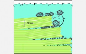
I’m sure Hansma is quite aware of what the ‘between the sheets’ phrase conjures and it works beautifully with her actual theory.  Here’s Hansma’s illustration of what might have happened (from the NSF site),

Diagram of biomolecules between sheets of mica in a primitive ocean. The green lines depict mica sheets and the grey structures depict various ancient biological molecules and fatty vesicles. In the 'between the sheets' mica hypothesis, water may have moved in and out of the spaces between stacks of sheets, thereby forcing the sheets to move up and down. This kind of energy may have ultimately pushed biological molecules and/or fatty acids together to form cells. Credit: Helen Greenwood Hansma, University of California, Santa Barbara

I’m thoroughly charmed.