Tag Archives: University of Florida

A labradoodle, gold nanoparticles, and cancer treatment for dogs and cats

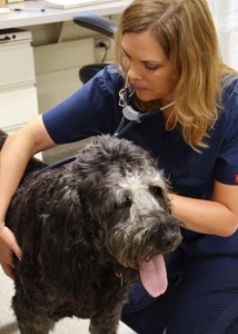
Here’s the labradoodle,

Caption: Dr. Shawna Klahn, an assistant professor of oncology at the Virginia-Maryland College of Veterinary Medicine, performs a checkup on "Grayton" four weeks after the animal's experimental cancer treatment involving gold nanoparticles and a targeted laser therapy. Credit: Virginia Tech

Caption: Dr. Shawna Klahn, an assistant professor of oncology at the Virginia-Maryland College of Veterinary Medicine, performs a checkup on “Grayton” four weeks after the animal’s experimental cancer treatment involving gold nanoparticles and a targeted laser therapy.
Credit: Virginia Tech

An Aug. 6, 2014 news item on Azonano outlines ‘Grayton’s’ story and how gold nanoparticles will factor in,

When Michael and Sandra Friedlander first came to the Virginia-Maryland College of Veterinary Medicine three years ago with their dog, Grayton, they learned some bad news: Grayton had nasal adenocarcinoma, a form of cancer with a short life expectancy.

“Most dogs with this form of cancer are with their owners no more than a few months after the diagnosis, but here Grayton is three years later,” said Michael Friedlander, who is the executive director of the Virginia Tech Carilion Research Institute and senior dean at the Virginia Tech Carilion School of Medicine.

No stranger to medical research, Friedlander was referred by Veterinary Teaching Hospital clinicians to an experimental treatment at the University of Florida called stereotactic radiation therapy, which delivers precise, high dosages of radiation to a tumor and can only be performed once.

“That shrunk the tumor down to almost nothing,” said Friedlander, who is also the associate provost for health sciences at Virginia Tech. “We knew when Grayton had the procedure that we couldn’t do it again, but now the cancer is back.”

An Aug. 4, 2014 Virginia Tech news release (also on EurekAlert) by Michael Sutphin, which originated the news item, explains what occasioned the release and how gold nanoparticles are being used in veterinary treatment for cancer,

Today [Aug. 4, 2014], the 11-year-old Labradoodle is the first patient at the Virginia-Maryland College of Veterinary Medicine in a new clinical trial that is testing the use of gold nanoparticles and a targeted laser treatment for solid tumors in dogs and cats. The study is one of several on new treatments for client-owned companion animals at the college. In January [2014], the college established the Veterinary Clinical Research Office to help facilitate this work.

“Clinical research at the veterinary college involves both primary research focused on advancing the treatment and diagnosis of veterinary diseases and translational research in which spontaneous diseases in animals can be used as models of human disease,” said Dr. Greg Daniel, head of the Department of Small Animal Clinical Sciences. “In the latter situation, we can provide our companion animal patients with treatment and diagnostic options that are not yet available in mainstream human medicine.”

Although medical researchers have tested gold nanoparticles with targeted laser treatments on human patients with some success, the treatment is still new to both human and veterinary medicine. The college is one of four current veterinary schools around the country testing the AuroLase therapy developed by Nanospectra Biosciences Inc., a startup company based in Houston, Texas. The others are Texas A&M University, the University of Wisconsin-Madison, and the University of Georgia.

Dr. Nick Dervisis, assistant professor of oncology in the Department of Small Animal Clinical Sciences, is leading the Nanospectra-funded study. Following a rhinoscopy performed on Grayton by Dr. David Grant, associate professor of internal medicine, Dervisis began the one-time, experimental therapy.

“The treatment involves two phases,” Dervisis said. “First, we infuse the patient with the gold nanoparticles. Although the nanoparticles distribute throughout the body, they tend to concentrate around blood vessels associated with tumors. Within 36 hours, they have cleared the bloodstream except for tumors. The gold nanoparticles are small enough to circulate freely in the bloodstream and become temporarily captured within the incomplete blood vessel walls common in solid tumors. Then, we use a non-ablative laser on the patient.”

Dervisis explained that a non-ablative laser is not strong enough to harm the skin or normal tissue, but “it does cause the remaining nanoparticles to absorb the laser energy and convert it into heat so that they damage the tumor cells.”

Like all clinical trials, the study involves many unknowns, including the treatment’s usefulness and effectiveness. One month after the AuroLase treatment, the nosebleeds that initially brought Grayton back to the Veterinary Teaching Hospital had stopped and Grayton has no other side effects.

“I’m delighted with the care and service that Grayton has received at the veterinary college,” said Friedlander, who explained that the treatment appears to be safe even though researchers do not know whether it is effective yet. “Grayton recently came with us on our annual vacation at the beach. We didn’t know if he would be able to come again, so it was great to have him with us swimming, catching fish and crabs, and doing what dogs do.”

Current clinical trials at the veterinary college range from the use of MRI to distinguish between benign and cancerous lymph nodes in dogs with oral melanoma, to a new chemotherapy drug for dogs with brain tumors, to the treatment of invasive skin cancer in horses with high-voltage, high-frequency electrical pulses. A complete list of current trials can be found at the college’s new clinical trials website.

Mindy Quigley, who oversees the college’s Veterinary Clinical Research Office, explained that veterinary trials, which follow a four-phase process and a variety of regulations similar to human medicine, have another layer of complexity that human trials do not.

“Variation among species means that a therapy that has proven safe and effective in, for example, humans or dogs, may not work for horses,” said Quigley, who comes to the college from the University of Edinburgh’s College of Medicine and Veterinary Medicine, where she helped set up a new neurology research clinic with funding from author J.K. Rowling. “Many veterinary clinical trials must therefore take therapies that have worked in one species and test them in other species with similar conditions. This is a necessary step to determine if a proposed treatment is safe and effective for our companion animals.”

Grayton may be the first companion animal in the AuroLase study at the veterinary college, but he certainly won’t be the last. Dervisis is continuing to enroll patients in the study and is seeking dogs and cats of a certain size with solid tumors who have not recently received radiation therapy or chemotherapy.

Interested parties can check this site for current clinical trials, including the Aurolase study,  being held by the Virginia-Maryland Regional College of Veterinary Medicine.

Cookies, ants, and a citizen science project plus a call for proposals for a 2015 Citizen Science Conference

My first citizen science item concerns summertime when the ants are out and about, oftentimes as uninvited participants to a picnic. Scientists at North Carolina State University (NCSU) and the University of Florida (UF) have decided to take advantage of this summer phenomenon as per a July 7, 2014 news item on ScienceDaily,

Scientists from North Carolina State University and the University of Florida have combined cookies, citizen science and robust research methods to track the diversity of ant species across the United States, and are now collaborating with international partners to get a global perspective on how ants are moving and surviving in the modern world.

“We think our School of Ants project serves as a good model for how citizen science can be used to collect more data, more quickly, from more places than a research team could do otherwise,” says Dr. Andrea Lucky, a researcher at the University of Florida who started work on the School of Ants while a postdoctoral researcher at NC State and now heads the project. Lucky is co-lead author of a paper describing the work and its early findings. “And our protocols help ensure that the data we are collecting are high quality.”

A July 7, 2014 NCSU news release (also on EurekAlert), which originated the news item, describes the various objectives for the project,

The School of Ants project was developed at NC State to help researchers get a handle on the diversity of ant species across the United States, with a particular focus on Chicago, Raleigh and New York City. In short, to discover which ant species are living where.

“But we also wanted to launch a citizen science project that both increased the public’s ecological literacy and addressed criticisms that public involvement made citizen science data unreliable,” says Dr. Amy Savage , a postdoctoral biological sciences researcher at NC State and the other co-lead author of the paper.

The research protocol, process, and outcomes are then described (from the news release),

The researchers developed a simple protocol involving Pecan Sandies cookies and sealable plastic bags, detailing precisely how the public should collect and label ant samples before shipping them to NC State or UF. [emphasis mine] This process was designed to engage the public in the aspect of the research that was easiest for non-scientists to enjoy and participate in, while also limiting the chances that the public could make mistakes that would skew the findings.

Once the samples arrive at NC State or UF, they are sorted, identified by a team of national experts and entered into a database. That information is then made publicly available in a user-friendly format on the project’s schoolofants.org site, allowing study participants to track the survey.

“This information is helping us tackle a variety of ecological and evolutionary questions, such as how ants may be evolving in urban environments, and how invasive species are spreading in the U.S.,” Savage says.

More than 1,000 participants, with samples from all 50 states, have taken part in the project since its 2011 launch – and there have already been some surprising findings.

For example, the researchers learned that a venomous invasive species, the Asian needle ant (Pachycondyla chinensis), had spread thousands of miles farther than anyone expected. Researchers knew the species had established itself in the Southeast, but study participants sent in Asian needle ant samples from as far afield as Wisconsin and Washington state.

To build on the School of Ants model, the researchers have launched collaborations with counterparts in Italy and Australia.

“We’re optimistic that this project will give us a broader view of ant diversity and how these species intersect with us, where we live and work around the world,” Lucky says.

The researchers are also working with teachers to incorporate the project into K-12 instruction modules that incorporate key elements of common core education standards. One early teacher collaboration has led to a research paper co-written by 4th and 5th graders.

“We also collaborated with a science writer to produce a free series of iBooks featuring natural history stories about the most common ants that our citizen science partners are collecting in their backyards and sidewalks,” Savage says.

“One of our big goals now is to move from collecting data and finding patterns to identifying ways that we can work with the public to figure out what is driving those patterns,” says Dr. Rob Dunn, an associate professor of biological sciences at NC State and co-author of the paper.

Not being familiar with Pecan Sandies cookies I went searching on the internet and found many recipes including this one from Martha Stewart’s website,

 Pecan Sandies

prep: 15 mins
total time: 30 mins
yield: Makes 18

Ingredients

1/2 cup (1 stick) unsalted butter, room temperature
1/2 cup packed light-brown sugar
1 1/2 teaspoons pure vanilla extract
1/4 teaspoon salt
1 cup all-purpose flour (spooned and leveled)
1 cup pecans, coarsely chopped

Cook’s Note
For best results, line cookie sheets with parchment prior to baking.
Directions

Step 1

Preheat oven to 350 degrees, with racks in upper and lower thirds. In a large bowl, using an electric mixer, beat butter and sugar until light and fluffy; beat in vanilla and salt. With mixer on low, gradually add flour, beating just until combined. Fold in pecans.

Step 2

Roll dough into 1 1/2-inch balls, and place on two baking sheets, 2 inches apart. With the dampened bottom of a glass, lightly flatten each ball.

Step 3

Bake until cookies are golden brown, 15 to 17 minutes, rotating sheets halfway through. Transfer to wire racks, and let cool.

This is what they look like (also from the Martha Stewart website),

[downloaded from http://www.marthastewart.com/342386/pecan-sandies]

[downloaded from http://www.marthastewart.com/342386/pecan-sandies]

I also checked out the School of Ants project website and found this,

The School of Ants project is a citizen-scientist driven study of the ants that live in urban areas, particularly around homes and schools. Participation is open to anyone interested!
Learn More!

Anyone can participate! Learn how to create your own sampling kit, sample your backyard or schoolyard, and get our collection back to us so that we can ID the ants and add your species list to the big School of Ants map. Together we’ll map ant diversity and species ranges across North America! Click here to get started!

There is at least one question you might want to ask before running off to collect ants, the researchers specify Keebler Pecan Sandies cookies are to be used as bait. I’m not sure how available those specific cookies and brand are in Canada, Mexico, Italy, or Australia. You may want to check with the organizers as to what alternatives might be acceptable. From the Participate webpage on the School of Ants website,

SAMPLING ANTS for the School of Ants involves placing cookie baits outdoors in green spaces (lawns, gardens, woods) and paved places (asphalt, concrete, cobblestone) for one hour on a warm day. We want to know what ants discover the baits in your neighborhood!(ALLERGY WARNING!: this activity uses Keebler Pecan Sandies cookies, which contain pecans, wheat, egg and whey).

Here’s a link to and a citation for the paper,

Ecologists, educators, and writers collaborate with the public to assess backyard diversity in The School of Ants Project [PDF] by Andrea Lucky, Amy M. Savage, Lauren M. Nichols, Leonora Shell, Robert R. Dunn, Cristina Castracani, Donato A. Grasso, and Alessandra Mori. Ecosphere 5(7):78. http://dx.doi.org/10.1890/ES13-00364.1 Published: online July 7, 2014,

Ecosphere is an open access journal. The PDF is 23 pp.

For my second citizen science item, I have a call for proposals for the Citizen Science 2015 Conference (CS2015), February 11 & 12, 2015 in San Jose, California (prior to the 2015 AAAS [American Association for the Advancement of Science] annual meeting February 12 -16, 2015 also in San Jose). Here’s more about the Citizen Science conference from the Overview page,

Anyone involved in citizen science is invited to attend this conference. Attendees will include citizen science participants, researchers, project leaders, educators, technology specialists, evaluators, and others – representing many disciplines including astronomy, molecular biology, human and environmental health, psychology, linguistics, environmental justice, biodiversity, conservation biology, public health, genetics, engineering, cyber technology, gaming, and more – at any level of expertise. There will be opportunities throughout the conference to make connections, share insights, and help move this field forward.

We have identified six main themes for this year’s conference:

  1. Tackling Grand Challenges and Everyday Problems with Citizen Science
  2. Broadening Engagement to Foster Diversity and Inclusion
  3. Making Education and Lifelong Learning Connections (K-12, university, informal)
  4. Digital Opportunities and Challenges in Citizen Science
  5. Research on and Evaluation of the Citizen Science Experience
  6. Best Practices for Designing, Implementing, and Managing Citizen Science Projects and Programs

Here are important dates for the conference (from a June 30, 2014 email announcement),

September 15, 2014          CS2015 Deadline to submit proposals* (talks, posters, etc)
October 6, 2014                 CS2015 Proposal selection notices sent out
November 10, 2014           CS2015 Early-bird registration discount ends
February 11 & 12, 2015     CS2015 Conference

Here’s more detail, from the Presentation Styles webpage,

… Several formats are available to choose from: three styles of oral presentations; symposia/panel discussions; and posters.

Audio-visual equipment will be provided as needed for all session types except posters.

Oral Presentations
Talks allow speakers to present their work in 12 minutes, with 3 additional minutes for audience questions. Talks with similar themes will be grouped together into sessions.

Speed Talks, as the name suggests, challenge each presenter to cover his or her topic in 5 minutes or less. Following a series of speed presentations, there will be time for audience members to gather with presenters for discussion.

Story Presentations (15 minutes) emphasize sharing valuable lessons through storytelling. We especially encourage telling stories of “what didn’t work and why” and strategies for addressing challenges and unintended consequences.

Symposium Sessions or Panel Discussions (1 to 2 hours)
Every symposium or panel has one convener (most likely the person submitting this proposal); that person is responsible for organizing the session and will act as the session’s contact person with conference organizers. Additionally, that person will moderate/guide the session. Symposia/Panels may be 1-to-2 hours in length, depending on the number of proposed talks, and must include at least 15 minutes for questions and discussion with the audience.

The proposal must (1) describe the symposium or panel’s objective, (2) how it will contribute to the overall theme of the conference, and (3) include a list of proposed speakers (and, in the case of a symposium, each speaker’s topic).

Posters
Posters are designed to visually display information and engage fellow attendees in an informal way. There will be two Poster Sessions—one each day—inviting attendees to discuss posters with authors. Posters will also be on display outside of formal poster-session times. All accepted posters will be given a display space measuring 4 x 4 feet (1.2 X 1.2 meters) in the Poster Hall (no additional audio-visual aids are permitted).

You can access a link to submit your proposal here.

CS2015 is being called a pre-conference to the AAAS meeting as per the Prepare for the Conference page,

Registration
Registration details, including the conference registration fee, are not yet finalized. We are seeking funding to help support the conference and keep it affordable to all. Check back for updates, or join the CSA to receive periodic updates.

Attend Two Great Conferences
CS2015 is a pre-conference of the Annual Meeting of the American Association for the Advancement of Science (AAAS), which immediately follows our meeting at the San Jose Convention Center. The AAAS theme for 2015 is “Innovations, Information, and Imaging.” Once you have completed your CS2015 registration, you will receive instructions on how to register for the AAAS Annual Meeting (February 12-16, 2015) at the discounted rate of $235. AAAS registration will open in August 2014.

Good luck with your proposal and with your ant-captures!

Good lignin, bad lignin: Florida researchers use plant waste to create lignin nanotubes while researchers in British Columbia develop trees with less lignin

An April 4, 2014 news item on Azonano describes some nanotube research at the University of Florida that reaches past carbon to a new kind of nanotube,

Researchers with the University of Florida’s [UF] Institute of Food and Agricultural Sciences took what some would consider garbage and made a remarkable scientific tool, one that could someday help to correct genetic disorders or treat cancer without chemotherapy’s nasty side effects.

Wilfred Vermerris, an associate professor in UF’s department of microbiology and cell science, and Elena Ten, a postdoctoral research associate, created from plant waste a novel nanotube, one that is much more flexible than rigid carbon nanotubes currently used. The researchers say the lignin nanotubes – about 500 times smaller than a human eyelash – can deliver DNA directly into the nucleus of human cells in tissue culture, where this DNA could then correct genetic conditions. Experiments with DNA injection are currently being done with carbon nanotubes, as well.

“That was a surprising result,” Vermerris said. “If you can do this in actual human beings you could fix defective genes that cause disease symptoms and replace them with functional DNA delivered with these nanotubes.”

An April 3, 2014 University of Florida’s Institute of Food and Agricultural Sciences news release, which originated the news item, describes the lignin nanotubes (LNTs) and future applications in more detail,

The nanotube is made up of lignin from plant material obtained from a UF biofuel pilot facility in Perry, Fla. Lignin is an integral part of the secondary cell walls of plants and enables water movement from the roots to the leaves, but it is not used to make biofuels and would otherwise be burned to generate heat or electricity at the biofuel plant. The lignin nanotubes can be made from a variety of plant residues, including sorghum, poplar, loblolly pine and sugar cane. [emphasis mine]

The researchers first tested to see if the nanotubes were toxic to human cells and were surprised to find that they were less so than carbon nanotubes. Thus, they could deliver a higher dose of medicine to the human cell tissue.  Then they researched if the nanotubes could deliver plasmid DNA to the same cells and that was successful, too. A plasmid is a small DNA molecule that is physically separate from, and can replicate independently of, chromosomal DNA within a cell.

“It’s not a very smooth road because we had to try different experiments to confirm the results,” Ten said. “But it was very fruitful.”

In cases of genetic disorders, the nanotube would be loaded with a functioning copy of a gene, and injected into the body, where it would target the affected tissue, which then makes the missing protein and corrects the genetic disorder.

Although Vermerris cautioned that treatment in humans is many years away, among the conditions that these gene-carrying nanotubes could correct include cystic fibrosis and muscular dystrophy. But, he added, that patients would have to take the corrective DNA via nanotubes on a continuing basis.

Another application under consideration is to use the lignin nanotubes for the delivery of chemotherapy drugs in cancer patients. The nanotubes would ensure the drugs only get to the tumor without affecting healthy tissues.

Vermerris said they created different types of nanotubes, depending on the experiment. They could also adapt nanotubes to a patient’s specific needs, a process called customization.

“You can think about it as a chest of drawers and, depending on the application, you open one drawer or use materials from a different drawer to get things just right for your specific application,” he said.  “It’s not very difficult to do the customization.”

The next step in the research process is for Vermerris and Ten to begin experiments on mice. They are in the application process for those experiments, which would take several years to complete.  If those are successful, permits would need to be obtained for their medical school colleagues to conduct research on human patients, with Vermerris and Ten providing the nanotubes for that research.

“We are a long way from that point,” Vermerris said. “That’s the optimistic long-term trajectory.”

I hope they have good luck with this work. I have emphasized the plant waste the University of Florida scientists studied due to the inclusion of poplar, which is featured in the University of British Columbia research work also being mentioned in this post.

Getting back to Florida for a moment, here’s a link to and a citation for the paper,

Lignin Nanotubes As Vehicles for Gene Delivery into Human Cells by Elena Ten, Chen Ling, Yuan Wang, Arun Srivastava, Luisa Amelia Dempere, and Wilfred Vermerris. Biomacromolecules, 2014, 15 (1), pp 327–338 DOI: 10.1021/bm401555p Publication Date (Web): December 5, 2013
Copyright © 2013 American Chemical Society

This is an open access paper.

Meanwhile, researchers at the University of British Columbia (UBC) are trying to limit the amount of lignin in trees (specifically poplars, which are not mentioned in this excerpt but in the next). From an April 3, 2014 UBC news release,

Researchers have genetically engineered trees that will be easier to break down to produce paper and biofuel, a breakthrough that will mean using fewer chemicals, less energy and creating fewer environmental pollutants.

“One of the largest impediments for the pulp and paper industry as well as the emerging biofuel industry is a polymer found in wood known as lignin,” says Shawn Mansfield, a professor of Wood Science at the University of British Columbia.

Lignin makes up a substantial portion of the cell wall of most plants and is a processing impediment for pulp, paper and biofuel. Currently the lignin must be removed, a process that requires significant chemicals and energy and causes undesirable waste.

Researchers used genetic engineering to modify the lignin to make it easier to break down without adversely affecting the tree’s strength.

“We’re designing trees to be processed with less energy and fewer chemicals, and ultimately recovering more wood carbohydrate than is currently possible,” says Mansfield.

Researchers had previously tried to tackle this problem by reducing the quantity of lignin in trees by suppressing genes, which often resulted in trees that are stunted in growth or were susceptible to wind, snow, pests and pathogens.

“It is truly a unique achievement to design trees for deconstruction while maintaining their growth potential and strength.”

The study, a collaboration between researchers at the University of British Columbia, the University of Wisconsin-Madison, Michigan State University, is a collaboration funded by Great Lakes Bioenergy Research Center, was published today in Science.

Here’s more about lignin and how a decrease would free up more material for biofuels in a more environmentally sustainable fashion, from the news release,

The structure of lignin naturally contains ether bonds that are difficult to degrade. Researchers used genetic engineering to introduce ester bonds into the lignin backbone that are easier to break down chemically.

The new technique means that the lignin may be recovered more effectively and used in other applications, such as adhesives, insolation, carbon fibres and paint additives.

Genetic modification

The genetic modification strategy employed in this study could also be used on other plants like grasses to be used as a new kind of fuel to replace petroleum.

Genetic modification can be a contentious issue, but there are ways to ensure that the genes do not spread to the forest. These techniques include growing crops away from native stands so cross-pollination isn’t possible; introducing genes to make both the male and female trees or plants sterile; and harvesting trees before they reach reproductive maturity.

In the future, genetically modified trees could be planted like an agricultural crop, not in our native forests. Poplar is a potential energy crop for the biofuel industry because the tree grows quickly and on marginal farmland. [emphasis mine] Lignin makes up 20 to 25 per cent of the tree.

“We’re a petroleum reliant society,” says Mansfield. “We rely on the same resource for everything from smartphones to gasoline. We need to diversify and take the pressure off of fossil fuels. Trees and plants have enormous potential to contribute carbon to our society.”

As noted earlier, the researchers in Florida mention poplars in their paper (Note: Links have been removed),

Gymnosperms such as loblolly pine (Pinus taeda L.) contain lignin that is composed almost exclusively of G-residues, whereas lignin from angiosperm dicots, including poplar (Populus spp.) contains a mixture of G- and S-residues. [emphasis mine] Due to the radical-mediated addition of monolignols to the growing lignin polymer, lignin contains a variety of interunit bonds, including aryl–aryl, aryl–alkyl, and alkyl–alkyl bonds.(3) This feature, combined with the association between lignin and cell-wall polysaccharides, which involves both physical and chemical interactions, make the isolation of lignin from plant cell walls challenging. Various isolation methods exist, each relying on breaking certain types of chemical bonds within the lignin, and derivatizations to solubilize the resulting fragments.(5) Several of these methods are used on a large scale in pulp and paper mills and biorefineries, where lignin needs to be removed from woody biomass and crop residues(6) in order to use the cellulose for the production of paper, biofuels, and biobased polymers. The lignin is present in the waste stream and has limited intrinsic economic value.(7)

Since hydroxyl and carboxyl groups in lignin facilitate functionalization, its compatibility with natural and synthetic polymers for different commercial applications have been extensively studied.(8-12) One of the promising directions toward the cost reduction associated with biofuel production is the use of lignin for low-cost carbon fibers.(13) Other recent studies reported development and characterization of lignin nanocomposites for multiple value-added applications. For example, cellulose nanocrystals/lignin nanocomposites were developed for improved optical, antireflective properties(14, 15) and thermal stability of the nanocomposites.(16) [emphasis mine] Model ultrathin bicomponent films prepared from cellulose and lignin derivatives were used to monitor enzyme binding and cellulolytic reactions for sensing platform applications.(17) Enzymes/“synthetic lignin” (dehydrogenation polymer (DHP)) interactions were also investigated to understand how lignin impairs enzymatic hydrolysis during the biomass conversion processes.(18)

The synthesis of lignin nanotubes and nanowires was based on cross-linking a lignin base layer to an alumina membrane, followed by peroxidase-mediated addition of DHP and subsequent dissolution of the membrane in phosphoric acid.(1) Depending upon monomers used for the deposition of DHP, solid nanowires, or hollow nanotubes could be manufactured and easily functionalized due to the presence of many reactive groups. Due to their autofluorescence, lignin nanotubes permit label-free detection under UV radiation.(1) These features make lignin nanotubes suitable candidates for numerous biomedical applications, such as the delivery of therapeutic agents and DNA to specific cells.

The synthesis of LNTs in a sacrificial template membrane is not limited to a single source of lignin or a single lignin isolation procedure. Dimensions of the LNTs and their cytotoxicity to HeLa cells appear to be determined primarily by the lignin isolation procedure, whereas the transfection efficiency is also influenced by the source of the lignin (plant species and genotype). This means that LNTs can be tailored to the application for which they are intended. [emphasis mine] The ability to design LNTs for specific purposes will benefit from a more thorough understanding of the relationship between the structure and the MW of the lignin used to prepare the LNTs, the nanomechanical properties, and the surface characteristics.

We have shown that DNA is physically associated with the LNTs and that the LNTs enter the cytosol, and in some case the nucleus. The LNTs made from NaOH-extracted lignin are of special interest, as they were the shortest in length, substantially reduced HeLa cell viability at levels above approximately 50 mg/mL, and, in the case of pine and poplar, were the most effective in the transfection [penetrating the cell with a bacterial plasmid to leave genetic material in this case] experiments. [emphasis mine]

As I see the issues presented with these two research efforts, there are environmental and energy issues with extracting the lignin while there seem to be some very promising medical applications possible with lignin ‘waste’. These two research efforts aren’t necessarily antithetical but they do raise some very interesting issues as to how we approach our use of resources and future policies.

ETA May 16, 2014: The beat goes on with the Georgia (US) Institute of Technology issues a roadmap for making money from lignin. From a Georgia Tech May 15, 2014 news release on EurekAlert,

When making cellulosic ethanol from plants, one problem is what to do with a woody agricultural waste product called lignin. The old adage in the pulp industry has been that one can make anything from lignin except money.

A new review article in the journal Science points the way toward a future where lignin is transformed from a waste product into valuable materials such as low-cost carbon fiber for cars or bio-based plastics. Using lignin in this way would create new markets for the forest products industry and make ethanol-to-fuel conversion more cost-effective.

“We’ve developed a roadmap for integrating genetic engineering with analytical chemistry tools to tailor the structure of lignin and its isolation so it can be used for materials, chemicals and fuels,” said Arthur Ragauskas, a professor in the School of Chemistry and Biochemistry at the Georgia Institute of Technology. Ragauskas is also part of the Institute for Paper Science and Technology at Georgia Tech.

The roadmap was published May 15 [2014] in the journal Science. …

Here’s a link to and citation for the ‘roadmap’,

Lignin Valorization: Improving Lignin Processing in the Biorefinery by  Arthur J. Ragauskas, Gregg T. Beckham, Mary J. Biddy, Richard Chandra, Fang Chen, Mark F. Davis, Brian H. Davison, Richard A. Dixon, Paul Gilna, Martin Keller, Paul Langan, Amit K. Naskar, Jack N. Saddler, Timothy J. Tschaplinski, Gerald A. Tuskan, and Charles E. Wyman. Science 16 May 2014: Vol. 344 no. 6185 DOI: 10.1126/science.1246843

This paper is behind a paywall.
Magical nanobots at University of Florida kill (almost) 100% of Hepatitis C virus—in the lab

I’ve always preferred the term nanobots but the folks at the University of Florida are calling them nanorobots, from the July 16, 2012 news item on phys.org,

University of Florida researchers have moved a step closer to treating diseases on a cellular level by creating a tiny particle that can be programmed to shut down the genetic production line that cranks out disease-related proteins.

In laboratory tests, these newly created “nanorobots” all but eradicated hepatitis C virus infection. The programmable nature of the particle makes it potentially useful against diseases such as cancer and other viral infections.

The research effort, led by Y. Charles Cao, a UF associate professor of chemistry, and Dr. Chen Liu, a professor of pathology and endowed chair in gastrointestinal and liver research in the UF College of Medicine, is described online this week in the Proceedings of the National Academy of Sciences.

The news item originated with a July 16, 2012 news release from the University of Florida which goes on to explain how the researchers succeeded,

The Holy Grail of nanotherapy is an agent so exquisitely selective that it enters only diseased cells, targets only the specified disease process within those cells and leaves healthy cells unharmed.

To demonstrate how this can work, Cao and colleagues, with funding from the National Institutes of Health, the Office of Naval Research and the UF [University of Florida] Research Opportunity Seed Fund, created and tested a particle that targets hepatitis C virus in the liver and prevents the virus from making copies of itself.

Hepatitis C infection causes liver inflammation, which can eventually lead to scarring and cirrhosis. The disease is transmitted via contact with infected blood, most commonly through injection drug use, needlestick injuries in medical settings, and birth to an infected mother. More than 3 million people in the United States are infected and about 17,000 new cases are diagnosed each year, according to the Centers for Disease Control and Prevention. Patients can go many years without symptoms, which can include nausea, fatigue and abdominal discomfort.

Current hepatitis C treatments involve the use of drugs that attack the replication machinery of the virus. But the therapies are only partially effective, on average helping less than 50 percent of patients, according to studies published in The New England Journal of Medicine and other journals. Side effects vary widely from one medication to another, and can include flu-like symptoms, anemia and anxiety.

Cao and colleagues, including graduate student Soon Hye Yang and postdoctoral associates Zhongliang Wang, Hongyan Liu and Tie Wang, wanted to improve on the concept of interfering with the viral genetic material in a way that boosted therapy effectiveness and reduced side effects.

The particle they created can be tailored to match the genetic material of the desired target of attack, and to sneak into cells unnoticed by the body’s innate defense mechanisms.

Recognition of genetic material from potentially harmful sources is the basis of important treatments for a number of diseases, including cancer, that are linked to the production of detrimental proteins. It also has potential for use in detecting and destroying viruses used as bioweapons.

The new virus-destroyer, called a nanozyme, has a backbone of tiny gold particles and a surface with two main biological components. The first biological portion is a type of protein called an enzyme that can destroy the genetic recipe-carrier, called mRNA, for making the disease-related protein in question. The other component is a large molecule called a DNA oligonucleotide that recognizes the genetic material of the target to be destroyed and instructs its neighbor, the enzyme, to carry out the deed. By itself, the enzyme does not selectively attack hepatitis C, but the combo does the trick.

“They completely change their properties,” Cao said.

In laboratory tests, the treatment led to almost a 100 percent decrease in hepatitis C virus levels. In addition, it did not trigger the body’s defense mechanism, and that reduced the chance of side effects. Still, additional testing is needed to determine the safety of the approach. [emphases mine]

This treatment builds on some previous research,

The UF nanoparticle design takes inspiration from the Nobel prize-winning discovery of a process in the body in which one part of a two-component complex destroys the genetic instructions for manufacturing protein, and the other part serves to hold off the body’s immune system attacks. This complex controls many naturally occurring processes in the body, so drugs that imitate it have the potential to hijack the production of proteins needed for normal function. The UF-developed therapy tricks the body into accepting it as part of the normal processes, but does not interfere with those processes.

Since there’s no mention of human clinical trials, I’m guessing that we are at least 10 years from seeing this therapeutic agent on the market.

After drafting this post yesterday (July 17, 2012) and while waiting to post it today, I found Dexter Johnson’s July 17 2012 posting where he makes some important points about this research (Note: I have removed a link),

Of course, this is a long way from becoming a treatment anytime soon. A major caveat is that the use of nanotreatments for the targeting and destroying of abnormal cells like cancer cells is always problematic since those cells are “still us” as George Whitesides noted some time back.  It’s always a bit of a tricky business to make sure that nanoparticles are targeting those biological processes within us that we want stopped and not the ones we want to keep.

Dexter goes on to comment about using the terms ‘nanobots’ or ‘nano robots’; he’s less sanguine about it than I am.

Using light to power movement and some philosophy

You know how sunflowers track the sun and move with it? They are powered by light. Well, researchers at the University of Florida have used a single molecule of DNA to create a molecular nanomotor powered only by particles of light (photons).  It’s not the first photon driven nanomotor but it is the first made entirely of a single DNA molecule. There’s an artist’s illustration and more detail about the work here.

On a more philosophical bent, a physicist at Canada’s Perimeter Institute, Lee Smolin who, based on his work with Roberto Mangabeira Unger, a Brazilian philospher, suggests that the timeless multiverse (beloved of physicists and science fiction writers) does not exist. The article by Smolin was written for Physics World and is available here.