Tag Archives: University of Fribourg

A snout weevil at the end of the rainbow

I’ve never heard of a snout weevil before but it seems to be a marvelous creature,

Caption: Left: A photograph of the ‘rainbow’ weevil, with the rainbow-colored spots on its thorax and elytra (wing casings). Right: A microscope image of the rim of a single rainbow spot, showing the different colors of individual scales. Credit: Dr Bodo D Wilts

From a Sept. 11, 2018 news item on Nanowerk,

Researchers from Yale [University]-NUS College and the University of Fribourg in Switzerland have discovered a novel colour-generation mechanism in nature, which if harnessed, has the potential to create cosmetics and paints with purer and more vivid hues, screen displays that project the same true image when viewed from any angle, and even reduce the signal loss in optical fibres.

Yale-NUS College Assistant Professor of Science (Life Science) Vinodkumar Saranathan led the study with Dr Bodo D Wilts from the Adolphe Merkle Institute at the University of Fribourg. Dr Saranathan examined the rainbow-coloured patterns in the elytra (wing casings) of a snout weevil from the Philippines, Pachyrrhynchus congestus pavonius, using high-energy X-rays, while Dr Wilts performed detailed scanning electron microscopy and optical modelling.

They discovered that to produce the rainbow palette of colours, the weevil utilised a colour-generation mechanism that is so far found only in squid, cuttlefish, and octopuses, which are renowned for their colour-shifting camouflage.

A Sept. 11, 2018 Yale-NUS College news release (also on EurekAlert), which originated the news item, offers more on the weevil and on the research,

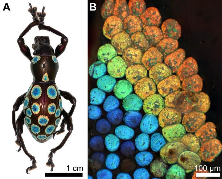
P. c. pavonius, or the “Rainbow” Weevil, is distinctive for its rainbow-coloured spots on its thorax and elytra (see attached image). These spots are made up of nearly-circular scales arranged in concentric rings of different hues, ranging from blue in the centre to red at the outside, just like a rainbow. While many insects have the ability to produce one or two colours, it is rare that a single insect can produce such a vast spectrum of colours. Researchers are interested to figure out the mechanism behind the natural formation of these colour-generating structures, as current technology is unable to synthesise structures of this size.

“The ultimate aim of research in this field is to figure out how the weevil self-assembles these structures, because with our current technology we are unable to do so,” Dr Saranathan said. “The ability to produce these structures, which are able to provide a high colour fidelity regardless of the angle you view it from, will have applications in any industry which deals with colour production. We can use these structures in cosmetics and other pigmentations to ensure high-fidelity hues, or in digital displays in your phone or tablet which will allow you to view it from any angle and see the same true image without any colour distortion. We can even use them to make reflective cladding for optical fibres to minimise signal loss during transmission.”

Dr Saranathan and Dr Wilts examined these scales to determine that the scales were composed of a three-dimensional crystalline structure made from chitin (the main ingredient in insect exoskeletons). They discovered that the vibrant rainbow colours on this weevil’s scales are determined by two factors: the size of the crystal structure which makes up each scale, as well as the volume of chitin used to make up the crystal structure. Larger scales have a larger crystalline structure and use a larger volume of chitin to reflect red light; smaller scales have a smaller crystalline structure and use a smaller volume of chitin to reflect blue light. According to Dr Saranathan, who previously examined over 100 species of insects and spiders and catalogued their colour-generation mechanisms, this ability to simultaneously control both size and volume factors to fine-tune the colour produced has never before been shown in insects, and given its complexity, is quite remarkable. “It is different from the usual strategy employed by nature to produce various different hues on the same animal, where the chitin structures are of fixed size and volume, and different colours are generated by orienting the structure at different angles, which reflects different wavelengths of light,” Dr Saranathan explained.

The research was partly supported though the National Centre of Competence in Research “Bio-Inspired Materials” and the Ambizione program of the Swiss National Science Foundation (SNSF) to Dr Wilts, and partly through a UK Royal Society Newton Fellowship, a Linacre College EPA Cephalosporin Junior Research Fellowship, and Yale-NUS College funds to Dr Saranathan. Dr Saranathan is currently part of a research team led by Yale-NUS College Associate Professor of Science Antonia Monteiro, which has recently been awarded a separate Competitive Research Programme (CRP) grant by Singapore’s National Research Foundation (NRF) to examine the genetic basis of the colour-generation mechanism in butterflies. Dr Saranathan and Dr Monteiro are both also from the Department of Biological Sciences at the National University of Singapore (NUS) Faculty of Science. In addition, Dr Saranathan is affiliated with the NUS Nanoscience and Nanotechnology Initiative.

Here’s a link to and a citation for the paper,

Literal Elytral Rainbow: Tunable Structural Colors Using Single Diamond Biophotonic Crystals in Pachyrrhynchus congestus Weevils by Bodo D. Wilts, Vinodkumar Saranathan. Samll https://doi.org/10.1002/smll.201802328 First published: 15 August 2018

This paper is behind a paywall.

More from PETA (People for the Ethical Treatment of Animals) about nanomaterials and lungs

Science progress by increments. First, there was this April 27, 2016 post featuring some recent work by the organization, People for the Ethical Treatment of Animals (PETA) focused on nanomaterials and lungs. Now approximately one month later, PETA announces a new paper on the topic according to a May 26, 2016 news item on phys.org,

A scientist from the PETA International Science Consortium Ltd. is the lead author of a review on pulmonary fibrosis that results from inhaling nanomaterials, which has been published in Archives of Toxicology. The coauthors are scientists from Health Canada, West Virginia University, and the University of Fribourg in Switzerland.

A May 26, 2016 PETA news release on EurekAlert, which originated the news item, provides more detail (Note: Links have been removed),

The increasing use of nanomaterials in consumer goods such as paint, building materials, and food products has increased the likelihood of human exposure. Inhalation is one of the most prominent routes by which exposure can occur, and because inhalation of nanomaterials may be linked to lung problems such as pulmonary fibrosis, testing is conducted to assess the safety of these materials.

The review is one part of the proceedings of a 2015 workshop [mentioned in my Sept. 3, 2015 posting] organized by the PETA International Science Consortium, at which scientists discussed recommendations for designing an in vitro approach to assessing the toxicity of nanomaterials in the human lung. The workshop also produced another report that was recently published in Archives of Toxicology (Clippinger et al. 2016) and a review published in Particle and Fibre Toxicology (Polk et al. 2016) [mentioned in my April 27, 2016 posting] on exposing nanomaterials to cells grown in vitro.

The expert recommendations proposed at the workshop are currently being used to develop an in vitro system to predict the development of lung fibrosis in humans, which is being funded by the Science Consortium.

“International experts who took part in last year’s workshop have advanced the understanding and application of non-animal methods of studying nanomaterial effects in the lung,” says Dr. Monita Sharma, nanotoxicology specialist at the Consortium and lead author of the review in Archives of Toxicology. “Good science is leading the way toward more humane testing of nanomaterials, which, in turn, will lead to better protection of human health.”

Here’s a link to and a citation for the paper,

Predicting pulmonary fibrosis in humans after exposure to multi-walled carbon nanotubes (MWCNTs) by Monita Sharma, Jake Nikota, Sabina Halappanavar, Vincent Castranova, Barbara Rothen-Rutishauser, Amy J. Clippinger. Archives of Toxicology pp 1-18 DOI: 10.1007/s00204-016-1742-7 First online: 23 May 2016

This paper is behind a paywall.

People for the Ethical Treatment of Animals (PETA) and a grant for in vitro nanotoxicity testing

This grant seems to have gotten its start at a workshop held at the US Environmental Protection Agency (EPA) in Washington, D.C., Feb. 24-25, 2015 as per this webpage on the People for Ethical Treatment of Animals (PETA) International Science Consortium Limited website,

The invitation-only workshop included experts from different sectors (government, industry, academia and NGO) and disciplines (in vitro and in vivo inhalation studies of NMs, fibrosis, dosimetry, fluidic models, aerosol engineering, and regulatory assessment). It focused on the technical details for the development and preliminary assessment of the relevance and reliability of an in vitro test to predict the development of pulmonary fibrosis in cells co-cultured at the air-liquid interface following exposure to aerosolized multi-walled carbon nanotubes (MWCNTs). During the workshop, experts made recommendations on cell types, exposure systems, endpoints and dosimetry considerations required to develop the in vitro model for hazard identification of MWCNTs.

The method is intended to be included in a non-animal test battery to reduce and eventually replace the use of animals in studies to assess the inhalation toxicity of engineered NMs. The long-term vision is to develop a battery of in silico and in vitro assays that can be used in an integrated testing strategy, providing comprehensive information on biological endpoints relevant to inhalation exposure to NMs which could be used in the hazard ranking of substances in the risk assessment process.

A September 1, 2015 news item on Azonano provides an update,

The PETA International Science Consortium Ltd. announced today the winners of a $200,000 award for the design of an in vitro test to predict the development of lung fibrosis in humans following exposure to nanomaterials, such as multi-walled carbon nanotubes.

Professor Dr. Barbara Rothen-Rutishauser of the Adolphe Merkle Institute at the University of Fribourg, Switzerland and Professor Dr. Vicki Stone of the School of Life Sciences at Heriot-Watt University, Edinburgh, U.K. will jointly develop the test method. Professor Rothen-Rutishauser co-chairs the BioNanomaterials research group at the Adolphe Merkle Institute, where her research is focused on the study of nanomaterial-cell interactions in the lung using three-dimensional cell models. Professor Vicki Stone is the Director of the Nano Safety Research Group at Heriot-Watt University and the Director of Toxicology for SAFENANO.

The Science Consortium is also funding MatTek Corporation for the development of a three-dimensional reconstructed primary human lung tissue model to be used in Professors Rothen-Rutishauser and Stone’s work. MatTek Corporation has extensive expertise in manufacturing human cell-based, organotypic in vitro models for use in regulatory and basic research applications. The work at MatTek will be led by Dr. Patrick Hayden, Vice President of Scientific Affairs, and Dr. Anna Maione, head of MatTek’s airway models research group.

I was curious about MatTek Corporation and found this on company’s About Us webpage,

MatTek Corporation was founded in 1985 by two chemical engineering professors from MIT. In 1991 the company leveraged its core polymer surface modification technology into the emerging tissue engineering market.

MatTek Corporation is at the forefront of tissue engineering and is a world leader in the production of innovative 3D reconstructed human tissue models. Our skin, ocular, and respiratory tissue models are used in regulatory toxicology (OECD, EU guidelines) and address toxicology and efficacy concerns throughout the cosmetics, chemical, pharmaceutical and household product industries.

EpiDerm™, MatTek’s first 3D human cell based in vitro model, was introduced in 1993 and became an immediate technical and commercial success.

I wish them good luck in their research on developing better ways to test toxicity.

The shorter, the better for cellulose nanofibres

Cellulose nanomaterials can be derived from any number of plants. In Canada, we tend to think of our trees first but there are other sources such as cotton, bananas, hemp, carrots, and more.

In anticipation that cellulose nanofibres will become increasingly important constituents of various products and having noticed a resemblance to carbon nanotubes, scientists in Switzerland have investigated the possible toxicity issues according to a May 7, 2015 news item on Nanowerk,

Plant-based cellulose nanofibres do not pose a short-term health risk, especially short fibres, shows a study conducted in the context of National Research Programme “Opportunities and Risks of Nanomaterials” (NRP 64). But lung cells are less efficient in eliminating longer fibres.

Similar to carbon nanotubes that are used in cycling helmets and tennis rackets, cellulose nanofibres are extremely light while being extremely tear-resistant. But their production is significantly cheaper because they can be manufactured from plant waste of cotton or banana plants. “It is only a matter of time before they prevail on the market,” says Christoph Weder of the Adolphe Merkle Institute at the University of Fribourg [Switzerland].

A May 7, 2015 Swiss National Science Foundation (SNSF) press release, which originated the news item, provides more detail,

In the context of the National Research Programme “Opportunities and Risks of Nanomaterials” (NRP 64), he collaborated with the team of Barbara Rothen-Rutishauser to examine whether these plant-based nanofibres are harmful to the lungs when inhaled. The investigation does not rely on animal testing; instead the group of Rothen-Rutishauser developped a complex 3D lung cell system to simulate the surface of the lungs by using various human cell cultures in the test tube.

The shorter, the better

Their results (*) show that cellulose nanofibres are not harmful: the analysed lung cells showed no signs of acute stress or inflammation. But there were clear differences between short and long fibres: the lung cell system efficiently eliminated short fibres while longer fibres stayed on the cell surface.

“The testing only lasted two days because we cannot grow the cell cultures for longer,” explains Barbara Rothen-Rutishauser. For this reason, she adds, they cannot say if the longer fibre may have a negative impact on the lungs in the long term. Tests involving carbon nanotubes have shown that lung cells lose their equilibrium when they are faced with long tubes because they try to incorporate them into the cell to no avail. “This frustrated phagocytosis can trigger an inflammatory reaction,” says Rothen-Rutishauser. To avoid potential harm, she recommends that companies developing products with nanofibres use fibres that are short and pliable instead of long and rigid.

National Research Programme “Opportunities and Risks of Nanomaterials” (NRP 64)

The National Research Programme “Opportunities and Risks of Nanomaterials” (NRP 64) hopes to be able to bridge the gaps in our current knowledge on nanomaterials. Opportunities and risks for human health and the environment in relation to the manufacture, use and disposal of synthetic nanomaterials need to be better understood. The projects started their research work in December 2010.

I have a link to and a citation for the paper (Note: They use the term cellulose nanocrystals in the paper’s title),

Fate of Cellulose Nanocrystal Aerosols Deposited on the Lung Cell Surface In Vitro by Carola Endes, Silvana Mueller, Calum Kinnear, Dimitri Vanhecke, E. Johan Foster, Alke Petri-Fink, Christoph Weder, Martin J. D. Clift, and Barbara Rothen-Rutishauser. Biomacromolecules, 2015, 16 (4), pp 1267–1275 DOI: 10.1021/acs.biomac.5b00055 Publication Date (Web): March 19, 2015

Copyright © 2015 American Chemical Society

While tracking down the 2015 paper, I found this from 2011,

Investigating the Interaction of Cellulose Nanofibers Derived from Cotton with a Sophisticated 3D Human Lung Cell Coculture by Martin J. D. Clift, E. Johan Foster, Dimitri Vanhecke, Daniel Studer, Peter Wick, Peter Gehr, Barbara Rothen-Rutishauser, and Christoph Weder. Biomacromolecules, 2011, 12 (10), pp 3666–3673 DOI: 10.1021/bm200865j Publication Date (Web): August 16, 2011

Copyright © 2011 American Chemical Society

Both papers are behind a paywall.