Tag Archives: University of Houston

Enhance or weaken memory with stretchy, bioinspired synaptic transistor

This news is intriguing since they usually want to enhance memory not weaken it. Interestingly, this October 3, 2022 news item on ScienceDaily doesn’t immediately answer why you might want to weaken memory,

Robotics and wearable devices might soon get a little smarter with the addition of a stretchy, wearable synaptic transistor developed by Penn State engineers. The device works like neurons in the brain to send signals to some cells and inhibit others in order to enhance and weaken the devices’ memories.

Led by Cunjiang Yu, Dorothy Quiggle Career Development Associate Professor of Engineering Science and Mechanics and associate professor of biomedical engineering and of materials science and engineering, the team designed the synaptic transistor to be integrated in robots or wearables and use artificial intelligence to optimize functions. The details were published on Sept. 29 [2022] in Nature Electronics.

“Mirroring the human brain, robots and wearable devices using the synaptic transistor can use its artificial neurons to ‘learn’ and adapt their behaviors,” Yu said. “For example, if we burn our hand on a stove, it hurts, and we know to avoid touching it next time. The same results will be possible for devices that use the synaptic transistor, as the artificial intelligence is able to ‘learn’ and adapt to its environment.”

A September 29, 2022 Pennsylvania State University (Penn State) news release (also on EurekAlert but published on October 3, 2022) by Mariah Chuprinski, which originated the news item, explains why you might want to weaken memory,

According to Yu, the artificial neurons in the device were designed to perform like neurons in the ventral tegmental area, a tiny segment of the human brain located in the uppermost part of the brain stem. Neurons process and transmit information by releasing neurotransmitters at their synapses, typically located at the neural cell ends. Excitatory neurotransmitters trigger the activity of other neurons and are associated with enhancing memories, while inhibitory neurotransmitters reduce the activity of other neurons and are associated with weakening memories.

“Unlike all other areas of the brain, neurons in the ventral tegmental area are capable of releasing both excitatory and inhibitory neurotransmitters at the same time,” Yu said. “By designing the synaptic transistor to operate with both synaptic behaviors simultaneously, fewer transistors are needed [emphasis mine] compared to conventional integrated electronics technology, which simplifies the system architecture and allows the device to conserve energy.”

To model soft, stretchy biological tissues, the researchers used stretchable bilayer semiconductor materials to fabricate the device, allowing it to stretch and twist while in use, according to Yu. Conventional transistors, on the other hand, are rigid and will break when deformed.

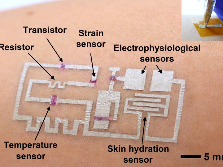
“The transistor is mechanically deformable and functionally reconfigurable, yet still retains its functions when stretched extensively,” Yu said. “It can attach to a robot or wearable device to serve as their outermost skin.”

In addition to Yu, other contributors include Hyunseok Shim and Shubham Patel, Penn State Department of Engineering Science and Mechanics; Yongcao Zhang, the University of Houston Materials Science and Engineering Program; Faheem Ershad, Penn State Department of Biomedical Engineering and University of Houston Department of Biomedical Engineering; Binghao Wang, School of Electronic Science and Engineering, Southeast University [Note: There’s one in Bangladesh, one in China, and there’s a Southeastern University in Florida, US] and Department of Chemistry and the Materials Research Center, Northwestern University; Zhihua Chen, Flexterra Inc.; Tobin J. Marks, Department of Chemistry and the Materials Research Center, Northwestern University; Antonio Facchetti, Flexterra Inc. and Northwestern University’s Department of Chemistry and Materials Research Center.

Here’s a link to and a citation for the paper,

An elastic and reconfigurable synaptic transistor based on a stretchable bilayer semiconductor by Hyunseok Shim, Faheem Ershad, Shubham Patel, Yongcao Zhang, Binghao Wang, Zhihua Chen, Tobin J. Marks, Antonio Facchetti & Cunjiang Yu. Nature Electronics (2022) DOI: DOI: https://doi.org/10.1038/s41928-022-00836-5 Published: 29 September 2022

This paper is behind a paywall.

You mean Fitbit makes mistakes? More accuracy with ‘drawn-on-skin’ electronics

A July 30, 2020 news item on ScienceDaily announces news about more accurate health monitoring with electronics applied directly to your skin,

A team of researchers led by Cunjiang Yu, Bill D. Cook Associate Professor of Mechanical Engineering at the University of Houston, has developed a new form of electronics known as “drawn-on-skin electronics,” allowing multifunctional sensors and circuits to be drawn on the skin with an ink pen.

The advance, the researchers report in Nature Communications, allows for the collection of more precise, motion artifact-free health data, solving the long-standing problem of collecting precise biological data through a wearable device when the subject is in motion.

The imprecision may not be important when your FitBit registers 4,000 steps instead of 4,200, but sensors designed to check heart function, temperature and other physical signals must be accurate if they are to be used for diagnostics and treatment.

A July 30, 2020 University of Houston news release (also on EurekAlert) by Jeannie Kever, which originated the news item, goes on to explain why you might want to have electronics ‘drawn on your skin’,

The drawn-on-skin electronics are able to seamlessly collect data, regardless of the wearer’s movements.  

They also offer other advantages, including simple fabrication techniques that don’t require dedicated equipment.

“It is applied like you would use a pen to write on a piece of paper,” said Yu. “We prepare several electronic materials and then use pens to dispense them. Coming out, it is liquid. But like ink on paper, it dries very quickly.”

Wearable bioelectronics – in the form of soft, flexible patches attached to the skin – have become an important way to monitor, prevent and treat illness and injury by tracking physiological information from the wearer. But even the most flexible wearables are limited by motion artifacts, or the difficulty that arises in collecting data when the sensor doesn’t move precisely with the skin.

The drawn-on-skin electronics can be customized to collect different types of information, and Yu said it is expected to be especially useful in situations where it’s not possible to access sophisticated equipment, including on a battleground.

The electronics are able to track muscle signals, heart rate, temperature and skin hydration, among other physical data, he said. The researchers also reported that the drawn-on-skin electronics have demonstrated the ability to accelerate healing of wounds.

In addition to Yu, researchers involved in the project include Faheem Ershad, Anish Thukral, Phillip Comeaux, Yuntao Lu, Hyunseok Shim, Kyoseung Sim, Nam-In Kim, Zhoulyu Rao, Ross Guevara, Luis Contreras, Fengjiao Pan, Yongcao Zhang, Ying-Shi Guan, Pinyi Yang, Xu Wang and Peng Wang, all from the University of Houston, and Jiping Yue and Xiaoyang Wu from the University of Chicago.

The drawn-on-skin electronics are actually comprised of three inks, serving as a conductor, semiconductor and dielectric.

“Electronic inks, including conductors, semiconductors, and dielectrics, are drawn on-demand in a freeform manner to develop devices, such as transistors, strain sensors, temperature sensors, heaters, skin hydration sensors, and electrophysiological sensors,” the researchers wrote.

This research is supported by the Office of Naval Research and National Institutes of Health.

Caption: A new form of electronics known as “drawn-on-skin electronics” allows multifunctional sensors and circuits to be drawn on the skin with an ink pen. Credit: University of Houston

Here’s a link to and a citation for the paper,

Ultra-conformal drawn-on-skin electronics for multifunctional motion artifact-free sensing and point-of-care treatment by Faheem Ershad, Anish Thukral, Jiping Yue, Phillip Comeaux, Yuntao Lu, Hyunseok Shim, Kyoseung Sim, Nam-In Kim, Zhoulyu Rao, Ross Guevara, Luis Contreras, Fengjiao Pan, Yongcao Zhang, Ying-Shi Guan, Pinyi Yang, Xu Wang, Peng Wang, Xiaoyang Wu & Cunjiang Yu. Nature Communications volume 11, Article number: 3823 (2020) DOI: https://doi.org/10.1038/s41467-020-17619-1

This paper is open access.

In the future your clothing may be a health monitor

It’s not ready for the COVID-19 pandemic but if I understand it properly, wearing this clothing will be a little like wearing a thermometer and that could be very useful. A March 4, 2020 news item on Nanowerk announces the research (Note: A link has been removed),

Researchers have reported a new material, pliable enough to be woven into fabric but imbued with sensing capabilities that can serve as an early warning system for injury or illness.

The material, described in a paper published by ACS Applied Nano Materials (“Poly(octadecyl acrylate)-Grafted Multiwalled Carbon Nanotube Composites for Wearable Temperature Sensors”), involves the use of carbon nanotubes and is capable of sensing slight changes in body temperature while maintaining a pliable disordered structure – as opposed to a rigid crystalline structure – making it a good candidate for reusable or disposable wearable human body temperature sensors. Changes in body heat change the electrical resistance, alerting someone monitoring that change to the potential need for intervention.

I think this is an artistic rendering of the research,

Caption: Researchers have reported a new material, pliable enough to be woven into fabric but imbued with sensing capabilities that could serve as an early warning system for injury or illness. Credit: University of Houston

A March 4, 2020 University of Houston (Texas, US) news release (also on EurekAlert) by Jeannie Kever, which originated the news item, describes the work in more detail,

“Your body can tell you something is wrong before it becomes obvious,” said Seamus Curran, a physics professor at the University of Houston and co-author on the paper. Possible applications range from detecting dehydration in an ultra-marathoner to the beginnings of a pressure sore in a nursing home patient.

The researchers said it is also cost-effective because the raw materials required are used in relatively low concentrations.

The discovery builds on work Curran and fellow researchers Kang-Shyang Liao and Alexander J. Wang began nearly a decade ago, when they developed a hydrophobic nanocoating for cloth, which they envisioned as a protective coating for clothing, carpeting and other fiber-based materials.

Wang is now a Ph.D. student at Technological University Dublin, currently working with Curran at UH, and is corresponding author for the paper. In addition to Curran and Liao, other researchers involved include Surendra Maharjan, Brian P. McElhenny, Ram Neupane, Zhuan Zhu, Shuo Chen, Oomman K. Varghese and Jiming Bao, all of UH; Kourtney D. Wright and Andrew R. Barron of Rice University, and Eoghan P. Dillon of Analysis Instruments in Santa Barbara.

The material, created using poly(octadecyl acrylate)-grafted multiwalled carbon nanotubes, is technically known as a nanocarbon-based disordered, conductive, polymeric nanocomposite, or DCPN, a class of materials increasingly used in materials science. But most DCPN materials are poor electroconductors, making them unsuitable for use in wearable technologies that require the material to detect slight changes in temperature.

The new material was produced using a technique called RAFT-polymerization, Wang said, a critical step that allows the attached polymer to be electronically and phononically coupled with the multiwalled carbon nanotube through covalent bonding. As such, subtle structural arrangements associated with the glass transition temperature of the system are electronically amplified to produce the exceptionally large electronic responses reported in the paper, without the negatives associated with solid-liquid phase transitions. The subtle structural changes associated with glass transition processes are ordinarily too small to produce large enough electronic responses.

Here’s a link to and a citation for the paper,

Poly(octadecyl acrylate)-Grafted Multiwalled Carbon Nanotube Composites for Wearable Temperature Sensors by Alexander J. Wang, Surendra Maharjan, Kang-Shyang Liao, Brian P. McElhenny, Kourtney D. Wright, Eoghan P. Dillon, Ram Neupane, Zhuan Zhu, Shuo Chen, Andrew R. Barron, Oomman K. Varghese, Jiming Bao, Seamus A. Curran. ACS Appl. Nano Mater. 2020, XXXX, XXX, XXX-XXX DOI: https://doi.org/10.1021/acsanm.9b02396 (Online) Publication Date:January 28, 2020 Copyright © 2020 American Chemical Society

This paper is behind a paywall.

So thin and soft you don’t notice it: new wearable tech

An August 2, 2019 news item on ScienceDaily features some new work on wearable technology that was a bit of a surprise to me,

Wearable human-machine interfaces — devices that can collect and store important health information about the wearer, among other uses — have benefited from advances in electronics, materials and mechanical designs. But current models still can be bulky and uncomfortable, and they can’t always handle multiple functions at one time.

Researchers reported Friday, Aug. 2 [2019], the discovery of a multifunctional ultra-thin wearable electronic device that is imperceptible to the wearer.

I expected this wearable technology to be a piece of clothing that somehow captured health data but it’s not,

While a health care application is mentioned early in the August 2, 2019 University of Houston news release (also on EurekAlert) by Jeannie Kever the primary interest seems to be robots and robotic skin (Note: This news release originated the news item on ScienceDaily),

The device allows the wearer to move naturally and is less noticeable than wearing a Band-Aid, said Cunjiang Yu, Bill D. Cook Associate Professor of Mechanical Engineering at the University of Houston and lead author for the paper, published as the cover story in Science Advances.

“Everything is very thin, just a few microns thick,” said Yu, who also is a principal investigator at the Texas Center for Superconductivity at UH. “You will not be able to feel it.”
It has the potential to work as a prosthetic skin for a robotic hand or other robotic devices, with a robust human-machine interface that allows it to automatically collect information and relay it back to the wearer.

That has applications for health care – “What if when you shook hands with a robotic hand, it was able to instantly deduce physical condition?” Yu asked – as well as for situations such as chemical spills, which are risky for humans but require human decision-making based on physical inspection.

While current devices are gaining in popularity, the researchers said they can be bulky to wear, offer slow response times and suffer a drop in performance over time. More flexible versions are unable to provide multiple functions at once – sensing, switching, stimulation and data storage, for example – and are generally expensive and complicated to manufacture.

The device described in the paper, a metal oxide semiconductor on a polymer base, offers manufacturing advantages and can be processed at temperatures lower than 300 C.

“We report an ultrathin, mechanically imperceptible, and stretchable (human-machine interface) HMI device, which is worn on human skin to capture multiple physical data and also on a robot to offer intelligent feedback, forming a closed-loop HMI,” the researchers wrote. “The multifunctional soft stretchy HMI device is based on a one-step formed, sol-gel-on-polymer-processed indium zinc oxide semiconductor nanomembrane electronics.”

In addition to Yu, the paper’s co-authors include first author Kyoseung Sim, Zhoulyu Rao, Faheem Ershad, Jianming Lei, Anish Thukral and Jie Chen, all of UH; Zhanan Zou and Jianliang Xiao, both of the University of Colorado; and Qing-An Huang of Southeast University in Nanjing, China.

Here’s a link to and a citation for the paper,

Metal oxide semiconductor nanomembrane–based soft unnoticeable multifunctional electronics for wearable human-machine interfaces by Kyoseung Sim, Zhoulyu Rao, Zhanan Zou, Faheem Ershad, Jianming Lei, Anish Thukral, Jie Chen, Qing-An Huang, Jianliang Xiao and Cunjiang Yu. Science Advances 02 Aug 2019: Vol. 5, no. 8, eaav9653 DOI: 10.1126/sciadv.aav9653

This paper appears to be open access.

Better and greener oil recovery

A June 27, 2016 news item on phys.org describes research on achieving better oil recovery,

As oil producers struggle to adapt to lower prices, getting as much oil as possible out of every well has become even more important, despite concerns from nearby residents that some chemicals used to boost production may pollute underground water resources.

Researchers from the University of Houston have reported the discovery of a nanotechnology-based solution that could address both issues – achieving 15 percent tertiary oil recovery at low cost, without the large volume of chemicals used in most commercial fluids.

A June 27, 2016 University of Houston news release (also on EurekAlert) by Jeannie Kever, which originated the news item, provides more detail,

The solution – graphene-based Janus amphiphilic nanosheets – is effective at a concentration of just 0.01 percent, meeting or exceeding the performance of both conventional and other nanotechnology-based fluids, said Zhifeng Ren, MD Anderson Chair professor of physics. Janus nanoparticles have at least two physical properties, allowing different chemical reactions on the same particle.

The low concentration and the high efficiency in boosting tertiary oil recovery make the nanofluid both more environmentally friendly and less expensive than options now on the market, said Ren, who also is a principal investigator at the Texas Center for Superconductivity at UH. He is lead author on a paper describing the work, published June 27 [2016] in the Proceedings of the National Academy of Sciences.

“Our results provide a novel nanofluid flooding method for tertiary oil recovery that is comparable to the sophisticated chemical methods,” they wrote. “We anticipate that this work will bring simple nanofluid flooding at low concentration to the stage of oilfield practice, which could result in oil being recovered in a more environmentally friendly and cost-effective manner.”

In addition to Ren, researchers involved with the project include Ching-Wu “Paul” Chu, chief scientist at the Texas Center for Superconductivity at UH; graduate students Dan Luo and Yuan Liu; researchers Feng Wang and Feng Cao; Richard C. Willson, professor of chemical and biomolecular engineering; and Jingyi Zhu, Xiaogang Li and Zhaozhong Yang, all of Southwest Petroleum University in Chengdu, China.

The U.S. Department of Energy estimates as much as 75 percent of recoverable reserves may be left after producers capture hydrocarbons that naturally rise to the surface or are pumped out mechanically, followed by a secondary recovery process using water or gas injection.

Traditional “tertiary” recovery involves injecting a chemical mix into the well and can recover between 10 percent and 20 percent, according to the authors.

But the large volume of chemicals used in tertiary oil recovery has raised concerns about potential environmental damage.

“Obviously simple nanofluid flooding (containing only nanoparticles) at low concentration (0.01 wt% or less) shows the greatest potential from the environmental and economic perspective,” the researchers wrote.

Previously developed simple nanofluids recover less than 5 percent of the oil when used at a 0.01 percent concentration, they reported. That forces oil producers to choose between a higher nanoparticle concentration – adding to the cost – or mixing with polymers or surfactants.

In contrast, they describe recovering 15.2 percent of the oil using their new and simple nanofluid at that concentration – comparable to chemical methods and about three times more efficient than other nanofluids.

Dan Luo, a UH graduate student and first author on the paper, said when the graphene-based fluid meets with the brine/oil mixture in the reservoir, the nanosheets in the fluid spontaneously go to the interface, reducing interfacial tension and helping the oil flow toward the production well.

Ren said the solution works in a completely new way.

“When it is injected, the solution helps detach the oil from the rock surface,” he said. Under certain hydrodynamic conditions, the graphene-based fluid forms a strong elastic and recoverable film at the oil and water interface, instead of forming an emulsion, he said.

Researchers said the difference is due to the asymmetric property of the 2-dimensional material. Nanoparticles are usually either hydrophobic – water-repelling, like oil – or hydrophilic, water-like, said Feng Wang, a post-doctoral researcher who shared first author-duties with Luo.

“Ours is both,” he said. “Ours is Janus and also strictly amphiphilic.”

Here’s a link to and a citation for the paper,

Nanofluid of graphene-based amphiphilic Janus nanosheets for tertiary or enhanced oil recovery: High performance at low concentration by Dan Luo, Feng Wang, Jingyi Zhu, Feng Cao, Yuan Liu, Xiaogang Li, Richard C. Willson, Zhaozhong Yang, Ching-Wu Chu, and Zhifeng Ren. PNAS 2016 doi: 10.1073/pnas.1608135113 published ahead of print June 27, 2016,

This paper is behind a paywall.

Turning gold into see-through rubber for an updated Rumpelstiltskin story

Rumpelstiltskin is a fairy tale whereby a young girl is trapped by her father’s lie that she can spin straw into gold. She is forced to spin gold by the King under pain of execution when an imp offers to help in exchange for various goods. As she succeeds each time, the King demands more until finally she has nothing left to trade for the imp’s help. Well, there is one last thing: her first-born child. She agrees to the bargain and she marries the King. On the birth of their first child, the imp reappears and under pressure of her pleas makes one last bargain. She must guess his name which she does, Rumplestiltskin. (The full story along with variants is here in this Wikipedia entry.)

With this latest research, we have a reverse Rumpelstiltskin story where gold is turned into something else according to a June 13, 2016 news item on Nanowerk (Note: A link has been removed),

Flexible solar panels that could be rolled up for easy transport and other devices would benefit from transparent metal electrodes that can conduct electricity, are stretchable, and resist damage following repeated stretching. Researchers found that topology and the adhesion between a metal nanomesh and the underlying substrate played key roles in creating such materials. The metal nanomesh can be stretched to three times its length while maintaining a transparency comparable to similar commercial materials used in solar cells and flat panel displays. Also, nanomeshes on pre-stretched slippery substrates led to electrodes that didn’t wear out, even after being stretched 50,000 times (Proceedings of the National Academy of Sciences, “Fatigue-free, superstretchable, transparent, and biocompatible metal electrodes”).

Tuning topology and adhesion of metal nanomeshes has led to super stretchable, transparent electrodes that don’t wear out. The scanning electron microscopy image shows the structure of a gold mesh created with a special lithographic technique that controlled the dimensions of the mesh structure. Optimizing this structure and its adhesion to the substrate was key to achieving super stretchability and long lifetimes in use—nanomeshes on pre-stretched slippery substrates did not show signs of wear even after repeated stretching, up to 50,000 cycles.

Tuning topology and adhesion of metal nanomeshes has led to super stretchable, transparent electrodes that don’t wear out. The scanning electron microscopy image shows the structure of a gold mesh created with a special lithographic technique that controlled the dimensions of the mesh structure. Optimizing this structure and its adhesion to the substrate was key to achieving super stretchability and long lifetimes in use—nanomeshes on pre-stretched slippery substrates did not show signs of wear even after repeated stretching, up to 50,000 cycles.

A June 9, 2016 US Dept. of Energy news release,which originated the news item, provides more detail,

Next-generation flexible electronics require highly stretchable and transparent electrodes. Fatigue, structural damage due to repeated use, is deadly in metals as it leads to poor conductivity and it commonly occurs in metals with repeated stretching—even with short elongations. However, few electronic conductors are transparent and stretchable, even fewer can be cyclically stretched to a large strain without causing fatigue. Now researchers led by the University of Houston found that optimizing topology of a metal nanomesh and its adhesion to an underlying substrate improved stretchability and eliminated fatigue, while maintaining transparency. A special lithographic technique called “grain boundary lithography” controlled the dimensions of the mesh structure. The metal nanomesh remained transparent after being stretched to three times its length. Gold nanomeshes on prestretched slippery substrates impressively showed no wear when stretched 50,000 times. The slippery surface advantageously allowed the structure of the nanomesh to reorient to relax the stress. Such electrically conductive, flexible, and transparent electrodes could lead to next-generation flexible electronics such as advanced solar cells.  The nanomesh electrodes are also promising for implantable electronics because the nanomeshes are biocompatible.

Here’s a link to and a citation for the paper,

Fatigue-free, superstretchable, transparent, and biocompatible metal electrodes by Chuan Fei Guo, Qihan Liu, Guohui Wang, Yecheng Wang, Zhengzheng Shi, Zhigang Suo, Ching-Wu Chu, and Zhifeng Ren. Proceedings of the National Academy of Sciences, vol. 112 no. 40,  12332–12337, doi: 10.1073/pnas.1516873112

This paper appears to be open access.

A fatigue-free stretchable conductor for foldable electronics

There’s been a lot of talk about foldable, stretchable, and/or bendable electronics, which is exciting in itself but I find this work on developing a fatigue-free conductor particularly intriguing. After all, who hasn’t purchased something that stretches, folds, etc. only to find that it becomes ‘fatigued’ and is now ‘stretched out’.

A Sept. 23, 2015 news item on Azonano describes the new conductors,

Researchers have discovered a new stretchable, transparent conductor that can be folded or stretched and released, resulting in a large curvature or a significant strain, at least 10,000 times without showing signs of fatigue.

This is a crucial step in creating a new generation of foldable electronics – think a flat-screen television that can be rolled up for easy portability – and implantable medical devices. The work, published Monday [Sept. 21, 2015] in the Proceedings of the National Academy of Sciences, pairs gold nanomesh with a stretchable substrate made with polydimethylsiloxane, or PDMS.

The research is the result of an international collaboration including the University of Houston (US), Harvard University (US), Methodist Research Institute (US), Zhengzhou University (China), Lawrence Berkeley National Laboratory (LBNL; US).

A Sept. 22, 2015 University of Houston news release by Jeannie Kever, which originated the news item, describes this -fatigue-free material in more detail,

The substrate is stretched before the gold nanomesh is placed on it – a process known as “prestretching” – and the material showed no sign of fatigue when cyclically stretched to a strain of more than 50 percent.

The gold nanomesh also proved conducive to cell growth, indicating it is a good material for implantable medical devices.

Fatigue is a common problem for researchers trying to develop a flexible, transparent conductor, making many materials that have good electrical conductivity, flexibility and transparency – all three are needed for foldable electronics – wear out too quickly to be practical, said Zhifeng Ren, a physicist at the University of Houston and principal investigator at the Texas Center for Superconductivity, who was the lead author for the paper.

The new material, produced by grain boundary lithography, solves that problem, he said.

In addition to Ren, other researchers on the project included Chuan Fei Guo and Ching-Wu “Paul” Chu, both from UH; Zhigang Suo, Qihan Liu and Yecheng Wang, all from Harvard University, and Guohui Wang and Zhengzheng Shi, both from the Houston Methodist Research Institute.

In materials science, “fatigue” is used to describe the structural damage to a material caused by repeated movement or pressure, known as “strain cycling.” Bend a material enough times, and it becomes damaged or breaks.    That means the materials aren’t durable enough for consumer electronics or biomedical devices.

“Metallic materials often exhibit high cycle fatigue, and fatigue has been a deadly disease for metals,” the researchers wrote.

“We weaken the constraint of the substrate by making the interface between the Au (gold) nanomesh and PDMS slippery, and expect the Au nanomesh to achieve superstretchability and high fatigue resistance,” they wrote in the paper. “Free of fatigue here means that both the structure and the resistance do not change or have little change after many strain cycles.”

As a result, they reported, “the Au nanomesh does not exhibit strain fatigue when it is stretched to 50 percent for 10,000 cycles.”

Many applications require a less dramatic stretch – and many materials break with far less stretching – so the combination of a sufficiently large range for stretching and the ability to avoid fatigue over thousands of cycles indicates a material that would remain productive over a long period of time, Ren said.

The grain boundary lithography involved a bilayer lift-off metallization process, which included an indium oxide mask layer and a silicon oxide sacrificial layer and offers good control over the dimensions of the mesh structure.

The researchers used mouse embryonic fibroblast cells to determine biocompatibility; that, along with the fact that the stretchability of gold nanomesh on a slippery substrate resembles the bioenvironment of tissue or organ surfaces, suggest the nanomesh “might be implanted in the body as a pacemaker electrode, a connection to nerve endings or the central nervous system, a beating heart, and so on,” they wrote.

Here’s a link to and citation for the paper,

Fatigue-free, superstretchable, transparent, and biocompatible metal electrodes by Chuan Fei Guo, Qihan Liu, Guohui Wang, Yecheng Wang, Zhengzheng Shi, Zhigang Suo, Ching-Wu Chu, and Zhifeng Ren. PNAS (Proceedings of the National Academy of Sciences)  doi: 10.1073/pnas.1516873112 Published online Sept. 21, 2015

This paper appears to be open access.

Following the sound of a nanoparticle through the body

I was hoping for some actual sound files of nanoparticles in the body but for some rason the researchers don’t seem to have made them freely available. However, there is this textual description in a Sept. 5, 2014 news item on Nanowerk,

Nanoparticles have become interesting means for biomedical applications. Thanks to their minute dimensions and large surface areas, they can often penetrate cellular membranes and deliver high payloads of targeting agents and drugs to achieve better specificity and therapeutic effects than non-targeted treatments. Yet, quantitative in vivo measurements of nanoparticle concentrations are essential for nanotechnology-based preclinical research.

To date, tedious ex vivo analysis of nanoparticle concentrations in organs of test animals remains a standard approach in such biodistribution studies. Most current imaging methods remain limited due to several disadvantages and/or high costs. Optoacoustic tomography (OAT), a method that utilizes ultrasound generated by absorption of nanosecond-scale laser pulses to recreate an image of the absorbing volume based on the spatial variation of optical absorption coefficients, is a potential alternative.

Usually, due to the unknown light distribution in a complex optical scattering environment, tomographic images of live animals contain only qualitative information and are not suitable for quantitative biodistribution analysis. …

A Sept. 3, 2014 Wiley-VCH publishers press release by K. Maedefessel-Herrmann, which originated the news item, provides more details about the work,

… A team of researchers from TomoWave Laboratories, Inc., Rice University, and the University of Houston now developed a methodology to correlate changes in optoacoustic signal intensity from organs of live animals detected with OAT in relation to changes of optical absorption coefficient in those organs caused by nanoparticle accumulation.

The researchers quantified localized OAT brightness changes induced by accumulation of single-walled carbon nanotubes (SWCNTs) in liver, kidney and spleen of nude mice. Using the intrinsic fluorescence properties of disaggregated nanotubes, they measured SWCNT concentrations in the parts-per-million range in the harvested organs and defined the corresponding changes in optical absorption coefficient. The observed increases in optoacoustic signal brightness in tissues were compared with the increases in optical absorption coefficients caused by SWCNT accumulation.
The combination of these methods allows one to perform sensitivity calibration of an OAT system for a selected type of animal and for a range of optical absorption coefficient values of their organs to enable non-invasive concentration measurements of optically absorbing nanoparticles and dyes in vivo.

Here’s a link to and a citation for the paper,

Enabling in vivo measurements of nanoparticle concentrations with three-dimensional optoacoustic tomography by Dmitri A. Tsyboulski, Anton V. Liopo, Richard Su, Sergey A. Ermilov, Sergei M. Bachilo, R. Bruce Weisman, and Alexander A. Oraevsky. Journal of Biophotonics, Volume 7, Issue 8, pages 581–588, August 2014. DOI: 10.1002/jbio.201200233  Article first published online: 2 APR 2013

This is an open access article.

Extracting biomolecules from live cells with carbon nanotubes

Being able to extract biomolecules from living cells means nondestruction of the rest of the cell and the ability to observe the consequences of the extraction. From a July 18, 2014 news item on Azonano,

University of Houston researchers have devised a new method for extracting molecules from live cells without disrupting cell development, work that could provide new avenues for the diagnosis of cancer and other diseases.

The researchers used magnetized carbon nanotubes to extract biomolecules from live cells, allowing them to retrieve molecular information without killing the individual cells. A description of the work appears this week in the Proceedings of the National Academy of Sciences.

A July 16, 2014 University of Houston news release by Jeannie Kever, which originated the news item, provides more detail,

Most current methods of identifying intracellular information result in the death of the individual cells, making it impossible to continue to gain information and assess change over time, said Zhifeng Ren, M.D. Anderson Chair professor of physics and principal investigator at the Center for Superconductivity at UH and lead author of the paper. The work was a collaboration between Ren’s lab and that of Paul Chu, T.L.L. Temple Chair of Science and founding director of the Texas Center for Superconductivity.

Chu, a co-author of the paper, said the new technique will allow researchers to draw fundamental information from a single cell.The researchers said the steps outlined in the paper offer proof of concept. Ren said the next step “will be more study of the biological and chemical processes of the cell, more analysis.”

The initial results hold promise for biomedicine, he said.  “This shows how nanoscience and nanoengineering can help the medical field.”

Cai said the new method will be helpful for cancer drug screening and carcinogenesis study, as well as for studies that allow researchers to obtain information from single cells, replacing previous sampling methods that average out cellular diversity and obscure the specificity of the biomarker profiles.

In the paper, the researchers explain their rationale for the work – most methods for extracting molecular information result in cell death, and those that do spare the cell carry special challenges, including limited efficiency.

This method is relatively straightforward, requiring the use of magnetized carbon nanotubes as the transporter and a polycarbonate filter as a collector, they report. Cells from a human embryonic kidney cancer cell line were used for the experiment.

The work builds on a 2005 paper published by Ren’s group in Nature Methods, which established that magnetized carbon nanotubes can deliver molecular payloads into cells. The current research takes that one step further to move molecules out of cells by magnetically driving them through the cell walls.

The carbon nanotubes were grown with a plasma-enhanced chemical vapor deposition system, with magnetic nickel particles enclosed at the tips. A layer of nickel was also deposited along the surface of individual nanotubes in order to make the nanotubes capable of penetrating a cell wall guided by a magnet.

Here’s a link to and a citation for the paper,

Molecular extraction in single live cells by sneaking in and out magnetic nanomaterials by Zhen Yang, Liangzi Deng, Yucheng Lan, Xiaoliu Zhang, Zhonghong Gao, Ching-Wu Chu, Dong Cai, and Zhifeng Ren. PNAS 2014 ; published ahead of print July 16, 2014, doi:10.1073/pnas.1411802111

This paper is behind a paywall.

Ferroelectric switching in the lung, heart, and arteries

A June 23, 2014 University of Washington (state) news release (also on EurekAlert) describes how the human body (and other biological tissue) is capable of generating ferroelectricity,

University of Washington researchers have shown that a favorable electrical property is present in a type of protein found in organs that repeatedly stretch and retract, such as the lungs, heart and arteries. These findings are the first that clearly track this phenomenon, called ferroelectricity, occurring at the molecular level in biological tissues.

The news release gives a brief description of ferroelectricity and describes the research team’s latest work with biological tissues,

Ferroelectricity is a response to an electric field in which a molecule switches from having a positive to a negative charge. This switching process in synthetic materials serves as a way to power computer memory chips, display screens and sensors. This property only recently has been discovered in animal tissues and researchers think it may help build and support healthy connective tissues in mammals.

A research team led by Li first discovered ferroelectric properties in biological tissues in 2012, then in 2013 found that glucose can suppress this property in the body’s connective tissues, wherever the protein elastin is present. But while ferroelectricity is a proven entity in synthetic materials and has long been thought to be important in biological functions, its actual existence in biology hasn’t been firmly established.

This study proves that ferroelectric switching happens in the biological protein elastin. When the researchers looked at the base structures within the protein, they saw similar behavior to the unit cells of solid-state materials, where ferroelectricity is well understood.

“When we looked at the smallest structural unit of the biological tissue and how it was organized into a larger protein fiber, we then were able to see similarities to the classic ferroelectric model found in solids,” Li said.

The researchers wanted to establish a more concrete, precise way of verifying ferroelectricity in biological tissues. They used small samples of elastin taken from a pig’s aorta and poled the tissues using an electric field at high temperatures. They then measured the current with the poling field removed and found that the current switched direction when the poling electric field was switched, a sign of ferroelectricity.

They did the same thing at room temperature using a laser as the heat source, and the current also switched directions.

Then, the researchers tested for this behavior on the smallest-possible unit of elastin, called tropoelastin, and again observed the phenomenon. They concluded that this switching property is “intrinsic” to the molecular make-up of elastin.

The next step is to understand the biological and physiological significance of this property, Li said. One hypothesis is that if ferroelectricity helps elastin stay flexible and functional in the body, a lack of it could directly affect the hardening of arteries.

“We may be able to use this as a very sensitive technique to detect the initiation of the hardening process at a very early stage when no other imaging technique will be able to see it,” Li said.

The team also is looking at whether this property plays a role in normal biological functions, perhaps in regulating the growth of tissue.

Co-authors are Pradeep Sharma at the University of Houston, Yanhang Zhang at Boston University, and collaborators at Nanjing University and the Chinese Academy of Sciences.

Here’s a link to and a citation for the research paper,

Ferroelectric switching of elastin by Yuanming Liu, Hong-Ling Cai, Matthew Zelisko, Yunjie Wang, Jinglan Sun, Fei Yan, Feiyue Ma, Peiqi Wang, Qian Nataly Chen, Hairong Zheng, Xiangjian Meng, Pradeep Sharma, Yanhang Zhang, and Jiangyu Li. Proceedings of the National Academy of Sciences (PNAS) doi: 10.1073/pnas.1402909111

This paper is behind a paywall.

I think this is a new practice. There is a paragraph on the significance of this work (follow the link to the paper),

Ferroelectricity has long been speculated to have important biological functions, although its very existence in biology has never been firmly established. Here, we present, to our knowledge, the first macroscopic observation of ferroelectric switching in a biological system, and we elucidate the origin and mechanism underpinning ferroelectric switching of elastin. It is discovered that the polarization in elastin is intrinsic at the monomer level, analogous to the unit cell level polarization in classical perovskite ferroelectrics. Our findings settle a long-standing question on ferroelectric switching in biology and establish ferroelectricity as an important biophysical property of proteins. We believe this is a critical first step toward resolving its physiological significance and pathological implications.