Tag Archives: University of Liverpool

A view to controversies about nanoparticle drug delivery, sticky-flares, and a PNAS surprise

Despite all the excitement and claims for nanoparticles as vehicles for drug delivery to ‘sick’ cells there is at least one substantive problem, the drug-laden nanoparticles don’t actually enter the interior of the cell. They are held in a kind of cellular ‘waiting room’.

Leonid Schneider in a Nov. 20, 2015 posting on his For Better Science blog describes the process in more detail,

A large body of scientific nanotechnology literature is dedicated to the biomedical aspect of nanoparticle delivery into cells and tissues. The functionalization of the nanoparticle surface is designed to insure their specificity at targeting only a certain type of cells, such as cancers cells. Other technological approaches aim at the cargo design, in order to ensure the targeted release of various biologically active agents: small pharmacological substances, peptides or entire enzymes, or nucleotides such as regulatory small RNAs or even genes. There is however a main limitation to this approach: though cells do readily take up nanoparticles through specific membrane-bound receptor interaction (endocytosis) or randomly (pinocytosis), these nanoparticles hardly ever truly reach the inside of the cell, namely its nucleocytoplasmic space. Solid nanoparticles are namely continuously surrounded by the very same membrane barrier they first interacted with when entering the cell. These outer-cell membrane compartments mature into endosomal and then lysosomal vesicles, where their cargo is subjected to low pH and enzymatic digestion. The nanoparticles, though seemingly inside the cell, remain actually outside. …

What follows is a stellar piece featuring counterclaims about and including Schneider’s own journalistic research into scientific claims that the problem of gaining entry to a cell’s true interior has been addressed by technologies developed in two different labs.

Having featured one of the technologies here in a July 24, 2015 posting titled: Sticky-flares nanotechnology to track and observe RNA (ribonucleic acid) regulation and having been contacted a couple of times by one of the scientists, Raphaël Lévy from the University of Liverpool (UK), challenging the claims made (Lévy’s responses can be found in the comments section of the July 2015 posting), I thought a followup of sorts was in order.

Scientific debates (then and now)

Scientific debates and controversies are part and parcel of the scientific process and what most outsiders, such as myself, don’t realize is how fraught it is. For a good example from the past, there’s Leviathan and the Air-Pump: Hobbes, Boyle, and the Experimental Life (from its Wikipedia entry), Note: Links have been removed),

Leviathan and the Air-Pump: Hobbes, Boyle, and the Experimental Life (published 1985) is a book by Steven Shapin and Simon Schaffer. It examines the debate between Robert Boyle and Thomas Hobbes over Boyle’s air-pump experiments in the 1660s.

The style seems more genteel than what a contemporary Canadian or US audience is accustomed to but Hobbes and Boyle (and proponents of both sides) engaged in bruising communication.

There was a lot at stake then and now. It’s not just the power, prestige, and money, as powerfully motivating as they are, it’s the research itself. Scientists work for years to achieve breakthroughs or to add more to our common store of knowledge. It’s painstaking and if you work at something for a long time, you tend to be invested in it. Saying you’ve wasted ten years of your life looking at the problem the wrong way or have misunderstood your data is not easy.

As for the current debate, Schneider’s description gives no indication that there is rancour between any of the parties but it does provide a fascinating view of two scientists challenging one of the US’s nanomedicine rockstars, Chad Mirkin. The following excerpt follows the latest technical breakthroughs to the interior portion of the cell through three phases of the naming conventions (Nano-Flares, also known by its trade name, SmartFlares, which is a precursor technology to Sticky-Flares), Note: Links have been removed,

The next family of allegedly nucleocytoplasmic nanoparticles which Lévy turned his attention to, was that of the so called “spherical nucleic acids”, developed in the lab of Chad Mirkin, multiple professor and director of the International Institute for Nanotechnology at the Northwestern University, USA. These so called “Nano-Flares” are gold nanoparticles, functionalized with fluorophore-coupled oligonucleotides matching the messenger RNA (mRNA) of interest (Prigodich et al., ACS Nano 3:2147-2152, 2009; Seferos et al., J Am. Chem.Soc. 129:15477-15479, 2007). The mRNA detection method is such that the fluorescence is initially quenched by the gold nanoparticle proximity. Yet when the oligonucleotide is displaced by the specific binding of the mRNA molecules present inside the cell, the fluorescence becomes detectable and serves thus as quantitative read-out for the intracellular mRNA abundance. Exactly this is where concerns arise. To find and bind mRNA, spherical nucleic acids must leave the endosomal compartments. Is there any evidence that Nano-Flares ever achieve this and reach intact the nucleocytoplasmatic space, where their target mRNA is?

Lévy’s lab has focused its research on the commercially available analogue of the Nano-Flares, based on the patent to Mirkin and Northwestern University and sold by Merck Millipore under the trade name of SmartFlares. These were described by Mirkin as “a powerful and prolific tool in biology and medical diagnostics, with ∼ 1,600 unique forms commercially available today”. The work, led by Lévy’s postdoctoral scientist David Mason, now available in post-publication process at ScienceOpen and on Figshare, found no experimental evidence for SmartFlares to be ever found outside the endosomal membrane vesicles. On the contrary, the analysis by several complementary approaches, i.e., electron, fluorescence and photothermal microscopy, revealed that the probes are retained exclusively within the endosomal compartments.

In fact, even Merck Millipore was apparently well aware of this problem when the product was developed for the market. As I learned, Merck performed a number of assays to address the specificity issue. Multiple hundred-fold induction of mRNA by biological cell stimulation (confirmed by quantitative RT-PCR) led to no significant changes in the corresponding SmartFlare signal. Similarly, biological gene downregulation or experimental siRNA knock-down had no effect on the corresponding SmartFlare fluorescence. Cell lines confirmed as negative for a certain biomarker proved highly positive in a SmartFlare assay.  Live cell imaging showed the SmartFlare signal to be almost entirely mitochondrial, inconsistent with reported patterns of the respective mRNA distributions.  Elsewhere however, cyanine dye-labelled oligonucleotides were found to unspecifically localise to mitochondria   (Orio et al., J. RNAi Gene Silencing 9:479-485, 2013), which might account to the often observed punctate Smart Flare signal.

More recently, Mirkin lab has developed a novel version of spherical nucleic acids, named Sticky-Flares (Briley et al., PNAS 112:9591-9595, 2015), which has also been patented for commercial use. The claim is that “the Sticky-flare is capable of entering live cells without the need for transfection agents and recognizing target RNA transcripts in a sequence-specific manner”. To confirm this, Lévy used the same approach as for the striped nanoparticles [not excerpted here]: he approached Mirkin by email and in person, requesting the original microscopy data from this publication. As Mirkin appeared reluctant, Lévy invoked the rules for data sharing by the journal PNAS, the funder NSF as well as the Northwestern University. After finally receiving Mirkin’s thin-optical microscopy data by air mail, Lévy and Mason re-analyzed it and determined the absence of any evidence for endosomal escape, while all Sticky-Flare particles appeared to be localized exclusively inside vesicular membrane compartments, i.e., endosomes (Mason & Levy, bioRxiv 2015).

I encourage you to read Schneider’s Nov. 20, 2015 posting in its entirety as these excerpts can’t do justice to it.

The PNAS surprise

PNAS (Proceedings of the National Academy of Science) published one of Mirkin’s papers on ‘Sticky-flares’ and is where scientists, Raphaël Lévy and David Mason, submitted a letter outlining their concerns with the ‘Sticky-flares’ research. Here’s the response as reproduced in Lévy’s Nov. 16, 2015 posting on his Rapha-Z-Lab blog

Dear Dr. Levy,

I regret to inform you that the PNAS Editorial Board has declined to publish your Letter to the Editor. After careful consideration, the Board has decided that your letter does not contribute significantly to the discussion of this paper.

Thank you for submitting your comments to PNAS.

Sincerely yours,
Inder Verma
Editor-in-Chief

Judge for yourself, Lévy’s and Mason’s letter can be found here (pdf) and here.

Conclusions

My primary interest in this story is in the view it provides of the scientific process and the importance of and difficulty associated with the debates.

I can’t venture an opinion about the research or the counterarguments other than to say that Lévy’s and Mason’s thoughtful challenge bears more examination than PNAS is inclined to accord. If their conclusions or Chad Mirkin’s are wrong, let that be determined in an open process.

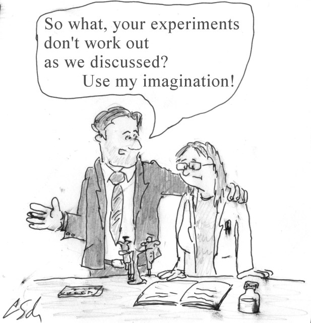
I’ll leave the very last comment to Schneider who is both writer and cartoonist, from his Nov. 20, 2015 posting,

LeonidSchneiderImagination

Nanotechnology research protocols for Environment, Health and Safety Studies in US and a nanomedicine characterization laboratory in the European Union

I have two items relating to nanotechnology and the development of protocols. The first item concerns the launch of a new web portal by the US National Institute of Standards and Technology.

US National Institute of Standards and Technology (NIST)

From a July 1, 2015 news item on Azonano,

As engineered nanomaterials increasingly find their way into commercial products, researchers who study the potential environmental or health impacts of those materials face a growing challenge to accurately measure and characterize them. These challenges affect measurements of basic chemical and physical properties as well as toxicology assessments.

To help nano-EHS (Environment, Health and Safety)researchers navigate the often complex measurement issues, the National Institute of Standards and Technology (NIST) has launched a new website devoted to NIST-developed (or co-developed) and validated laboratory protocols for nano-EHS studies.

A July 1, 2015 NIST news release on EurekAlert, which originated the news item, offers more details about the information available through the web portal,

In common lab parlance, a “protocol” is a specific step-by-step procedure used to carry out a measurement or related activity, including all the chemicals and equipment required. Any peer-reviewed journal article reporting an experimental result has a “methods” section where the authors document their measurement protocol, but those descriptions are necessarily brief and condensed, and may lack validation of any sort. By comparison, on NIST’s new Protocols for Nano-EHS website the protocols are extraordinarily detailed. For ease of citation, they’re published individually–each with its own unique digital object identifier (DOI).

The protocols detail not only what you should do, but why and what could go wrong. The specificity is important, according to program director Debra Kaiser, because of the inherent difficulty of making reliable measurements of such small materials. “Often, if you do something seemingly trivial–use a different size pipette, for example–you get a different result. Our goal is to help people get data they can reproduce, data they can trust.”

A typical caution, for example, notes that if you’re using an instrument that measures the size of nanoparticles in a solution by how they scatter light, it’s important also to measure the transmission spectrum of the particles if they’re colored, because if they happen to absorb light strongly at the same frequency as your instrument, the result may be biased.

“These measurements are difficult because of the small size involved,” explains Kaiser. “Very few new instruments have been developed for this. People are adapting existing instruments and methods for the job, but often those instruments are being operated close to their limits and the methods were developed for chemicals or bulk materials and not for nanomaterials.”

“For example, NIST offers a reference material for measuring the size of gold nanoparticles in solution, and we report six different sizes depending on the instrument you use. We do it that way because different instruments sense different aspects of a nanoparticle’s dimensions. An electron microscope is telling you something different than a dynamic light scattering instrument, and the researcher needs to understand that.”

The nano-EHS protocols offered by the NIST site, Kaiser says, could form the basis for consensus-based, formal test methods such as those published by ASTM and ISO.

NIST’s nano-EHS protocol site currently lists 12 different protocols in three categories: sample preparation, physico-chemical measurements and toxicological measurements. More protocols will be added as they are validated and documented. Suggestions for additional protocols are welcome at nanoprotocols@nist.gov.

The next item concerns European nanomedicine.

CEA-LETI and Europe’s first nanomedicine characterization laboratory

A July 1, 2015 news item on Nanotechnology Now describes the partnership which has led to launch of the new laboratory,

CEA-Leti today announced the launch of the European Nano-Characterisation Laboratory (EU-NCL) funded by the European Union’s Horizon 2020 research and innovation programm[1]e. Its main objective is to reach a level of international excellence in nanomedicine characterisation for medical indications like cancer, diabetes, inflammatory diseases or infections, and make it accessible to all organisations developing candidate nanomedicines prior to their submission to regulatory agencies to get the approval for clinical trials and, later, marketing authorization.

“As reported in the ETPN White Paper[2], there is a lack of infrastructure to support nanotechnology-based innovation in healthcare,” said Patrick Boisseau, head of business development in nanomedicine at CEA-Leti and chairman of the European Technology Platform Nanomedicine (ETPN). “Nanocharacterisation is the first bottleneck encountered by companies developing nanotherapeutics. The EU-NCL project is of most importance for the nanomedicine community, as it will contribute to the competiveness of nanomedicine products and tools and facilitate regulation in Europe.”

EU-NCL is partnered with the sole international reference facility, the Nanotechnology Characterization Lab of the National Cancer Institute in the U.S. (US-NCL)[3], to get faster international harmonization of analytical protocols.

“We are excited to be part of this cooperative arrangement between Europe and the U.S.,” said Scott E. McNeil, director of U.S. NCL. “We hope this collaboration will help standardize regulatory requirements for clinical evaluation and marketing of nanomedicines internationally. This venture holds great promise for using nanotechnologies to overcome cancer and other major diseases around the world.”

A July 2, 2015 EMPA (Swiss Federal Laboratories for Materials Science and Technology) news release on EurekAlert provides more detail about the laboratory and the partnerships,

The «European Nanomedicine Characterization Laboratory» (EU-NCL), which was launched on 1 June 2015, has a clear-cut goal: to help bring more nanomedicine candidates into the clinic and on the market, for the benefit of patients and the European pharmaceutical industry. To achieve this, EU-NCL is partnered with the sole international reference facility, the «Nanotechnology Characterization Laboratory» (US-NCL) of the US-National Cancer Institute, to get faster international harmonization of analytical protocols. EU-NCL is also closely connected to national medicine agencies and the European Medicines Agency to continuously adapt its analytical services to requests of regulators. EU-NCL is designed, organized and operated according to the highest EU regulatory and quality standards. «We are excited to be part of this cooperative project between Europe and the U.S.,» says Scott E. McNeil, director of US-NCL. «We hope this collaboration will help standardize regulatory requirements for clinical evaluation and marketing of nanomedicines internationally. This venture holds great promise for using nanotechnologies to overcome cancer and other major diseases around the world.»

Nine partners from eight countries

EU-NCL, which is funded by the EU for a four-year period with nearly 5 million Euros, brings together nine partners from eight countries: CEA-Tech in Leti and Liten, France, the coordinator of the project; the Joint Research Centre of the European Commission in Ispra, Italy; European Research Services GmbH in Münster Germany; Leidos Biomedical Research, Inc. in Frederick, USA; Trinity College in Dublin, Ireland; SINTEF in Oslo, Norway; the University of Liverpool in the UK; Empa, the Swiss Federal Laboratories for Materials Science and Technology in St. Gallen, Switzerland; Westfälische Wilhelms-Universität (WWU) and Gesellschaft für Bioanalytik, both in Münster, Germany. Together, the partnering institutions will provide a trans-disciplinary testing infrastructure covering a comprehensive set of preclinical characterization assays (physical, chemical, in vitro and in vivo biological testing), which will allow researchers to fully comprehend the biodistribution, metabolism, pharmacokinetics, safety profiles and immunological effects of their medicinal nano-products. The project will also foster the use and deployment of standard operating procedures (SOPs), benchmark materials and quality management for the preclinical characterization of medicinal nano-products. Yet another objective is to promote intersectoral and interdisciplinary communication among key drivers of innovation, especially between developers and regulatory agencies.

The goal: to bring safe and efficient nano-therapeutics faster to the patient

Within EU-NCL, six analytical facilities will offer transnational access to their existing analytical services for public and private developers, and will also develop new or improved analytical assays to keep EU-NCL at the cutting edge of nanomedicine characterization. A complementary set of networking activities will enable EU-NCL to deliver to European academic or industrial scientists the high-quality analytical services they require for accelerating the industrial development of their candidate nanomedicines. The Empa team of Peter Wick at the «Particles-Biology Interactions» lab will be in charge of the quality management of all analytical methods, a key task to guarantee the best possible reproducibility and comparability of the data between the various analytical labs within the consortium. «EU-NCL supports our research activities in developing innovative and safe nanomaterials for healthcare within an international network, which will actively shape future standards in nanomedicine and strengthen Empa as an enabler to facilitate the transfer of novel nanomedicines from bench to bedside», says Wick.

You can find more information about the laboratory on the Horizon 2020 (a European Union science funding programme) project page for the EU-NCL laboratory. For anyone curious about CEA-Leti, it’s a double-layered organization. CEA is France’s Commission on Atomic Energy and Alternative Energy (Commissariat à l’énergie atomique et aux énergies alternatives); you can go here to their French language site (there is an English language clickable option on the page). Leti is one of the CEA’s institutes and is known as either Leti or CEA-Leti. I have no idea what Leti stands for. Here’s the Leti website (this is the English language version).

Trapping gases left from nuclear fuels

A July 20, 2014 news item on ScienceDaily provides some insight into recycling nuclear fuel,

When nuclear fuel gets recycled, the process releases radioactive krypton and xenon gases. Naturally occurring uranium in rock contaminates basements with the related gas radon. A new porous material called CC3 effectively traps these gases, and research appearing July 20 in Nature Materials shows how: by breathing enough to let the gases in but not out.

The CC3 material could be helpful in removing unwanted or hazardous radioactive elements from nuclear fuel or air in buildings and also in recycling useful elements from the nuclear fuel cycle. CC3 is much more selective in trapping these gases compared to other experimental materials. Also, CC3 will likely use less energy to recover elements than conventional treatments, according to the authors.

A July 21, 2014 US Department of Energy (DOE) Pacific Northwest National Laboratory (PNNL) news release (also on EurekAlert), which originated the news item despite the difference in dates, provides more details (Note: A link has been removed),

The team made up of scientists at the University of Liverpool in the U.K., the Department of Energy’s Pacific Northwest National Laboratory, Newcastle University in the U.K., and Aix-Marseille Universite in France performed simulations and laboratory experiments to determine how — and how well — CC3 might separate these gases from exhaust or waste.

“Xenon, krypton and radon are noble gases, which are chemically inert. That makes it difficult to find materials that can trap them,” said coauthor Praveen Thallapally of PNNL. “So we were happily surprised at how easily CC3 removed them from the gas stream.”

Noble gases are rare in the atmosphere but some such as radon come in radioactive forms and can contribute to cancer. Others such as xenon are useful industrial gases in commercial lighting, medical imaging and anesthesia.

The conventional way to remove xenon from the air or recover it from nuclear fuel involves cooling the air far below where water freezes. Such cryogenic separations are energy intensive and expensive. Researchers have been exploring materials called metal-organic frameworks, also known as MOFs, that could potentially trap xenon and krypton without having to use cryogenics. Although a leading MOF could remove xenon at very low concentrations and at ambient temperatures admirably, researchers wanted to find a material that performed better.

Thallapally’s collaborator Andrew Cooper at the University of Liverpool and others had been researching materials called porous organic cages, whose molecular structures are made up of repeating units that form 3-D cages. Cages built from a molecule called CC3 are the right size to hold about three atoms of xenon, krypton or radon.

To test whether CC3 might be useful here, the team simulated on a computer CC3 interacting with atoms of xenon and other noble gases. The molecular structure of CC3 naturally expands and contracts. The researchers found this breathing created a hole in the cage that grew to 4.5 angstroms wide and shrunk to 3.6 angstroms. One atom of xenon is 4.1 angstroms wide, suggesting it could fit within the window if the cage opens long enough. (Krypton and radon are 3.69 angstroms and 4.17 angstroms wide, respectively, and it takes 10 million angstroms to span a millimeter.)

The computer simulations revealed that CC3 opens its windows big enough for xenon about 7 percent of the time, but that is enough for xenon to hop in. In addition, xenon has a higher likelihood of hopping in than hopping out, essentially trapping the noble gas inside.

The team then tested how well CC3 could pull low concentrations of xenon and krypton out of air, a mix of gases that included oxygen, argon, carbon dioxide and nitrogen. With xenon at 400 parts per million and krypton at 40 parts per million, the researchers sent the mix through a sample of CC3 and measured how long it took for the gases to come out the other side.

Oxygen, nitrogen, argon and carbon dioxide — abundant components of air — traveled through the CC3 and continued to be measured for the experiment’s full 45 minute span. Xenon however stayed within the CC3 for 15 minutes, showing that CC3 could separate xenon from air.

In addition, CC3 trapped twice as much xenon as the leading MOF material. It also caught xenon 20 times more often than it caught krypton, a characteristic known as selectivity. The leading MOF only preferred xenon 7 times as much. These experiments indicated improved performance in two important characteristics of such a material, capacity and selectivity.

“We know that CC3 does this but we’re not sure why. Once we understand why CC3 traps the noble gases so easily, we can improve on it,” said Thallapally.

To explore whether MOFs and porous organic cages offer economic advantages, the researchers estimated the cost compared to cryogenic separations and determined they would likely be less expensive.

“Because these materials function well at ambient or close to ambient temperatures, the processes based on them are less energy intensive to use,” said PNNL’s Denis Strachan.

The material might also find use in pharmaceuticals. Most molecules come in right- and left-handed forms and often only one form works in people. In additional experiments, Cooper and colleagues in the U.K. tested CC3’s ability to distinguish and separate left- and right-handed versions of an alcohol. After separating left- and right-handed forms of CC3, the team showed in biochemical experiments that each form selectively trapped only one form of the alcohol.

The researchers have provided an image illustrating a CC3 cage,

Breathing room: In this computer simulation, light and dark purple highlight the cavities within the 3D pore structure of CC3. Courtesy:  PNNL

Breathing room: In this computer simulation, light and dark purple highlight the cavities within the 3D pore structure of CC3. Courtesy: PNNL

Here’s a link to and a citation for the paper,

Separation of rare gases and chiral molecules by selective binding in porous organic cages by Linjiang Chen, Paul S. Reiss, Samantha Y. Chong, Daniel Holden, Kim E. Jelfs, Tom Hasell, Marc A. Little, Adam Kewley, Michael E. Briggs, Andrew Stephenson, K. Mark Thomas, Jayne A. Armstrong, Jon Bell, Jose Busto, Raymond Noel, Jian Liu, Denis M. Strachan, Praveen K. Thallapally, & Andrew I. Cooper. Nature Material (2014) doi:10.1038/nmat4035 Published online 20 July 2014

This paper is behind a paywall.

Single-element quasicrystal created in laboratory for the first time

There’s a background story which gives this breakthrough a fabulous aspect but, first, here’s the research breakthrough from a Dec. 24, 2013 news item on Nanowerk (Note: A link has been removed),

A research group led by Assistant Professor Kazuki Nozawa and Professor Yasushi Ishii from the Department of Physics, Faculty of Science and Engineering, Chuo University, Chief Researcher Masahiko Shimoda from the National Institute for Materials Science (NIMS) and Professor An-Pang Tsai from the Institute of Multidisciplinary Research for Advanced Materials (IMRAM), Tohoku University, succeeded for the first time in the world in fabricating a three-dimensional structure of a quasicrystal composed of a single element, through joint research with a group led by Dr. Hem Raj Sharma from the University of Liverpool, the United Kingdom.

The Dec. 2, 2013 National Institute for Materials Science (NIMS; Japan) press release, which originated the news item, describes quasicrystals and the reasons why this particular achievement is such a breakthrough,

Quasicrystals are substances discovered in 1984 by Dr. Dan Shechtman (who was awarded the Nobel Prize in Chemistry in 2011). [emphasis mine] To date, quasicrystals have been found in more than one hundred kinds of alloy, polymer and nanoparticle systems. However, a quasicrystal composed of a single element has not been found yet. Quasicrystals have a beautiful crystalline structure which is closely related to the golden ratio, called a quasiperiodic structure. This structure is made of a pentagonal or decagonal atomic arrangement that is not found in ordinary periodic crystals (see the reference illustrations). Due to the complexity of the crystalline structure and chemical composition, much about quasicrystals is still veiled in mystery, including the mechanism for stabilizing a quasiperiodic structure and the novel properties of this unique type of crystalline structure. For these reasons, efforts have been made for a long time in the quest for a chemically simple type of quasicrystal composed only of a single element. The joint research group has recently succeeded in growing a crystal of lead with a quasiperiodic structure which is modeled on the structure of a substrate quasicrystal, by vapor-depositing lead atoms on the quasicrystal substrate of an existing alloy made of silver (Ag), indium (In) and ytterbium (Yb). Success using this approach had been reported for fabricating a single-element quasiperiodic film consisting of a single atomic layer (two-dimensional structure), but there had been no successful case of fabricating a single-element quasiperiodic structure consisting of multiple atomic layers (three-dimensional structure). This recent success by the joint research group is a significant step forward toward achieving single-element quasicrystals. It is also expected to lead to advancement in various fields, such as finding properties unique to quasiperiodic structures that cannot be found in periodic crystals and elucidating the mechanism of stabilization of quasiperiodic structures.

Here’s an image illustrating the researchers’ achievement,

Illustrations of the deposition structure of lead. The Tsai cluster in the substrate quasicrystal which is near the surface of the substrate is cut at the point where it contacts the surface. While lead usually has a face-centered cubic structure, it is deposited on the quasicrystal substrate in a manner that it recovers Tsai clusters which are cut near the surface of the substrate. This indicates that a crystal of lead is grown with the same structure as the structure of the quasicrystal substrate. (Courtesy National Institute for Materials Science, Japan)

Illustrations of the deposition structure of lead. The Tsai cluster in the substrate quasicrystal which is near the surface of the substrate is cut at the point where it contacts the surface. While lead usually has a face-centered cubic structure, it is deposited on the quasicrystal substrate in a manner that it recovers Tsai clusters which are cut near the surface of the substrate. This indicates that a crystal of lead is grown with the same structure as the structure of the quasicrystal substrate. (Courtesy National Institute for Materials Science, Japan)

I suggested earlier that this achievement has a fabulous quality and the Daniel Schechtman backstory is the reason. The winner of the 2011 Nobel Prize for Chemistry, Schechtman was reviled for years within his scientific community as Ian Sample notes in his Oct. 5, 2011 article on the announcement of Schechtman’s Nobel win written for the Guardian newspaper (Note: A link has been removed),

A scientist whose work was so controversial he was ridiculed and asked to leave his research group has won the Nobel Prize in Chemistry.

Daniel Shechtman, 70, a researcher at Technion-Israel Institute of Technology in Haifa, received the award for discovering seemingly impossible crystal structures in frozen gobbets of metal that resembled the beautiful patterns seen in Islamic mosaics.

Images of the metals showed their atoms were arranged in a way that broke well-establised rules of how crystals formed, a finding that fundamentally altered how chemists view solid matter.

On the morning of 8 April 1982, Shechtman saw something quite different while gazing at electron microscope images of a rapidly cooled metal alloy. The atoms were packed in a pattern that could not be repeated. Shechtman said to himself in Hebrew, “Eyn chaya kazo,” which means “There can be no such creature.”

The bizarre structures are now known as “quasicrystals” and have been seen in a wide variety of materials. Their uneven structure means they do not have obvious cleavage planes, making them particularly hard.

In an interview this year with the Israeli newspaper, Haaretz, Shechtman said: “People just laughed at me.” He recalled how Linus Pauling, a colossus of science and a double Nobel laureate, mounted a frightening “crusade” against him. After telling Shechtman to go back and read a crystallography textbook, the head of his research group asked him to leave for “bringing disgrace” on the team. “I felt rejected,” Shachtman said.

It takes a lot to persevere when most, if not all, of your colleagues are mocking and rejecting your work so bravo to Schechtman! And,bravo to the Japan-UK project researchers who have persevered to help solve at least part of a complex problem requiring that our basic notions of matter be rethought.

I encourage you to read Sample’s article in its entirety as it is well written and I’ve excerpted only bits of the story as it relates to a point I’m making in this post, i.e., perseverance in the face of extreme resistance.

University of Liverpool announces work on HIV/AIDS nanomedicines

Given that Vancouver (Canada) is a world centre for HIV/AIDS  research (courtesy of Dr. Julio Montaner‘s work), the Aug. 30, 2012 news item on Nanowerk  about nanomedicines being developed at the University of Liverpool, which are less toxic therapeutic alternatives to current HIV/AIDS medications, caught my eye. From the news item,

Scientists at the University of Liverpool are leading a £1.65 million project to produce and test the first nanomedicines for treating HIV/AIDS.

There aren’t many details about how they are going to produce these nanomedicines other than what’s in these paragraphs in the Aug. 30, 2012 University of Liverpool news release,

The research project, funded by the [UK] Engineering and Physical Sciences Research Council (EPSRC), aims to produce cheaper, more effective medicines which have fewer side effects and are easier to give to newborns and children.

The new therapy options were generated by modifying existing HIV treatments, called antiretrovirals (ARVs). The University has recently produced ARV drug particles at the nanoscale which potentially reduce the toxicity and variability in the response different patients have to therapies. Drug nanoparticles have been shown to allow smaller doses in other disease areas which opens up possibilities to reduce drug side-effects and the risk of drug resistance. Nanoscale objects are less than one micron in size – a human hair is approximately 80 microns in diameter.

If I read the news release for this project rightly, there aren’t any immediate plans for making these nanomedicines widely available for treatment (from the University of Liverpool news release),

The project aims to deliver highly valuable data within three years and provide a platform for continual development and testing during that time

Elsewhere in the news release they do mention clinical trials,

Professor Andrew Owen, from the University’s Department of Molecular and Clinical Pharmacology, added: “We have integrated an assessment of pharmacology and safety early in the research and this has allowed us to rapidly progress lead options for clinical trials. The work has been conducted with the Medical Research Council (MRC) Centre for Drug Safety Science also based at the University.”

“Our data so far looks really exciting, offering the potential to reduce the doses required to control the HIV virus.  This work builds on initiatives by Médecins Sans Frontières and other groups to seek ways to improve ARV therapy and could have real benefits for the safety of ARVs globally. Importantly we also hope to reduce the costs of therapy for resource-limited countries where the burden of disease is highest.”

Interestingly, the other mention of taking this medicine into the field is in a  photo caption for the research team’s other featured member,

Professor Steve Rannard: “This project is the first step towards taking nanomedicine options out of our labs and into the clinic”

Good luck to them all!