Researchers have created a small device that ‘sees’ and creates memories in a similar way to humans, in a promising step towards one day having applications that can make rapid, complex decisions such as in self-driving cars.
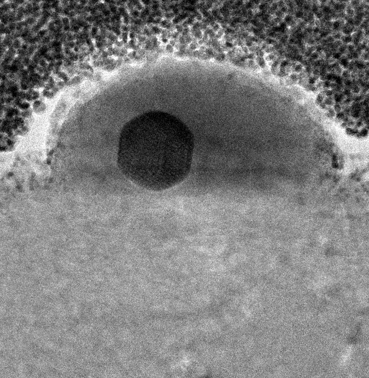
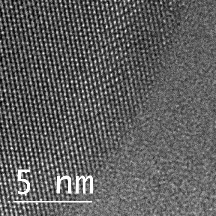
The neuromorphic invention is a single chip enabled by a sensing element, doped indium oxide, that’s thousands of times thinner than a human hair and requires no external parts to operate.
RMIT University engineers in Australia led the work, with contributions from researchers at Deakin University and the University of Melbourne.
The team’s research demonstrates a working device that captures, processes and stores visual information. With precise engineering of the doped indium oxide, the device mimics a human eye’s ability to capture light, pre-packages and transmits information like an optical nerve, and stores and classifies it in a memory system like the way our brains can.
Collectively, these functions could enable ultra-fast decision making, the team says.
Team leader Professor Sumeet Walia said the new device can perform all necessary functions – sensing, creating and processing information, and retaining memories – rather than relying on external energy-intensive computation, which prevents real-time decision making.
“Performing all of these functions on one small device had proven to be a big challenge until now,” said Walia from RMIT’s School of Engineering.
“We’ve made real-time decision making a possibility with our invention, because it doesn’t need to process large amounts of irrelevant data and it’s not being slowed down by data transfer to separate processors.”
What did the team achieve and how does the technology work?
The new device was able to demonstrate an ability to retain information for longer periods of time, compared to previously reported devices, without the need for frequent electrical signals to refresh the memory. This ability significantly reduces energy consumption and enhances the device’s performance.
Their findings and analysis are published in Advanced Functional Materials.
First author and RMIT PhD researcher Aishani Mazumder said the human brain used analog processing, which allowed it to process information quickly and efficiently using minimal energy.
“By contrast, digital processing is energy and carbon intensive, and inhibits rapid information gathering and processing,” she said.
“Neuromorphic vision systems are designed to use similar analog processing to the human brain, which can greatly reduce the amount of energy needed to perform complex visual tasks compared with today’s technologies
What are the potential applications?
The team used ultraviolet light as part of their experiments, and are working to expand this technology even further for visible and infrared light – with many possible applications such as bionic vision, autonomous operations in dangerous environments, shelf-life assessments of food and advanced forensics.
“Imagine a self-driving car that can see and recognise objects on the road in the same way that a human driver can or being able to able to rapidly detect and track space junk. This would be possible with neuromorphic vision technology.”
Walia said neuromorphic systems could adapt to new situations over time, becoming more efficient with more experience.
“Traditional computer vision systems – which cannot be miniaturised like neuromorphic technology – are typically programmed with specific rules and can’t adapt as easily,” he said.
“Neuromorphic robots have the potential to run autonomously for long periods, in dangerous situations where workers are exposed to possible cave-ins, explosions and toxic air.”
The human eye has a single retina that captures an entire image, which is then processed by the brain to identify objects, colours and other visual features.
The team’s device mimicked the retina’s capabilities by using single-element image sensors that capture, store and process visual information on one platform, Walia said.
“The human eye is exceptionally adept at responding to changes in the surrounding environment in a faster and much more efficient way than cameras and computers currently can,” he said.
“Taking inspiration from the eye, we have been working for several years on creating a camera that possesses similar abilities, through the process of neuromorphic engineering.”
What are the ethics of incorporating human cells into computer chips? That’s the question that Julian Savulescu (Visiting Professor in biomedical Ethics, University of Melbourne and Uehiro Chair in Practical Ethics, University of Oxford), Christopher Gyngell (Research Fellow in Biomedical Ethics, The University of Melbourne), and Tsutomu Sawai (Associate Professor, Humanities and Social Sciences, Hiroshima University) discuss in a May 24, 2022 essay on The Conversation (Note: A link has been removed),
The year is 2030 and we are at the world’s largest tech conference, CES in Las Vegas. A crowd is gathered to watch a big tech company unveil its new smartphone. The CEO comes to the stage and announces the Nyooro, containing the most powerful processor ever seen in a phone. The Nyooro can perform an astonishing quintillion operations per second, which is a thousand times faster than smartphone models in 2020. It is also ten times more energy-efficient with a battery that lasts for ten days.
A journalist asks: “What technological advance allowed such huge performance gains?” The chief executive replies: “We created a new biological chip using lab-grown human neurons. These biological chips are better than silicon chips because they can change their internal structure, adapting to a user’s usage pattern and leading to huge gains in efficiency.”
Another journalist asks: “Aren’t there ethical concerns about computers that use human brain matter?”
Although the name and scenario are fictional, this is a question we have to confront now. In December 2021, Melbourne-based Cortical Labs grew groups of neurons (brain cells) that were incorporated into a computer chip. The resulting hybrid chip works because both brains and neurons share a common language: electricity.
…
The authors explain their comment that brains and neurons share the common language of electricity (Note: Links have been removed),
In silicon computers, electrical signals travel along metal wires that link different components together. In brains, neurons communicate with each other using electric signals across synapses (junctions between nerve cells). In Cortical Labs’ Dishbrain system, neurons are grown on silicon chips. These neurons act like the wires in the system, connecting different components. The major advantage of this approach is that the neurons can change their shape, grow, replicate, or die in response to the demands of the system.
Dishbrain could learn to play the arcade game Pong faster than conventional AI systems. The developers of Dishbrain said: “Nothing like this has ever existed before … It is an entirely new mode of being. A fusion of silicon and neuron.”
Cortical Labs believes its hybrid chips could be the key to the kinds of complex reasoning that today’s computers and AI cannot produce. Another start-up making computers from lab-grown neurons, Koniku, believes their technology will revolutionise several industries including agriculture, healthcare, military technology and airport security. Other types of organic computers are also in the early stages of development.
…
Ethics issues arise (Note: Links have been removed),
… this raises questions about donor consent. Do people who provide tissue samples for technology research and development know that it might be used to make neural computers? Do they need to know this for their consent to be valid?
People will no doubt be much more willing to donate skin cells for research than their brain tissue. One of the barriers to brain donation is that the brain is seen as linked to your identity. But in a world where we can grow mini-brains from virtually any cell type, does it make sense to draw this type of distinction?
…
…
… Consider the scandal regarding Henrietta Lacks, an African-American woman whose cells were used extensively in medical and commercial research without her knowledge and consent.
Henrietta’s cells are still used in applications which generate huge amounts of revenue for pharmaceutical companies (including recently to develop COVID vaccines. The Lacks family still has not received any compensation. If a donor’s neurons end up being used in products like the imaginary Nyooro, should they be entitled to some of the profit made from those products?
Another key ethical consideration for neural computers is whether they could develop some form of consciousness and experience pain. Would neural computers be more likely to have experiences than silicon-based ones? …
This May 24, 2022 essay is fascinating and, if you have the time, I encourage you to read it all.
If you’re curious, you can find out about Cortical Labs here, more about Dishbrain in a February 22, 2022 article by Brian Patrick Green for iai (Institute for Art and Ideas) news, and more about Koniku in a May 31, 2018 posting about ‘wetware’ by Alissa Greenberg on Medium.
*HeLa cells are named for Henrietta Lacks who unknowingly donated her immortal cell line to medical research. You can find more about the story on the Oprah Winfrey website, which features an excerpt from the Rebecca Skloot book “The Immortal Life of Henrietta Lacks.”’ …
I checked; the excerpt is still on the Oprah Winfrey site.
There have been only two people who have tested the device from Australia but the research raises hope, from an Oct, 28, 2020 news item on ScienceDaily,
A tiny device the size of a small paperclip has been shown to help patients with upper limb paralysis to text, email and even shop online in the first human trial.
The device, Stentrode™, has been implanted successfully in two patients, who both suffer from severe paralysis due to amyotrophic lateral sclerosis (ALS) — also known as motor neuron disease (MND) — and neither had the ability to move their upper limbs.
Published in the Journal of NeuroInterventional Surgery, the results found the Stentrode™ was able to wirelessly restore the transmission of brain impulses out of the body. This enabled the patients to successfully complete daily tasks such as online banking, shopping and texting, which previously had not been available to them.
The Royal Melbourne Hospital’s Professor Peter Mitchell, Neurointervention Service Director and principal investigator on the trial, said the findings were promising and demonstrate the device can be safely implanted and used within the patients.
“This is the first time an operation of this kind has been done, so we couldn’t guarantee there wouldn’t be problems, but in both cases the surgery has gone better than we had hoped,” Professor Mitchell said.
Professor Mitchell implanted the device on the study participants through their blood vessels, next to the brain’s motor cortex, in a procedure involving a small ‘keyhole’ incision in the neck.
“The procedure isn’t easy, in each surgery there were differences depending on the patient’s anatomy, however in both cases the patients were able to leave the hospital only a few days later, which also demonstrates the quick recovery from the surgery,” Professor Mitchell said.
Neurointerventionalist and CEO of Synchron – the research commercial partner – Associate Professor Thomas Oxley, said this was a breakthrough moment for the field of brain-computer interfaces.
“We are excited to report that we have delivered a fully implantable, take home, wireless technology that does not require open brain surgery, which functions to restore freedoms for people with severe disability,” Associate Professor Oxley, who is also co-head of the Vascular Bionics Laboratory at the University of Melbourne, said.
The two patients used the Stentrode™ to control the computer-based operating system, in combination with an eye-tracker for cursor navigation. This meant they did not need a mouse or keyboard.
They also undertook machine learning-assisted training to control multiple mouse click actions, including zoom and left click. The first two patients achieved an average click accuracy of 92 per cent and 93 per cent, respectively, and typing speeds of 14 and 20 characters per minute with predictive text disabled.
University of Melbourne Associate Professor Nicholas Opie, co-head of the Vascular Bionics Laboratory at the University and founding chief technology officer of Synchron said the developments were exciting and the patients involved had a level of freedom restored in their lives.
“Observing the participants use the system to communicate and control a computer with their minds, independently and at home, is truly amazing,” Associate Professor Opie said.
“We are thankful to work with such fantastic participants, and my colleagues and I are honoured to make a difference in their lives. I hope others are inspired by their success.
“Over the last eight years we have drawn on some of the world’s leading medical and engineering minds to create an implant that enables people with paralysis to control external equipment with the power of thought. We are pleased to report that we have achieved this.”
The researchers caution that while it is some years away before the technology, capable of returning independence to complete everyday tasks is publicly available, the global, multidisciplinary team is working tirelessly to make this a reality.
The trial recently received a $AU1.48 million grant from the Australian commonwealth government to expand the trial to hospitals in New South Wales and Queensland, with hopes to enrol more patients.
###
About Stentrode™
Stentrode™ was developed by researchers from the University of Melbourne, the Royal Melbourne Hospital, the Florey Institute of Neuroscience and Mental Health, Monash University and the company Synchron Australia – the corporate vehicle established by Associate Professors Thomas Oxley (CEO) and Nicholas Opie (CTO) that aims to develop and commercialise neural bionics technology and products. It draws on some of the world’s leading medical and engineering minds
Researchers demonstrated the success of a fully implantable wireless medical device, the Stentrode™ brain-computer interface (BCI), designed to allow patients with severe paralysis to resume daily tasks — including texting, emailing, shopping and banking online — without the need for open brain surgery. The first-in-human study was published in the Journal of NeuroInterventional Surgery™, the leading international peer-reviewed journal for the clinical field of neurointerventional surgery.
The patients enrolled in the study utilized the Stentrode neuroprosthesis to control the Microsoft Windows 10 operating system in combination with an eye-tracker for cursor navigation, without a mouse or keyboard. The subjects undertook machine learning-assisted training to control multiple mouse-click actions, including zoom and left click.
“This is a breakthrough moment for the field of brain-computer interfaces. We are excited to report that we have delivered a fully implantable, take home, wireless technology that does not require open brain surgery, which functions to restore freedoms for people with severe disability,” said Thomas Oxley, MD, PhD, and CEO of Synchron, a neurovascular bioelectronics medicine company that conducted the research. “Seeing these first heroic patients resume important daily tasks that had become impossible, such as using personal devices to connect with loved ones, confirms our belief that the Stentrode will one day be able to help millions of people with paralysis.”[1]
Graham Felstead, a 75-year-old man living at home with his wife, has experienced severe paralysis due to amyotrophic lateral sclerosis (ALS). He was the first patient enrolled in the first Stentrode clinical study and the first person to have any BCI implanted via the blood vessels. He received the Stentrode implant in August 2019. With the Stentrode, Felstead was able to remotely contact his spouse, increasing his autonomy and reducing her burden of care. Philip O’Keefe, a 60-year-old man with ALS who works part time, was able to control computer devices to conduct work-related tasks and other independent activities after receiving the Stentrode in April 2020. Functional impairment to his fingers, elbows and shoulders had previously inhibited his ability to engage in these efforts.
The Stentrode device is small and flexible enough to safely pass through curving blood vessels, so the implantation procedure is similar to that of a pacemaker and does not require open brain surgery. Entry through the blood vessels may reduce risk of brain tissue inflammation and rejection of the device, which has been an issue for techniques that require direct brain penetration. Implantation is conducted using well-established neurointerventional techniques that do not require any novel automated robotic assistance.
Here’s a link to and a citation for the paper,
Motor neuroprosthesis implanted with neurointerventional surgery improves capacity for activities of daily living tasks in severe paralysis: first in-human experience by Thomas J Oxley, Peter E Yoo, Gil S Rind, Stephen M Ronayne, C M Sarah Lee, Christin Bird, Victoria Hampshire, Rahul P Sharma, Andrew Morokoff, Daryl L Williams, Christopher MacIsaac, Mark E Howard, Lou Irving, Ivan Vrljic, Cameron Williams, Sam E John, Frank Weissenborn, Madeleine Dazenko, Anna H Balabanski, David Friedenberg, Anthony N Burkitt, Yan T Wong, Katharine J Drummond, Patricia Desmond, Douglas Weber, Timothy Denison, Leigh R Hochberg, Susan Mathers, Terence J O’Brien, Clive N May, J Mocco, David B Grayden, Bruce C V Campbell, Peter Mitchell, Nicholas L Opie. Journal of Neurointerventional Surgery, DOI: http://dx.doi.org/10.1136/neurintsurg-2020-016862 Published Online First: 28 October 2020
Duke University researchers along with their international collaborators have made an extraordinary observation. From an Aug. 3, 2016 news item on ScienceDaily,
Imagine pouring a glass of ice water and having the ice cubes remain unchanged hours later, even under a broiler’s heat or in the very back corner of the freezer.
That’s fundamentally the surprising discovery recently made by an international group of researchers led by an electrical engineering professor at Duke University in a paper published online in Nature Matter on July 25, 2016. But instead of a refreshing mixture of H2O in a pint glass, the researchers were working with the chemical element gallium on a nanoscopic scale.
This image shows a single gallium nanoparticle sitting on top of a sapphire base. The black sphere in the center reveals the presence of solid gallium within the liquid drop exterior. The sapphire base is important, as it is rigid with a relatively high surface energy. As the nanoparticle and sapphire try to minimize their total energy, this combination of properties drives the formation and coexistence of the two phases. Courtesy: Duke University
Gallium is a soft, silvery bluish metal at room temperature. Raise the heat to 86 degrees Fahrenheit, however, and it melts. Drop the temperature to subzero levels, and it becomes hard and brittle. But when gallium nanoparticles sit on top of a sapphire surface, they form a solid core surrounded by a liquid outer layer. The discovery marks the first time that this stable phase coexistence phenomenon at the nanoscale has ever been directly observed.
“This odd combination of a liquid and solid state existing together has been predicted theoretically and observed indirectly in other materials in narrow bands of specific temperatures,” said April Brown, the John Cocke Professor of Electrical and Computer Engineering at Duke. “But this finding was very unexpected, especially because of its stability over such a large temperature range.”
The temperature range Brown is referring to covers more than 1,000 degrees Fahrenheit, all the way from -135 to 980 degrees.
“At a fundamental level, this finding reveals the need to reconsider all our presumptions about solid–liquid equilibrium,” wrote Andrés Aguado, professor of theoretical, atomic and optical physics at the University of Valladolid in Spain, in a News and Views piece appearing in the same edition of Nature Matter. “At a more applied level, the results hold much promise for future nanotechnology applications.”
Gallium is an important element in electronics and is used in microwave circuits, high-speed switching circuits and infrared circuits. The discovery of this novel part-solid, part-liquid nanoparticle phase could be useful in ultraviolet sensors, molecular sensing devices and enhanced photodetectors.
Brown hopes this work is just the tip of the iceberg, as she is planning on creating a facility at Duke to investigate what other nanoparticles might have similar unexpected phase qualities.
The research was conducted in conjunction with researchers at the Institute of Nanotechnology-CNR-Italy, the University of Western Australia, the University of Melbourne and Johannes Kepler University Linz.
This is an atomic view of liquid and solid gallium coexisting in a single nanoparticle taken by a transmission electron microscope. The circular shape on the left-hand side shows gallium atoms in an organized, crystalline, solid structure, while the atoms on the right are in liquid form, showing no organized structure at all. Courtesy: Duke University
“Getting into” as used in the headline is slang for exploring a topic in more depth which is what an international team of researchers did when they ‘got into’ cellulose. From a June 9, 2016 news item on phys.org (Note: Links have been removed),
In the search for low emission plant-based fuels, new research may help avoid having to choose between growing crops for food or fuel.
Scientists have identified new steps in the way plants produce cellulose, the component of plant cell walls that provides strength, and forms insoluble fibre in the human diet.
The findings could lead to improved production of cellulose and guide plant breeding for specific uses such as wood products and ethanol fuel, which are sustainable alternatives to fossil fuel-based products.
Published in the journal Nature Communications today, the work was conducted by an international team of scientists, led by the University of Cambridge and the University of Melbourne.
“Our research identified several proteins that are essential in the assembly of the protein machinery that makes cellulose”, said Melbourne’s Prof Staffan Persson.
“We found that these assembly factors control how much cellulose is made, and so plants without them can not produce cellulose very well and the defect substantially impairs plant biomass production. The ultimate aim of this research would be breed plants that have altered activity of these proteins so that cellulose production can be improved for the range of applications that use cellulose including paper, timber and ethanol fuels.”
The newly discovered proteins are located in an intracellular compartment called the Golgi where proteins are sorted and modified.
“If the function of this protein family is abolished the cellulose synthesizing complexes become stuck in the Golgi and have problems reaching the cell surface where they normally are active” said the lead authors of the study, Drs. Yi Zhang (Max-Planck Institute for Molecular Plant Physiology) and Nino Nikolovski (University of Cambridge).
“We therefore named the new proteins STELLO, which is Greek for to set in place, and deliver.”
“The findings are important to understand how plants produce their biomass”, said Professor Paul Dupree from the University of Cambridge’s Department of Biochemistry.
“Greenhouse-gas emissions from cellulosic ethanol, which is derived from the biomass of plants, are estimated to be roughly 85 percent less than from fossil fuel sources. Research to understand cellulose production in plants is therefore an important part of climate change mitigation.”
“In addition, by using cellulosic plant materials we get around the problem of food-versus-fuel scenario that is problematic when using corn as a basis for bioethanol.”
“It is therefore of great importance to find genes and mechanisms that can improve cellulose production in plants so that we can tailor cellulose production for various needs.”
Previous studies by Profs. Persson’s and Dupree’s research groups have, together with other scientists, identified many proteins that are important for cellulose synthesis and for other cell wall polymers.
With the newly presented research they substantially increase our understanding for how the bulk of a plant’s biomass is produced and is therefore of vast importance to industrial applications.
Here’s a link to and a citation for the paper,
Golgi-localized STELLO proteins regulate the assembly and trafficking of cellulose synthase complexes in Arabidopsis by Yi Zhang, Nino Nikolovski, Mathias Sorieul, Tamara Vellosillo, Heather E. McFarlane, Ray Dupree, Christopher Kesten, René Schneider, Carlos Driemeier, Rahul Lathe, Edwin Lampugnani, Xiaolan Yu, Alexander Ivakov, Monika S. Doblin, Jenny C. Mortimer, Steven P. Brown, Staffan Persson, & Paul Dupree. Nature Communications 7,
Article number: 11656 doi:10.1038/ncomms11656 Published 09 June 2016
Researchers at the University of Adelaide (Australia) have found a way to embed luminiscent nanoparticles in glass, according to a June 8, 2016 news item on Nanotechnology,
This new “hybrid glass” successfully combines the properties of these special luminescent (or light-emitting) nanoparticles with the well-known aspects of glass, such as transparency and the ability to be processed into various shapes including very fine optical fibres.
The research, in collaboration with Macquarie University and University of Melbourne, has been published online in the journal Advanced Optical Materials.
“These novel luminescent nanoparticles, called upconversion nanoparticles, have become promising candidates for a whole variety of ultra-high tech applications such as biological sensing, biomedical imaging and 3D volumetric displays,” says lead author Dr Tim Zhao, from the University of Adelaide’s School of Physical Sciences and Institute for Photonics and Advanced Sensing (IPAS).
“Integrating these nanoparticles into glass, which is usually inert, opens up exciting possibilities for new hybrid materials and devices that can take advantage of the properties of nanoparticles in ways we haven’t been able to do before. For example, neuroscientists currently use dye injected into the brain and lasers to be able to guide a glass pipette to the site they are interested in. If fluorescent nanoparticles were embedded in the glass pipettes, the unique luminescence of the hybrid glass could act like a torch to guide the pipette directly to the individual neurons of interest.”
Although this method was developed with upconversion nanoparticles, the researchers believe their new ‘direct-doping’ approach can be generalised to other nanoparticles with interesting photonic, electronic and magnetic properties. There will be many applications – depending on the properties of the nanoparticle.
“If we infuse glass with a nanoparticle that is sensitive to radiation and then draw that hybrid glass into a fibre, we could have a remote sensor suitable for nuclear facilities,” says Dr Zhao.
To date, the method used to integrate upconversion nanoparticles into glass has relied on the in-situ growth of the nanoparticles within the glass.
“We’ve seen remarkable progress in this area but the control over the nanoparticles and the glass compositions has been limited, restricting the development of many proposed applications,” says project leader Professor Heike Ebendorff-Heideprem, Deputy Director of IPAS.
“With our new direct doping method, which involves synthesizing the nanoparticles and glass separately and then combining them using the right conditions, we’ve been able to keep the nanoparticles intact and well dispersed throughout the glass. The nanoparticles remain functional and the glass transparency is still very close to its original quality. We are heading towards a whole new world of hybrid glass and devices for light-based technologies.”
Here’s a link to and a citation for the paper,
Upconversion Nanocrystal-Doped Glass: A New Paradigm for Photonic Materials by Jiangbo Zhao, Xianlin Zheng, Erik P. Schartner, Paul Ionescu, Run Zhang, Tich-Lam Nguyen, Dayong Jin, and Heike Ebendorff-Heidepriem. Advanced Optical Materials DOI: 10.1002/adom.201600296 Version of Record online: 30 MAY 2016
This research from Singapore could make neuroprosthetics and exoskeletons a little easier to manage as long as you don’t mind having a neural implant. From a Feb. 11, 2016 news item on ScienceDaily,
A versatile chip offers multiple applications in various electronic devices, report researchers, suggested that there is now hope that a low-powered, wireless neural implant may soon be a reality. Neural implants when embedded in the brain can alleviate the debilitating symptoms of Parkinson’s disease or give paraplegic people the ability to move their prosthetic limbs.
Caption: NTU Asst Prof Arindam Basu is holding his low-powered smart chip. Credit: NTU Singapore
Scientists at Nanyang Technological University, Singapore (NTU Singapore) have developed a small smart chip that can be paired with neural implants for efficient wireless transmission of brain signals.
Neural implants when embedded in the brain can alleviate the debilitating symptoms of Parkinson’s disease or give paraplegic people the ability to move their prosthetic limbs.
However, they need to be connected by wires to an external device outside the body. For a prosthetic patient, the neural implant is connected to a computer that decodes the brain signals so the artificial limb can move.
These external wires are not only cumbersome but the permanent openings which allow the wires into the brain increases the risk of infections.
The new chip by NTU scientists can allow the transmission of brain data wirelessly and with high accuracy.
Assistant Professor Arindam Basu from NTU’s School of Electrical and Electronic Engineering said the research team have tested the chip on data recorded from animal models, which showed that it could decode the brain’s signal to the hand and fingers with 95 per cent accuracy.
“What we have developed is a very versatile smart chip that can process data, analyse patterns and spot the difference,” explained Prof Basu.
“It is about a hundred times more efficient than current processing chips on the market. It will lead to more compact medical wearable devices, such as portable ECG monitoring devices and neural implants, since we no longer need large batteries to power them.”
Different from other wireless implants
To achieve high accuracy in decoding brain signals, implants require thousands of channels of raw data. To wirelessly transmit this large amount of data, more power is also needed which means either bigger batteries or more frequent recharging.
This is not feasible as there is limited space in the brain for implants while frequent recharging means the implants cannot be used for long-term recording of signals.
Current wireless implant prototypes thus suffer from a lack of accuracy as they lack the bandwidth to send out thousands of channels of raw data.
Instead of enlarging the power source to support the transmission of raw data, Asst Prof Basu tried to reduce the amount of data that needs to be transmitted.
Designed to be extremely power-efficient, NTU’s patented smart chip will analyse and decode the thousands of signals from the neural implants in the brain, before compressing the results and sending it wirelessly to a small external receiver.
This invention and its findings were published last month [December 2015] in the prestigious journal, IEEE Transactions on Biomedical Circuits & Systems, by the Institute of Electrical and Electronics Engineers, the world’s largest professional association for the advancement of technology.
Its underlying science was also featured in three international engineering conferences (two in Atlanta, USA and one in China) over the last three months.
Versatile smart chip with multiple uses
This new smart chip is designed to analyse data patterns and spot any abnormal or unusual patterns.
For example, in a remote video camera, the chip can be programmed to send a video back to the servers only when a specific type of car or something out of the ordinary is detected, such as an intruder.
This would be extremely beneficial for the Internet of Things (IOT), where every electrical and electronic device is connected to the Internet through a smart chip.
With a report by marketing research firm Gartner Inc predicting that 6.4 billion smart devices and appliances will be connected to the Internet by 2016, and will rise to 20.8 billion devices by 2020, reducing network traffic will be a priority for most companies.
Using NTU’s new chip, the devices can process and analyse the data on site, before sending back important details in a compressed package, instead of sending the whole data stream. This will reduce data usage by over a thousand times.
Asst Prof Basu is now in talks with Singapore Technologies Electronics Limited to adapt his smart chip that can significantly reduce power consumption and the amount of data transmitted by battery-operated remote sensors, such as video cameras.
The team is also looking to expand the applications of the chip into commercial products, such as to customise it for smart home sensor networks, in collaboration with a local electronics company.
The chip, measuring 5mm by 5mm can now be licensed by companies from NTU’s commercialisation arm, NTUitive.
Earlier this month there was a Feb. 9, 2016 announcement about a planned human clinical trial in Australia for a new brain-machine interface (neural implant). Before proceeding with the news, here’s what this implant looks like,
Caption: This tiny device, the size of a small paperclip, is implanted in to a blood vessel next to the brain and can read electrical signals from the motor cortex, the brain’s control centre. These signals can then be transmitted to an exoskeleton or wheelchair to give paraplegic patients greater mobility. Users will need to learn how to communicate with their machinery, but over time, it is thought it will become second nature, like driving or playing the piano. The first human trials are slated for 2017 in Melbourne, Australia. Credit: The University of Melbourne.
Melbourne medical researchers have created a new minimally invasive brain-machine interface, giving people with spinal cord injuries new hope to walk again with the power of thought.
The brain machine interface consists of a stent-based electrode (stentrode), which is implanted within a blood vessel next to the brain, and records the type of neural activity that has been shown in pre-clinical trials to move limbs through an exoskeleton or to control bionic limbs.
The new device is the size of a small paperclip and will be implanted in the first in-human trial at The Royal Melbourne Hospital in 2017.
The results published today in Nature Biotechnology show the device is capable of recording high-quality signals emitted from the brain’s motor cortex, without the need for open brain surgery.
Principal author and Neurologist at The Royal Melbourne Hospital and Research Fellow at The Florey Institute of Neurosciences and the University of Melbourne, Dr Thomas Oxley, said the stentrode was revolutionary.
“The development of the stentrode has brought together leaders in medical research from The Royal Melbourne Hospital, The University of Melbourne and the Florey Institute of Neuroscience and Mental Health. In total 39 academic scientists from 16 departments were involved in its development,” Dr Oxley said.
“We have been able to create the world’s only minimally invasive device that is implanted into a blood vessel in the brain via a simple day procedure, avoiding the need for high risk open brain surgery.
“Our vision, through this device, is to return function and mobility to patients with complete paralysis by recording brain activity and converting the acquired signals into electrical commands, which in turn would lead to movement of the limbs through a mobility assist device like an exoskeleton. In essence this a bionic spinal cord.”
Stroke and spinal cord injuries are leading causes of disability, affecting 1 in 50 people. There are 20,000 Australians with spinal cord injuries, with the typical patient a 19-year old male, and about 150,000 Australians left severely disabled after stroke.
Co-principal investigator and biomedical engineer at the University of Melbourne, Dr Nicholas Opie, said the concept was similar to an implantable cardiac pacemaker – electrical interaction with tissue using sensors inserted into a vein, but inside the brain.
“Utilising stent technology, our electrode array self-expands to stick to the inside wall of a vein, enabling us to record local brain activity. By extracting the recorded neural signals, we can use these as commands to control wheelchairs, exoskeletons, prosthetic limbs or computers,” Dr Opie said.
“In our first-in-human trial, that we anticipate will begin within two years, we are hoping to achieve direct brain control of an exoskeleton for three people with paralysis.”
“Currently, exoskeletons are controlled by manual manipulation of a joystick to switch between the various elements of walking – stand, start, stop, turn. The stentrode will be the first device that enables direct thought control of these devices”
Neurophysiologist at The Florey, Professor Clive May, said the data from the pre-clinical study highlighted that the implantation of the device was safe for long-term use.
“Through our pre-clinical study we were able to successfully record brain activity over many months. The quality of recording improved as the device was incorporated into tissue,” Professor May said.
“Our study also showed that it was safe and effective to implant the device via angiography, which is minimally invasive compared with the high risks associated with open brain surgery.
“The brain-computer interface is a revolutionary device that holds the potential to overcome paralysis, by returning mobility and independence to patients affected by various conditions.”
Professor Terry O’Brien, Head of Medicine at Departments of Medicine and Neurology, The Royal Melbourne Hospital and University of Melbourne said the development of the stentrode has been the “holy grail” for research in bionics.
“To be able to create a device that can record brainwave activity over long periods of time, without damaging the brain is an amazing development in modern medicine,” Professor O’Brien said.
“It can also be potentially used in people with a range of diseases aside from spinal cord injury, including epilepsy, Parkinsons and other neurological disorders.”
The development of the minimally invasive stentrode and the subsequent pre-clinical trials to prove its effectiveness could not have been possible without the support from the major funding partners – US Defense Department DARPA [Defense Advanced Research Projects Agency] and Australia’s National Health and Medical Research Council.
So, DARPA is helping fund this, eh? Interesting but not a surprise given the agency’s previous investments in brain research and neuroprosthetics.
For those who like to get their news via video,
Here’s a link to and a citation for the paper,
Minimally invasive endovascular stent-electrode array for high-fidelity, chronic recordings of cortical neural activity by Thomas J Oxley, Nicholas L Opie, Sam E John, Gil S Rind, Stephen M Ronayne, Tracey L Wheeler, Jack W Judy, Alan J McDonald, Anthony Dornom, Timothy J H Lovell, Christopher Steward, David J Garrett, Bradford A Moffat, Elaine H Lui, Nawaf Yassi, Bruce C V Campbell, Yan T Wong, Kate E Fox, Ewan S Nurse, Iwan E Bennett, Sébastien H Bauquier, Kishan A Liyanage, Nicole R van der Nagel, Piero Perucca, Arman Ahnood et al. Nature Biotechnology (2016) doi:10.1038/nbt.3428 Published online 08 February 2016
This paper is behind a paywall.
I wish the researchers in Singapore, Australia, and elsewhere, good luck!
*’Sinagpore’ in head changed to ‘Singapore’ on May 14, 2019.
A new method for building “drawbridges” between metal nanoparticles may allow electronics makers to build full-color displays using light-scattering nanoparticles that are similar to the gold materials that medieval artisans used to create red stained-glass.
“Wouldn’t it be interesting if we could create stained-glass windows that changed colors at the flip of a switch?” said Christy Landes, associate professor of chemistry at Rice and the lead researcher on a new study about the drawbridge method that appears this week in the open-access journal Science Advances.
The research by Landes and other experts at Rice University’s Smalley-Curl Institute could allow engineers to use standard electrical switching techniques to construct color displays from pairs of nanoparticles that scatter different colors of light.
For centuries, stained-glass makers have tapped the light-scattering properties of tiny gold nanoparticles to produce glass with rich red tones. Similar types of materials could increasingly find use in modern electronics as manufacturers work to make smaller, faster and more energy-efficient components that operate at optical frequencies.
Though metal nanoparticles scatter bright light, researchers have found it difficult to coax them to produce dramatically different colors, Landes said.
Rice’s new drawbridge method for color switching incorporates metal nanoparticles that absorb light energy and convert it into plasmons, waves of electrons that flow like a fluid across a particle’s surface. Each plasmon scatters and absorbs a characteristic frequency of light, and even minor changes in the wave-like sloshing of a plasmon shift that frequency. The greater the change in plasmonic frequency, the greater the difference between the colors observed.
“Engineers hoping to make a display from optically active nanoparticles need to be able to switch the color,” Landes said. “That type of switching has proven very difficult to achieve with nanoparticles. People have achieved moderate success using various plasmon-coupling schemes in particle assemblies. What we’ve shown though is variation of the coupling mechanism itself, which can be used to produce huge color changes both rapidly and reversibly.”
To demonstrate the method, Landes and study lead author Chad Byers, a graduate student in her lab, anchored pairs of gold nanoparticles to a glass surface covered with indium tin oxide (ITO), the same conductor that’s used in many smartphone screens. By sealing the particles in a chamber filled with a saltwater electrolyte and a silver electrode, Byers and Landes were able form a device with a complete circuit. They then showed they could apply a small voltage to the ITO to electroplate silver onto the surface of the gold particles. In that process, the particles were first coated with a thin layer of silver chloride. By later applying a negative voltage, the researchers caused a conductive silver “drawbridge” to form. Reversing the voltage caused the bridge to withdraw.
“The great thing about these chemical bridges is that we can create and eliminate them simply by applying or reversing a voltage,” Landes said. “This is the first method yet demonstrated to produce dramatic, reversible color changes for devices built from light-activated nanoparticles.”
This research has its roots in previous work (from the news release),
Byers said his research into the plasmonic behavior of gold dimers began about two years ago.
“We were pursuing the idea that we could make significant changes in optical properties of individual particles simply by altering charge density,” he said. “Theory predicts that colors can be changed just by adding or removing electrons, and we wanted to see if we could do that reversibly, simply by turning a voltage on or off.”
The experiments worked. The color shift was observed and reversible, but the change in the color was minute.
“It wasn’t going to get anybody excited about any sort of switchable display applications,” Landes said.
But she and Byers also noticed that their results differed from the theoretical predictions.
Landes said that was because the predictions were based upon using an inert electrode made of a metal like palladium that isn’t subject to oxidation. But silver is not inert. It reacts easily with oxygen in air or water to form a coat of unsightly silver oxide. This oxidizing layer can also form from silver chloride, and Landes said that is what was occurring when the silver counter electrode was used in Byers’ first experiments.
The scientists decided to embrace imperfection (from the news release),
“It was an imperfection that was throwing off our results, but rather than run away from it, we decided to use it to our advantage,” Landes said.
Rice plasmonics pioneer and study co-author Naomi Halas, director of the Smalley-Curl Institute, said the new research shows how plasmonic components could be used to produce electronically switchable color-displays.
“Gold nanoparticles are particularly attractive for display purposes,” said Halas, Rice’s Stanley C. Moore Professor of Electrical and Computer Engineering and professor of chemistry, bioengineering, physics and astronomy, and materials science and nanoengineering. “Depending upon their shape, they can produce a variety of specific colors. They are also extremely stable, and even though gold is expensive, very little is needed to produce an extremely bright color.”
In designing, testing and analyzing the follow-up experiments on dimers, Landes and Byers engaged with a brain trust of Rice plasmonics experts that included Halas, physicist and engineer Peter Nordlander, chemist Stephan Link, materials scientist Emilie Ringe and their students, as well as Paul Mulvaney of the University of Melbourne in Australia.
Together, the team confirmed the composition and spacing of the dimers and showed how metal drawbridges could be used to induce large color shifts based on voltage inputs.
Nordlander and Hui Zhang, the two theorists in the group, examined the device’s “plasmonic coupling,” the interacting dance that plasmons engage in when they are in close contact. For instance, plasmonic dimers are known to act as light-activated capacitors, and prior research has shown that connecting dimers with nanowire bridges brings about a new state of resonance known as a “charge-transfer plasmon,” which has its own distinct optical signature.
“The electrochemical bridging of the interparticle gap enables a fully reversible transition between two plasmonic coupling regimes, one capacitive and the other conductive,” Nordlander said. “The shift between these regimes is evident from the dynamic evolution of the charge transfer plasmon.”
Halas said the method provides plasmonic researchers with a valuable tool for precisely controlling the gaps between dimers and other multiparticle plasmonic configurations.
“In an applied sense, gap control is important for the development of active plasmonic devices like switches and modulators, but it is also an important tool for basic scientists who are conducting curiosity-driven research in the emerging field of quantum plasmonics.”
I’m glad the news release writer included the background work leading to this new research and to hint at the level of collaboration needed to achieve the scientists’ new understanding of color switching.
The accuracy and reliability of expert advice is often compromised by “cognitive frailties”, and needs to be interrogated with the same tenacity as research data to avoid weak and ill-informed policy, warn two leading risk analysis and conservation researchers in the journal Nature today.
While many governments aspire to evidence-based policy [emphasis mine], the researchers say the evidence on experts themselves actually shows that they are highly susceptible to “subjective influences” – from individual values and mood, to whether they stand to gain or lose from a decision – and, while highly credible, experts often vastly overestimate their objectivity and the reliability of peers.
They appear to be conflating evidence and expertise. Evidence usually means data while expertise is a more ephemeral concept. (Presumably, an expert is someone whose opinion is respected for one reason or another and who has studied the evidence and drawn some conclusions from it.)
The study described in the press release notes that one of the weaknesses of relying on experts is that they are subject to bias. They don’t mention that evidence or data can also be subject to bias but perhaps that’s why they suggest the experts should provide and assess the evidence on which they are basing their advice,
The researchers caution that conventional approaches of informing policy by seeking advice from either well-regarded individuals or assembling expert panels needs to be balanced with methods that alleviate the effects of psychological and motivational bias.
They offer a straightforward framework for improving expert advice, and say that experts should provide and assess [emphasis mine] evidence on which decisions are made – but not advise decision makers directly, which can skew impartiality.
“We are not advocating replacing evidence with expert judgements, rather we suggest integrating and improving them,” write professors William Sutherland and Mark Burgman from the universities of Cambridge and Melbourne respectively.
“Policy makers use expert evidence as though it were data. So they should treat expert estimates with the same critical rigour that must be applied to data,” they write.
“Experts must be tested, their biases minimised, their accuracy improved, and their estimates validated with independent evidence. Put simply, experts should be held accountable for their opinions.”
Sutherland and Burgman point out that highly regarded experts are routinely shown to be no better than novices at making judgements.
However, several processes have been shown to improve performances across the spectrum, they say, such as ‘horizon scanning’ – identifying all possible changes and threats – and ‘solution scanning’ – listing all possible options, using both experts and evidence, to reduce the risk of overlooking valuable alternatives.
To get better answers from experts, they need better, more structured questions, say the authors. “A seemingly straightforward question, ‘How many diseased animals are there in the area?’ for example, could be interpreted very differently by different people. Does it include those that are infectious and those that have recovered? What about those yet to be identified?” said Sutherland, from Cambridge’s Department of Zoology.
“Structured question formats that extract upper and lower boundaries, degrees of confidence and force consideration of alternative theories are important for shoring against slides into group-think, or individuals getting ascribed greater credibility based on appearance or background,” he said.
When seeking expert advice, all parties must be clear about what they expect of each other, says Burgman, Director of the Centre of Excellence for Biosecurity Risk Analysis. “Are policy makers expecting estimates of facts, predictions of the outcome of events, or advice on the best course of action?”
“Properly managed, experts can help with estimates and predictions, but providing advice assumes the expert shares the same values and objectives as the decision makers. Experts need to stick to helping provide and assess evidence on which such decisions are made,” he said.
Sutherland and Burgman have created a framework of eight key ways to improve the advice of experts. These include using groups – not individuals – with diverse, carefully selected members well within their expertise areas.
They also caution against being bullied or “starstruck” by the over-assertive or heavyweight. “People who are less self-assured will seek information from a more diverse range of sources, and age, number of qualifications and years of experience do not explain an expert’s ability to predict future events – a finding that applies in studies from geopolitics to ecology,” said Sutherland.
Added Burgman: “Some experts are much better than others at estimation and prediction. However, the only way to tell a good expert from a poor one is to test them. Qualifications and experience don’t help to tell them apart.”
“The cost of ignoring these techniques – of using experts inexpertly – is less accurate information and so more frequent, and more serious, policy failures,” write the researchers.
Here’s a link to and a citation for the paper,
Policy advice: Use experts wisely by William J. Sutherland & Mark Burgman. Nature 526, 317–318 (15 October 2015) doi:10.1038/526317a
It’s good to see a nuanced attempt to counteract mindless adherence to expert opinion. I hope they will include evidence and data as needing to be approached cautiously in future work.
A multi-institutional team of scientists has taken an important step in understanding where atoms are located on the surfaces of rough materials, information that could be very useful in diverse commercial applications, such as developing green energy and understanding how materials rust.
Researchers from Northwestern University, Brookhaven National Laboratory, Lawrence Berkeley National Laboratory and the University of Melbourne, Australia, have developed a new imaging technique that uses atomic resolution secondary electron images in a quantitative way to determine the arrangement of atoms on the surface.
Many important processes take place at surfaces, ranging from the catalysis used to generate energy-dense fuels from sunlight and carbon dioxide to how bridges and airplanes corrode, or rust. Every material interacts with the world through its surface, which is often different in both structure and chemistry from the bulk of the material.
The real focus of the work is on corrosion, according to the news release,
“We are excited by the possibilities of applying our imaging technique to corrosion and catalysis problems,” said Laurence Marks, a co-author of the paper and a professor of materials science and engineering at Northwestern’s McCormick School of Engineering and Applied Science. “The cost of corrosion to industry and the military is enormous, and we do not understand everything that is taking place. We must learn more, so we can produce materials that will last longer.”
To understand these processes and improve material performance, it is vital to know how the atoms are arranged on surfaces. While there are many good methods for obtaining this information for rather flat surfaces, most currently available tools are limited in what they can reveal when the surfaces are rough.
Scanning electron microscopes are widely used to produce images of many different materials, and roughness of the surface is not that important. Until very recently, instruments could not obtain clear atomic images of surfaces until a group at Brookhaven managed in 2011 to get the first images that seemed to show the surfaces very clearly. However, it was not clear to what extent they really were able to image the surface, as there was no theory for the imaging and many uncertainties.
The new work has answered all these questions, Marks said, providing a definitive way of understanding the surfaces in detail. What was needed was to use a carefully controlled sample of strontium titanate and perform a large range of different types of imaging to unravel the precise details of how secondary electron images are produced.
“We started this work by investigating a well-studied material,” said Jim Ciston, a staff scientist at Lawrence Berkeley National Laboratory and the lead author of the paper, who obtained the experimental images. “This new technique is so powerful that we had to revise much of what was already thought to be well-known. This is an exciting prospect because the surface of every material can act as its own nanomaterial coating, which can greatly change the chemistry and behavior.”
“The beauty of the technique is that we can image surface atoms and bulk atoms simultaneously,” said Yimei Zhu, a scientist at Brookhaven National Laboratory. “Currently, no existing methods can achieve that.”
Les Allen, who led the theoretical and modeling aspects of the new imaging technique in Melbourne, said, “We now have a sophisticated understanding of what the images mean. It now will be full steam ahead to apply them to many different types of problems.”