Tag Archives: University of Nebraska-Lincoln

Fish biofluorescence evolved more than 100 times in 112 million years

Before launching into the news announcements, a word about biofluorescence and bioluminesccnce. Biofluorescence is due to optical properties in fish scales, fur, butterfly wings, etc., with these properties usually being due to nanoscale structures on the wings, in the fur, etc. Bioluminescence is due to a chemical reaction, e.g., a reaction in fireflies’ stomachs.

A June 17, 2025 news item on ScienceDaily announced new information about the evolution of biofluorescence in fish,

New research led by scientists at the American Museum of Natural History sheds light on the ancient origins of biofluorescence in fishes and the range of brilliant colors involved in this biological phenomenon. Detailed in two complementary studies recently published in Nature Communications and PLOS One, the findings suggest that biofluorescence dates back at least 112 million years and, since then, has evolved independently more than 100 times, with the majority of that activity happening among fish that live on coral reefs.

A tropical striped triplefin (Helcogramma striata). © John Sparks and David Gruber Courtesy: American Museum of Natural History

A June 17, 2025 American Museum of Natural History news release (also on EurekAlert but published June 16, 2025), which originated the news item, delves further into biofluorescent fish and evolution,

The new work also reveals that in marine fishes, biofluorescence—which occurs when an organism absorbs light, transforms it, and emits it as a different color—involves a greater variety of colors than previously reported, spanning multiple wavelengths of green, yellow, orange, and red.

“Researchers have known for a while that biofluorescence is quite widespread in marine animals, from sea turtles to corals, and especially among fishes,” said Emily Carr, a Ph.D. student in the Museum’s Richard Gilder Graduate School and the lead author on the two new studies. “But to really get to the root of why and how these species use this unique adaptation—whether for camouflage, predation, or reproduction—we need to understand the underlying evolutionary story as well as the scope of biofluorescence as it currently exists.”

For the Nature Communications study, Carr led a comprehensive survey of all known biofluorescent teleosts—a type of bony fish that make up by far the largest group of vertebrates alive today. This resulted in a list of 459 biofluorescent species, including 48 species that were previously unknown to be biofluorescent. The researchers found that biofluorescence evolved more than 100 times in marine teleosts and is estimated to date back about 112 million years, with the first instance occurring in eels.

The team also found that fish species that live in or around coral reefs evolve biofluorescence at about 10 times the rate of non-reef species, with an increase in the number of fluorescent species following the Cretaceous-Paleogene (K-Pg) extinction about 66 million years ago, when all of the non-avian dinosaurs died off.

“This trend coincides with the rise of modern coral-dominated reefs and the rapid colonization of reefs by fishes, which occurred following a significant loss of coral diversity in the K-Pg extinction,” Carr said. “These correlations suggest that the emergence of modern coral reefs could have facilitated the diversification of fluorescence in reef-associated teleost fishes.”

Of the 459 known biofluorescent teleosts reported in this study, the majority are associated with coral reefs.

For the PLOS One study, Carr and colleagues used a specialized photography setup with ultraviolet and blue excitation lights and emission filters to look at the wavelengths of light emitted by fishes in the Museum’s Ichthyology collection. Collected over the last decade and a half on Museum expeditions to the Solomon Islands, Greenland, and Thailand, the specimens in the study were previously observed fluorescing, but the full range of their biofluorescent emissions was unknown.

The new work reveals far more diversity in colors emitted by teleosts—some families of which exhibit at least six distinct fluorescent emission peaks, which correspond with wavelengths across multiple colors—than had previously been reported.

“The remarkable variation we observed across a wide array of these fluorescent fishes could mean that these animals use incredibly diverse and elaborate signaling systems based on species-specific fluorescent emission patterns,” said Museum Curator John Sparks, an author on the new studies and Carr’s advisor. “As these studies show, biofluorescence is both pervasive and incredibly phenotypically variable among marine fishes. What we would really like to understand better is how fluorescence functions in these highly variable marine lineages, as well as its role in diversification.”

The researchers also note that the numerous wavelengths of fluorescent emissions found in this study could have implications for identifying novel fluorescent molecules, which are routinely used in biomedical applications, including fluorescence-guided disease diagnosis and therapy.

Other authors involved in this work include Rene Martin, from the Museum and the University of Nebraska-Lincoln; Mason Thurman, from Clemson University; Karly Cohen, from California State University; Jonathan Huie, from George Washington University; David Gruber, from Baruch College and The Graduate Center, City University of New York; and Tate Sparks, Rutgers University.

Research in the Solomon Islands was supported by the National Science Foundation under Grant Number DEB-1257555.

The Museum greatly acknowledges the Dalio Foundation for its generous support of the inaugural Explore21 Expedition.

The Museum’s Exlopre21 initiative is generously supported by the leadership contributions of Katheryn P. and Thomas L. Kempner, Jr. 

The 2019 Constantine S. Niarchos Expedition to Greenland was generously supported by the Stavros Niarchos Foundation. 

Research in Thailand was funded by the Museum and the National Science Foundation Graduate Research Fellowship Program under Grant Number DEB-1938103.

Additional funding for this work was provided by the National Science Foundation under Grant Number DGE-1746914.

I have links and citations for both papers mentioned in the news release. They will be listed in the order in which they were published earliest to latest: first (Nature Communications) and last (PLOS One).

Repeated and widespread evolution of biofluorescence in marine fishes by Emily M. Carr, Rene P. Martin, Mason A. Thurman, Karly E. Cohen, Jonathan M. Huie, David F. Gruber & John S. Sparks. Nature Communications volume 16, Article number: 4826 (2025) DOI: https://doi.org/10.1038/s41467-025-59843-7 Published: 24 May 2025

This paper is open access.

Marine fishes exhibit exceptional variation in biofluorescent emission spectra by Emily M. Carr, Mason A. Thurman, Rene P. Martin, Tate S. Sparks, John S. Sparks. PLOS One DOI: https://doi.org/10.1371/journal.pone.0316789 Published: June 16, 2025

This paper is open access.

Nanostructured materials and radiation

If you’re planning on using nanostructured materials in a nuclear facility, you might want to check out this work (from a June 8, 2018 Purdue University (Indiana, US) news release by Brian L. Huchel,

A professor in the Purdue College of Engineering examined the potential use of various materials in nuclear reactors in an extensive review article in the journal Progress in Materials Science.

The article, titled “Radiation Damage in Nanostructured Materials,” was led by Xinghang Zhang, a professor of materials engineering. It will be published in the July issue of the journal.

Zhang said there is a significant demand for advanced materials that can survive high temperature and high doses of radiation. These materials contain significant amount of internal changes, called defect sinks, which are too small to be seen with the naked eye, but may form the next generation of materials used in nuclear reactors.

“Nanostructured materials with abundant internal defect sinks are promising as these materials have shown significantly improved radiation tolerance,” he said. “However, there are many challenges and fundamental science questions that remain to be solved before these materials can have applications in advanced nuclear reactors.”

The 100-page article, which took two years to write, focuses on metallic materials and metal-ceramic compounds and reviews types of internal material defects on the reduction of radiation damage in nanostructured materials.

Under the extreme radiation conditions, a large number of defects and their clusters are generated inside materials, and such significant microstructure damage often leads to degradation of the mechanical and physical properties of the materials

The article discusses the usage of a combination of defect sink networks to collaboratively improve the radiation tolerance of nanomaterials, while pointing out the need to improve the thermal and radiation stabilities of the defect sinks.

“The field of radiation damage in nanostructured materials is an exciting and rapidly evolving arena, enriched with challenges and opportunities,” Zhang said. “The integration of extensive research effort, resources and expertise in various fields may eventually lead to the design of advanced nanomaterials with unprecedented radiation tolerance.”

Jin Li, co-author of the review article and a postdoctoral fellow in the School of Materials Engineering, said researchers with different expertise worked collaboratively on the article, which contains more than 100 pages, 100 figures and 700 references.

The team involved in the research article included researchers from Purdue, Texas A&M University, Drexel University, the University of Nebraska-Lincoln and China University of Petroleum-Beijing, as well as Sandia National Laboratory, Los Alamos National Laboratory and Idaho National Laboratory.

Here’s an image illustrating the work,

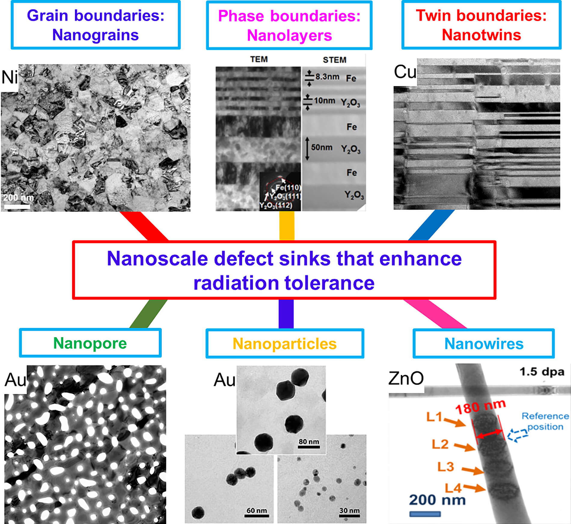
Various imperfections in nanostructures, call defect sinks, can enhance the material’s tolerance to radiation. (Photo/Xinghang Zhang)

Here’s a link to and a citation for the paper,

Radiation damage in nanostructured materials by Xinghang Zhang, Khalid Hattar, Youxing Chen, Lin Shao, Jin Li, Cheng Sun, Kaiyuan Yu, Nan Li, Mitra L.Taheri, Haiyan Wang, Jian Wang, Michael Nastasi. Progress in Materials Science Volume 96, July 2018, Pages 217-321 https://doi.org/10.1016/j.pmatsci.2018.03.002

This paper is behind a paywall.

ht/ to June 8, 2018 Nanowerk news item.

Titanium dioxide nanoparticles and the brain

This research into titanium dioxide nanoparticles and possible effects on your brain should they pass the blood-brain barrier comes from the University of Nebraska-Lincoln (US) according to a Dec. 15, 2015 news item on Nanowerk (Note: A link has been removed),

Even moderate concentrations of a nanoparticle used to whiten certain foods, milk and toothpaste could potentially compromise the brain’s most numerous cells, according to a new study from the University of Nebraska-Lincoln (Nanoscale, “Mitochondrial dysfunction and loss of glutamate uptake in primary astrocytes exposed to titanium dioxide nanoparticles”).

A Dec. 14, 2015 University of Nebraska-Lincoln news release, which originated the news item, provides more detail (Note: Links have been removed),

The researchers examined how three types of titanium dioxide nanoparticles [rutile, anatase, and commercially available P25 TiO2 nanoparticles], the world’s second-most abundant nanomaterial, affected the functioning of astrocyte cells. Astrocytes help regulate the exchange of signal-carrying neurotransmitters in the brain while also supplying energy to the neurons that process those signals, among many other functions.

The team exposed rat-derived astrocyte cells to nanoparticle concentrations well below the extreme levels that have been shown to kill brain cells but are rarely encountered by humans. At the study’s highest concentration of 100 parts per million, or PPM, two of the nanoparticle types still killed nearly two-thirds of the astrocytes within a day. That mortality rate fell to between half and one-third of cells at 50 PPM, settling to about one-quarter at 25 PPM.

Yet the researchers found evidence that even surviving cells are severely impaired by exposure to titanium dioxide nanoparticles. Astrocytes normally take in and process a neurotransmitter called glutamate that plays wide-ranging roles in cognition, memory and learning, along with the formation, migration and maintenance of other cells.

When allowed to accumulate outside cells, however, glutamate becomes a potent toxin that kills neurons and may increase the risk of neurodegenerative diseases such as Alzheimer’s and Parkinson’s. The study reported that one of the nanoparticle types reduced the astrocytes’ uptake of glutamate by 31 percent at concentrations of just 25 PPM. Another type decreased that uptake by 45 percent at 50 PPM.

The team further discovered that the nanoparticles upset the intricate balance of protein dynamics occurring within astrocytes’ mitochondria, the cellular organelles that help regulate energy production and contribute to signaling among cells. Titanium dioxide exposure also led to other signs of mitochondrial distress, breaking apart a significant proportion of the mitochondrial network at 100 PPM.

“These events are oftentimes predecessors of cell death,” said Oleh Khalimonchuk, a UNL assistant professor of biochemistry who co-authored the study. “Usually, people are looking at those ultimate consequences, but what happens before matters just as much. Those little damages add up over time. Ultimately, they’re going to cause a major problem.”

Khalimonchuk and fellow author Srivatsan Kidambi, assistant professor of chemical and biomolecular engineering, cautioned that more research is needed to determine whether titanium dioxide nanoparticles can avoid digestion and cross the blood-brain barrier that blocks the passage of many substances. [emphasis mine]

However, the researchers cited previous studies that have discovered these nanoparticles in the brain tissue of animals with similar blood-brain barriers. [emphasis mine] The concentrations of nanoparticles found in those specimens served as a reference point for the levels examined in the new study.

“There’s evidence building up now that some of these particles can actually cross the (blood-brain) barrier,” Khalimonchuk said. “Few molecules seem to be able to do so, but it turns out that there are certain sites in the brain where you can get this exposure.”

Kidambi said the team hopes the study will help facilitate further research on the presence of nanoparticles in consumer and industrial products.

“We’re hoping that this study will get some discussion going, because these nanoparticles have not been regulated,” said Kidambi, who also holds a courtesy appointment with the University of Nebraska Medical Center. “If you think about anything white – milk, chewing gum, toothpaste, powdered sugar – all these have nanoparticles in them.

“We’ve found that some nanoparticles are safe and some are not, so we are not saying that all of them are bad. Our reasoning is that … we need to have a classification of ‘safe’ versus ‘not safe,’ along with concentration thresholds (for each type). It’s about figuring out how the different forms affect the biology of cells.

I notice the researchers are being careful about alarming anyone unduly while emphasizing the importance of this research. For anyone curious enough to read the paper, here’s a link to and a citation for it,

Mitochondrial dysfunction and loss of glutamate uptake in primary astrocytes exposed to titanium dioxide nanoparticles by Christina L. Wilson, Vaishaali Natarajan, Stephen L. Hayward, Oleh Khalimonchuk and   Srivatsan Kidambi. Nanoscale, 2015,7, 18477-18488 DOI: 10.1039/C5NR03646A First published online 31 Jul 2015

This is paper is open access although you may need to register on the site.

Final comment, I note this was published online way back in July 2015. Either the paper version of the journal was just published and that’s what’s being promoted or the media people thought they’d try to get some attention for this work by reissuing the publicity. Good on them! It’s hard work getting people to notice things when there is so much information floating around.

$81M for US National Nanotechnology Coordinated Infrastructure (NNCI)

Academics, small business, and industry researchers are the big winners in a US National Science Foundation bonanza according to a Sept. 16, 2015 news item on Nanowerk,

To advance research in nanoscale science, engineering and technology, the National Science Foundation (NSF) will provide a total of $81 million over five years to support 16 sites and a coordinating office as part of a new National Nanotechnology Coordinated Infrastructure (NNCI).

The NNCI sites will provide researchers from academia, government, and companies large and small with access to university user facilities with leading-edge fabrication and characterization tools, instrumentation, and expertise within all disciplines of nanoscale science, engineering and technology.

A Sept. 16, 2015 NSF news release provides a brief history of US nanotechnology infrastructures and describes this latest effort in slightly more detail (Note: Links have been removed),

The NNCI framework builds on the National Nanotechnology Infrastructure Network (NNIN), which enabled major discoveries, innovations, and contributions to education and commerce for more than 10 years.

“NSF’s long-standing investments in nanotechnology infrastructure have helped the research community to make great progress by making research facilities available,” said Pramod Khargonekar, assistant director for engineering. “NNCI will serve as a nationwide backbone for nanoscale research, which will lead to continuing innovations and economic and societal benefits.”

The awards are up to five years and range from $500,000 to $1.6 million each per year. Nine of the sites have at least one regional partner institution. These 16 sites are located in 15 states and involve 27 universities across the nation.

Through a fiscal year 2016 competition, one of the newly awarded sites will be chosen to coordinate the facilities. This coordinating office will enhance the sites’ impact as a national nanotechnology infrastructure and establish a web portal to link the individual facilities’ websites to provide a unified entry point to the user community of overall capabilities, tools and instrumentation. The office will also help to coordinate and disseminate best practices for national-level education and outreach programs across sites.

New NNCI awards:

Mid-Atlantic Nanotechnology Hub for Research, Education and Innovation, University of Pennsylvania with partner Community College of Philadelphia, principal investigator (PI): Mark Allen
Texas Nanofabrication Facility, University of Texas at Austin, PI: Sanjay Banerjee

Northwest Nanotechnology Infrastructure, University of Washington with partner Oregon State University, PI: Karl Bohringer

Southeastern Nanotechnology Infrastructure Corridor, Georgia Institute of Technology with partners North Carolina A&T State University and University of North Carolina-Greensboro, PI: Oliver Brand

Midwest Nano Infrastructure Corridor, University of  Minnesota Twin Cities with partner North Dakota State University, PI: Stephen Campbell

Montana Nanotechnology Facility, Montana State University with partner Carlton College, PI: David Dickensheets
Soft and Hybrid Nanotechnology Experimental Resource,

Northwestern University with partner University of Chicago, PI: Vinayak Dravid

The Virginia Tech National Center for Earth and Environmental Nanotechnology Infrastructure, Virginia Polytechnic Institute and State University, PI: Michael Hochella

North Carolina Research Triangle Nanotechnology Network, North Carolina State University with partners Duke University and University of North Carolina-Chapel Hill, PI: Jacob Jones

San Diego Nanotechnology Infrastructure, University of California, San Diego, PI: Yu-Hwa Lo

Stanford Site, Stanford University, PI: Kathryn Moler

Cornell Nanoscale Science and Technology Facility, Cornell University, PI: Daniel Ralph

Nebraska Nanoscale Facility, University of Nebraska-Lincoln, PI: David Sellmyer

Nanotechnology Collaborative Infrastructure Southwest, Arizona State University with partners Maricopa County Community College District and Science Foundation Arizona, PI: Trevor Thornton

The Kentucky Multi-scale Manufacturing and Nano Integration Node, University of Louisville with partner University of Kentucky, PI: Kevin Walsh

The Center for Nanoscale Systems at Harvard University, Harvard University, PI: Robert Westervelt

The universities are trumpeting this latest nanotechnology funding,

NSF-funded network set to help businesses, educators pursue nanotechnology innovation (North Carolina State University, Duke University, and University of North Carolina at Chapel Hill)

Nanotech expertise earns Virginia Tech a spot in National Science Foundation network

ASU [Arizona State University] chosen to lead national nanotechnology site

UChicago, Northwestern awarded $5 million nanotechnology infrastructure grant

That is a lot of excitement.